- Physical Examination
- Surgical Examination
- Ophthalmology
- Clinical Skills
- Orthopedics
- Surgery Videos
- Laparoscopy
- Pediatrics
- Funny Videos
- Cardiothoracic Surgery
- Nursing Videos
- Plastic Surgery
- Otorhinolaryngology
- Histology and Histopathology
- Neurosurgery
- Dermatology
- Pediatric Surgery
- Urology
- Dentistry
- Oncology and Cancers
- Anatomy Videos
- Health and Fitness
- Radiology
- Anaesthesia
- Physical Therapy
- Pharmacology
- Interventional Radiology
- Cardiology
- Endocrinology
- Gynecology
- Emergency Medicine
- Psychiatry and Psychology
- Childbirth Videos
- General Medical Videos
- Nephrology
- Physiology
- Diet and Food Health
- Diabetes Mellitus
- Neurology
- Women Health
- Osteoporosis
- Gastroenterology
- Pulmonology
- Hematology
- Rheumatology
- Toxicology
- Nuclear Medicine
- Infectious Diseases
- Vascular Disease
- Reproductive Health
- Burns and Wound Healing
- Other
Cardiothoracic Surgery
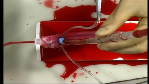
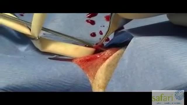
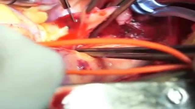
Cardiac Surgical Skills LaboratoryTraining Procedures:/n Aortic Cannulation and Decannulation/nCardiac surgery training
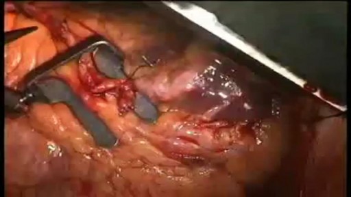
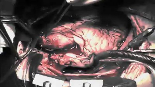
When your arteries cannot supply enough blood to your heart, your doctor may recommend coronary artery bypass graft (CABG) surgery. One of the most common heart surgeries in the United States, CABG surgery restores blood flow to your heart. Approximately every 10 minutes, someone has beating heart or "off-pump" bypass surgery1. Beating heart bypass surgery is — in simple terms — bypass surgery that is performed on your heart while it is beating. Your heart will not be stopped during surgery. You will not need a heart-lung machine. Your heart and lungs will continue to perform during your surgery. Surgeons use a tissue stabilization system to immobilize the area of the heart where they need to work. Beating heart bypass surgery is also called Off Pump Coronary Artery Bypass Surgery (OPCAB). Both OPCAB and conventional on-pump surgery restore blood flow to the heart. However, off-pump bypass surgery has proven to reduce side effects in certain types of patients.
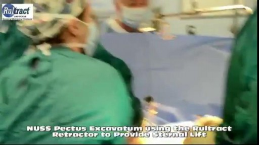
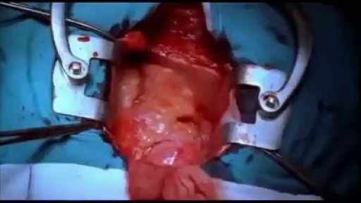
Pectus excavatum repair is surgery to correct pectus excavatum. This is a congenital (present at birth) deformity of the front of the chest wall that causes a sunken breastbone (sternum) and ribs. Pectus excavatum is also called funnel or sunken chest. It may worsen during the teen years.
Could this be a viable alternative to open heart surgery?
The first operation is harvesting the heart from the donor. The donor is usually an unfortunate person who has suffered irreversible brain injury, called "brain death". Very often these are patients who have had major trauma to the head, for example, in an automobile accident. The victim's organs, other than the brain, are working well with the help of medications and other "life support" that may include a respirator or other devices. A team of physicians, nurses, and technicians goes to the hospital of the donor to remove donated organs once brain death of the donor has been determined. The removed organs are transported on ice to keep them alive until they can be implanted. For the heart, this is optimally less than six hours. So, the organs are often flown by airplane or helicopter to the recipient's hospital.
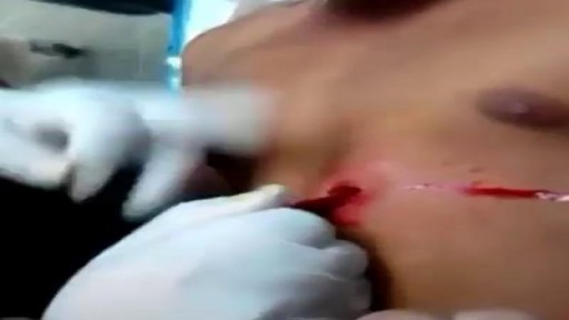
Gunshot wounds have become increasing common in urban cities and many such cases can lead to undesirable outcomes. While gunshot wounds to the head are considered most lethal, gunshot wounds to the chest too may be dangerous. Gunshot wound to the chest is challenging owing to the presence of vital organs like lungs, heart and their surrounding structures including major blood vessels. Gunshot wound is caused by penetration of the bullet, which travels through a projectile path after being shot from a firearm. The bullet, on hitting the chest, punctures the tissue it first encounters with, the bones or the muscular chest wall. The extent and severity of the injury depends on the characteristics of the bullet and the firearm, the position and the distance of the victim, the projectile path and the nature of the tissue penetrated.
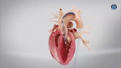
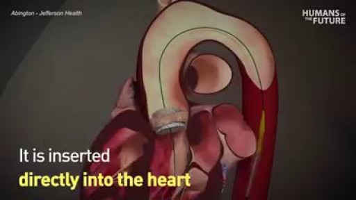
This minimally invasive surgical procedure repairs the valve without removing the old, damaged valve. Instead, it wedges a replacement valve into the aortic valve’s place. The surgery may be called a transcatheter aortic valve replacement (TAVR) or transcatheter aortic valve implantation (TAVI). Valve-within-valve — How does it work? Somewhat similar to a stent placed in an artery, the TAVR approach delivers a fully collapsible replacement valve to the valve site through a catheter. Once the new valve is expanded, it pushes the old valve leaflets out of the way and the tissue in the replacement valve takes over the job of regulating blood flow.
Beating Heart during surgery
Coronary artery bypass grafting (CABG) is a type of surgery that improves blood flow to the heart. Surgeons use CABG to treat people who have severe coronary heart disease (CHD). CHD is a disease in which a waxy substance called plaque (plak) builds up inside the coronary arteries.
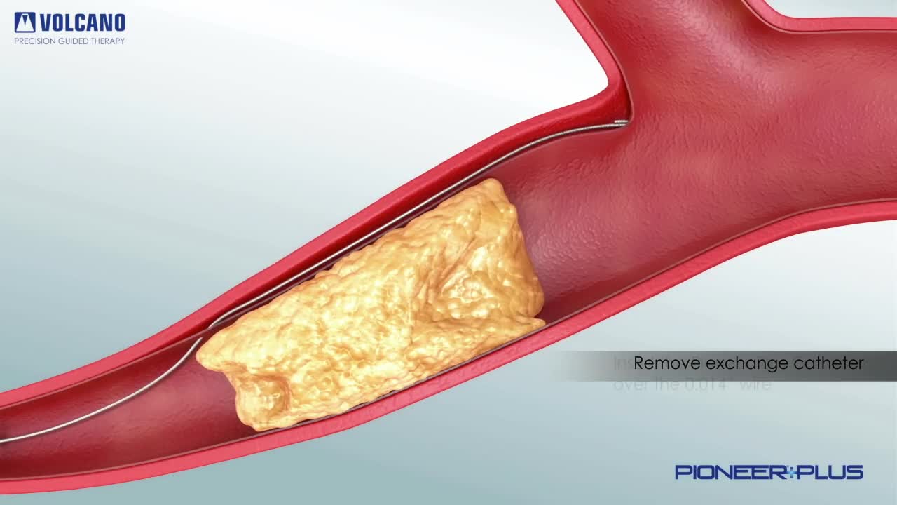
Pioneer Plus IVUS Re-Entry Catheter plaque removal
The first operation is harvesting the heart from the donor. The donor is usually an unfortunate person who has suffered irreversible brain injury, called "brain death". Very often these are patients who have had major trauma to the head, for example, in an automobile accident. The victim's organs, other than the brain, are working well with the help of medications and other "life support" that may include a respirator or other devices. A team of physicians, nurses, and technicians goes to the hospital of the donor to remove donated organs once brain death of the donor has been determined. The removed organs are transported on ice to keep them alive until they can be implanted. For the heart, this is optimally less than six hours. So, the organs are often flown by airplane or helicopter to the recipient's hospital.
remove a staggering number of heartworms. If you don't have your pet on heartworm prevention, go to your vet, get your pet tested and put on heartworm prevention right away!
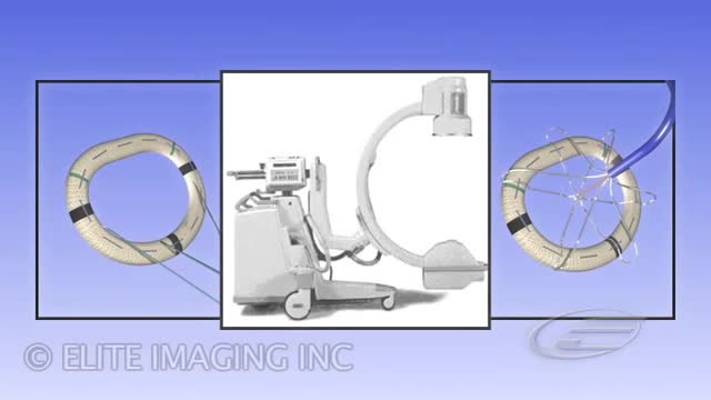
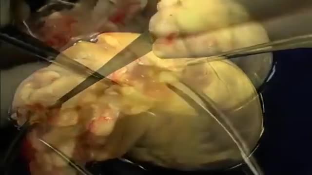
Ring System is used to correct mitral regurgitation via implantation of an annuloplasty ring
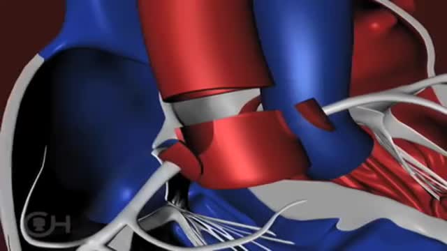
The "great arteries" in this anomaly refer to the aorta and the pulmonary artery, the two major arteries carrying blood away from the heart. In cases of transposition of the great arteries, these vessels arise from the wrong ventricle. They are "transposed" from their normal position so that the aorta arises from the right ventricle and the pulmonary artery from the left ventricle. Other heart defects may occur along with transposition of the great arteries. About 25 percent of children with transposition will also have a ventricular septal defect (VSD) . In nearly a third, the branching pattern of the coronary arteries as they leave the transposed aorta is unusual. Infants may also have narrowing below the pulmonary valve that blocks blood flow from the left ventricle to the lungs.
Surgical repair of a coexisting ruptured sinus of Valsalva into the RV along with closure of a VSD
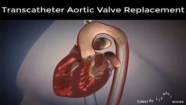
Transcatheter aortic valve replacement (TAVR) has recently emerged as a therapeutic option for patients with severe aortic stenosis
Dr. Chadrick Denlinger, MUSC transplant surgeon, describes what must take place when a patient receives a lung transplant.
If you need heart bypass surgery, the procedure is pretty similar. A surgeon takes blood vessels from another part of your body to go around, or bypass, a blocked artery. The result is that more blood and oxygen can flow to your heart again. ... Bypass surgery is also known as coronary artery bypass grafting (CABG).Dec 12, 2015
Surgery to replace an aortic valve is done for aortic valve stenosis and aortic valve regurgitation. During this surgery, the damaged valve is removed and replaced with an artificial valve. The valve replacement is typically an open-heart surgery.
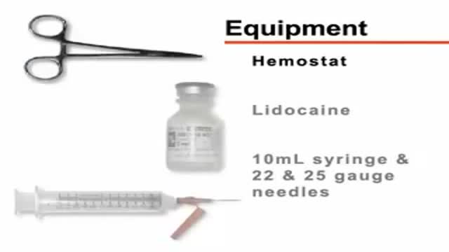
Thoracentesis is a procedure to remove fluid or air from around the lungs. A needle is put through the chest wall into the pleural space. The pleural space is the thin gap between the pleura. The pleura are a double layer of membranes that surrounds the lungs.
