
- Physical Examination
- Surgical Examination
- Ophthalmology
- Clinical Skills
- Orthopedics
- Surgery Videos
- Laparoscopy
- Pediatrics
- Funny Videos
- Cardiothoracic Surgery
- Nursing Videos
- Plastic Surgery
- Otorhinolaryngology
- Histology and Histopathology
- Neurosurgery
- Dermatology
- Pediatric Surgery
- Urology
- Dentistry
- Oncology and Cancers
- Anatomy Videos
- Health and Fitness
- Radiology
- Anaesthesia
- Physical Therapy
- Pharmacology
- Interventional Radiology
- Cardiology
- Endocrinology
- Gynecology
- Emergency Medicine
- Psychiatry and Psychology
- Childbirth Videos
- General Medical Videos
- Nephrology
- Physiology
- Diet and Food Health
- Diabetes Mellitus
- Neurology
- Women Health
- Osteoporosis
- Gastroenterology
- Pulmonology
- Hematology
- Rheumatology
- Toxicology
- Nuclear Medicine
- Infectious Diseases
- Vascular Disease
- Reproductive Health
- Burns and Wound Healing
- Kidney Zone Podcast
- Other
Cardiothoracic Surgery
Cardiothoracic surgery videos: open-heart surgery, bypass (CABG), valve replacement and lung procedures performed step by step. Free for students and surgeons.
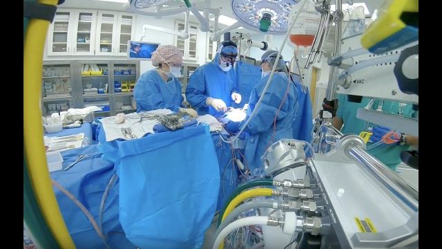

"I’m essentially taking care of the baby right now to give them 60 or 70 or 80 years of life so I have to perform my best every time. Every single time. That is a commitment that I have to the parents."
The highest standard. That’s what cardiothoracic surgeon Sergio Carrillo demands of himself every time he steps into the OR. Dr. Carrillo and his Heart Center team at Nationwide Children’s Hospital treat patients with congenital heart disease with the simplest to the most complex procedures.
Connect with a specialist: http://bit.ly/2LU2kJn
The Heart Center at Nationwide Children's: http://bit.ly/2LTQmPR
Advancing cardiac care through research: http://bit.ly/2LXFqAD
Tissue Engineering Research & Innovation: http://bit.ly/2LUD0Ts
Heart & Chest Surgery, What to Expect: http://bit.ly/2LVQr5J
Meet our Heart Center Team: http://bit.ly/2LUvdF9
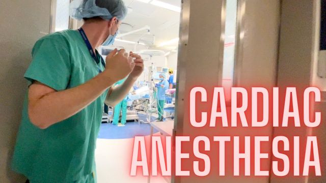
What goes into providing anesthesia for cardiac surgery where a patient's heart is completely arrested? In this video, I take you into the operating room during a surgery and talk with Dr. Benji Salter, program director for Mt. Sinai Hospital's cardiothoracic anesthesiology fellowship program.
While no patient information is shown in this video, the patient did provide written consent for filming to occur during surgery. Permission was also obtained from Mount Sinai Hospital's Department of Anesthesiology as well as the hospital's Press Office.
Chapters
0:00 Start
0:44 Surgery background
1:40 Case preparation
2:45 Anesthesia equipment
6:21 Echocardiography
7:16 Preparing for bypass
8:34 Stopping the heart
9:06 Fellowship
10:46 Why cardiac anesthesia?
11:52 Coming off of bypass
13:06 Post-op recovery
The information in this video is not intended nor implied to be a substitute for professional medical advice, diagnosis or treatment. All content, including text, graphics, images, and information, contained in this video is for general information purposes only and does not replace a consultation with your own doctor/health professional.
#Anesthesiology #Residency #MedicalSchool
Dr. Ailawadi, M.D., the Chair of Cardiac Surgery at Michigan Medicine, specializes in minimally invasive valve surgery as well as complex cardiac operations. This video shows step by step footage of a Coronary Artery Bypass Graft (CABG) in a complex patient. In this case, CABG was performed through a sternotomy (through the breast bone) using the internal thoracic artery and saphenous leg veins to bypass obstructed coronary arteries. In this video, Dr. Ailawadi will perform a triple vessel bypass (CABG) which has been shown to minimize the risk of future heart attack and help patients live longer in the setting of complex coronary artery disease.
To learn more about cardiac surgery at Michigan Medicine, visit: https://medicine.umich.edu/dept/cardiac-surgery
To learn more about Frankel Cardiovascular Center, visit: https://www.umcvc.org/
To watch the full playlist, visit: https://www.youtube.com/playli....st?list=PLNxqP-XbH8B
-------------------------------------------------------
Subscribe to Michigan Medicine’s YouTube channel for upcoming videos and future live streams featuring our experts answering your questions.
-------------------------------------------------------
Follow Michigan Medicine on Social:
Twitter: https://twitter.com/umichmedicine
Instagram: https://www.instagram.com/umichmedicine/
Facebook: https://www.facebook.com/MichiganMedicine/
Follow the U-M Frankel Cardiovascular Center on Social:
Twitter: https://twitter.com/umichcvc
Facebook: https://www.facebook.com/Unive....rsityofMichiganCardi
#MichiganMedicine #MedEd #CardiacSurgery #UniversityOfMichiganHealth #FrankelCardiovascularCenter #Cardiology #CardiacSurgeon
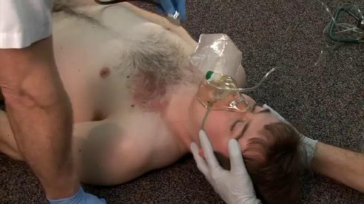
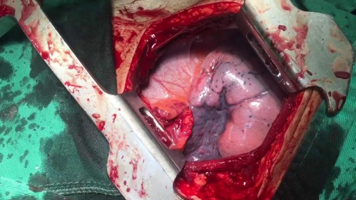

Know About Cardiothoracic Surgery in 60 Seconds About what cardiothoracic surgery is, why it is done and what is the result of such surgery. A Major Session on #cardiothoracic #surgery at #Congress #2018HCC 2018 Healthcare and Cardiology Conference #BANGKOK http://cosmicseries.org/cardiology-conferences/
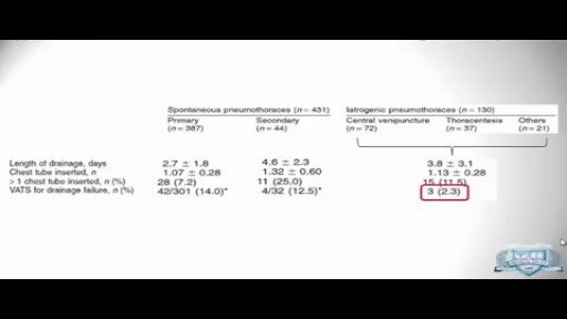
The surgeon may use treatment options such as thoracoscopy, electrocautery, laser treatment, resection of blebs or pleura, or open thoracotomy. Other surgical indications are as follows: Persistent air leak for longer than 7 days. Recurrent, ipsilateral pneumothorax.
Bullae resection for right pneumothrax via VATS
Empyema can develop after you have pneumonia. Many different types of bacteria may cause pneumonia, but the two most common are Streptococcus pneumoniae and Staphylococcus aureus. Occasionally, empyema may happen after you've had surgery on your chest. Medical instruments can transfer bacteria into your pleural cavity
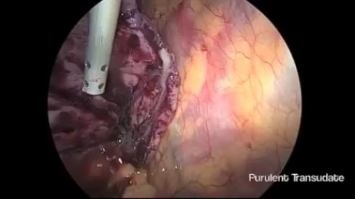
Thoracoscopic Management of Lung Abscess Before Empyema
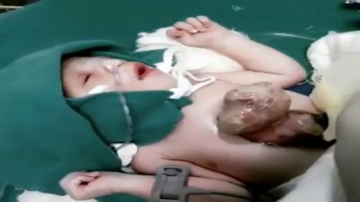
Ectopia cordis is a rare genetic defect. During a baby’s development in utero, their chest wall doesn’t form correctly. It also doesn’t fuse together as it normally would. This prevents the heart from developing where it should, leaving it defenseless and exposed outside of the protection of the chest wall. The defect affects about one in 126,000 births. In partial ectopia cordis, the heart is located outside the chest wall, but just under the skin. The heart can be seen beating through the skin.
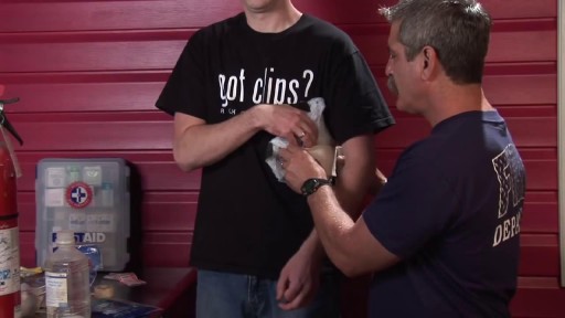
A fractured rib is usually a result of a fall or accident. Prolonged coughing and sports with repetitive movement, such as golf, also can cause a rib fracture. Symptoms include pain when taking a deep breath, pressing on the injured area, or bending or twisting the body. In most cases, fractured ribs usually heal on their own in one or two months. Pain relievers can make it easier to breathe deeply.
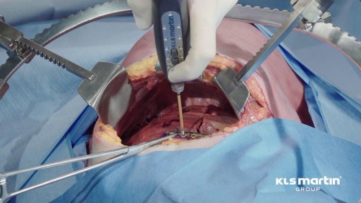
A fractured rib is usually a result of a fall or accident. Prolonged coughing and sports with repetitive movement, such as golf, also can cause a rib fracture. Symptoms include pain when taking a deep breath, pressing on the injured area, or bending or twisting the body. In most cases, fractured ribs usually heal on their own in one or two months. Pain relievers can make it easier to breathe deeply.
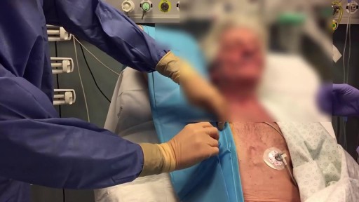
For blunt trauma patients lying supine, drains should be placed anteriorly in the chest. This pevents a tension pneumothorax developing if the chest tube is blocked by dependent lung tissue. Normal movement of the lungs will allow drainage of a basal haemothorax through an anterior chest tube
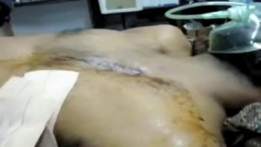
Paradoxical movement is an obvious sign that the portion of the chest wall is not assisting with the breathing function. Other symptoms of flail chest can include: Bruises, grazes, and/or discoloration in the chest area. Telltale markings from a seat belt.
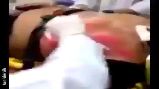
A flail chest occurs when a segment of the thoracic cage is separated from the rest of the chest wall. This is usually defined as at least two fractures per rib (producing a free segment), in at least two ribs. A segment of the chest wall that is flail is unable to contribute to lung expansion. Large flail segments will involve a much greater proportion of the chest wall and may extend bilaterally or involve the sternum. In these cases the disruption of normal pulmonary mechanics may be large enough to require mechanical ventilation.
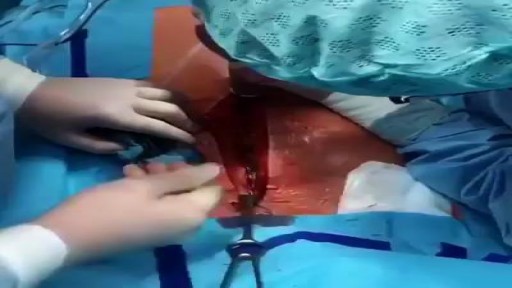
Median Sternotomy performed before open heart surgery !
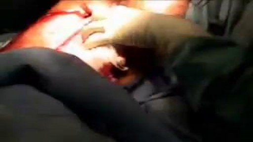
Surgeons Open Chest of Stabbed Patient to Pull Knife Out of His Heart

tretment
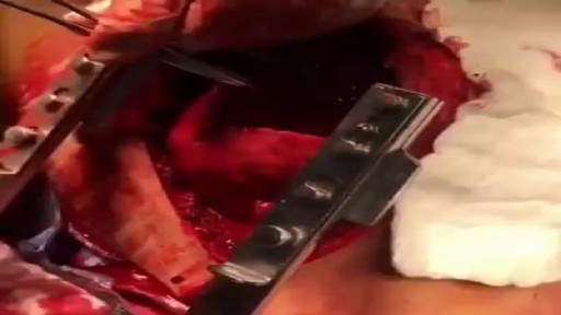
CARDIAC TAMPONADE!!! The surgeon in this video is performing a pericardiotomy in a 30-year-old man that presented to ER after a motor vehicle crash....
Medical Videos
Free medical and surgery videos for patients, students and professionals — trusted since 2007.
Founded by Dr. Mohamed Ibrahim, MD, PhD — physician-scientist.
Follow us on Facebook · Instagram