- Physical Examination
- Surgical Examination
- Ophthalmology
- Clinical Skills
- Orthopedics
- Surgery Videos
- Laparoscopy
- Pediatrics
- Funny Videos
- Cardiothoracic Surgery
- Nursing Videos
- Plastic Surgery
- Otorhinolaryngology
- Histology and Histopathology
- Neurosurgery
- Dermatology
- Pediatric Surgery
- Urology
- Dentistry
- Oncology and Cancers
- Anatomy Videos
- Health and Fitness
- Radiology
- Anaesthesia
- Physical Therapy
- Pharmacology
- Interventional Radiology
- Cardiology
- Endocrinology
- Gynecology
- Emergency Medicine
- Psychiatry and Psychology
- Childbirth Videos
- General Medical Videos
- Nephrology
- Physiology
- Diet and Food Health
- Diabetes Mellitus
- Neurology
- Women Health
- Osteoporosis
- Gastroenterology
- Pulmonology
- Hematology
- Rheumatology
- Toxicology
- Nuclear Medicine
- Infectious Diseases
- Vascular Disease
- Reproductive Health
- Burns and Wound Healing
- Other
Surgical Examination
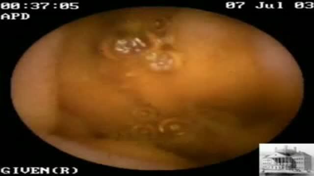
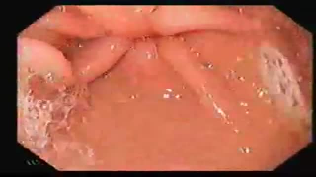
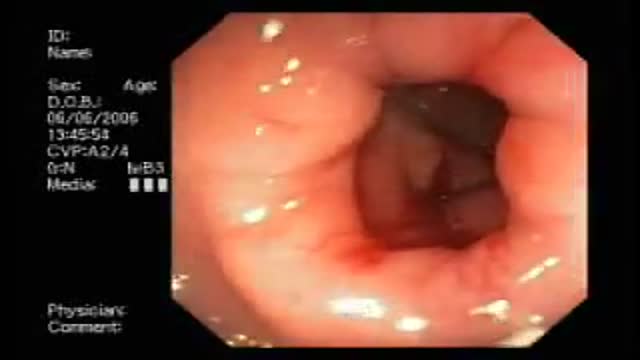
The colonoscope is slowly withdrawn during this screening colonoscopy down from the transverse colon, back around the splenic flexure, and down the descending colon, and reveals this finding a colonic diverticula. Diverticulosis is a common, acquired, age-related occurrence affecting over 50% of the... western adult population over the age of 50. It is seen rarely in Africa and Asia where the dietary fiber content is traditionally higher. Thus most investigators feel that low fiber diets are related to the development of this condition. Ironically, colonic diverticula are not true diverticula but rather pseudodiverticula in that the sac includes layers of the mucosa and submucosa that push through rather than include the outer muscular layer. As with the small bowel the colon has an inner circular muscular layer, but the outer longitudinal layer is composed of three bands of muscle that run the length of the colon known as teniae. Diverticula occur in rows between the mesenteric and two antimesenteric teniae where the colonic wall is further weakened by the defect caused by the perforating vasa recti artery which supplies the colonic mucosa. Occasionally, the anatomic propensity of diverticula to form in rows is quite apparent as seen when this clip is replayed in slow motion. Most often, however, the arrangement of the diverticula appears random due to the angulation of the bowel and thickening of the semi lunar folds. The conditions that cause these pulsion diverticula are not know with certainty but may include high intrahaustral pressures, muscular hypertrophy, and age related alterations in collagen cross linking. Diverticula can bleed or can abscess and perforate. The incidence of diverticulitis or diverticular bleeding is in the range of 1:1,000 patients with diverticulosis.
This 81 year old man with severe CAD and CHF was referred for VCE following a negative endoscopic workup for chronic guaiac positive stools. Seen on only three frames, this sequence reveals a single mid small bowel telangectasia, a possible source for his chronic GI blood loss. He has been managed c...onservatively and continues to require intermittent transfusions despite oral iron therapy.
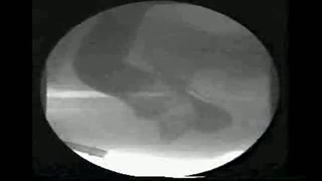
Defecography showing Rectocele
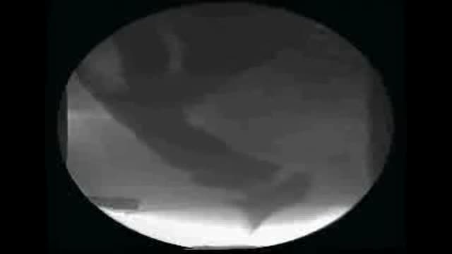
Defecography showing Internal Rectal Prolapse
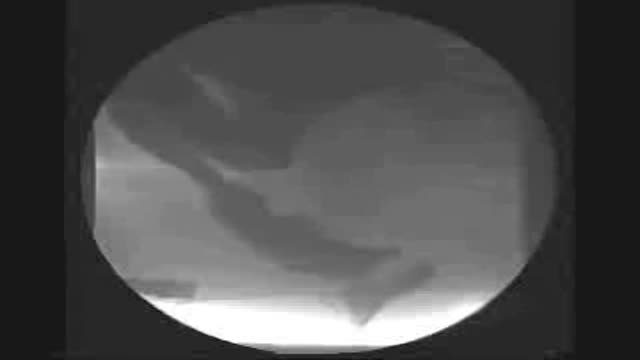
Defecography showing Anterior Rectal Wall Prolapse
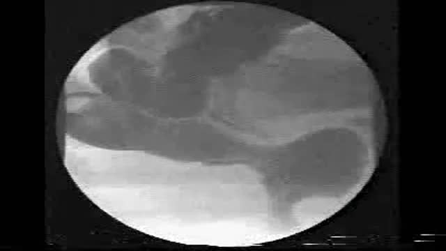
Defecography showing Enterocele

Defecography showing Normal Defecation
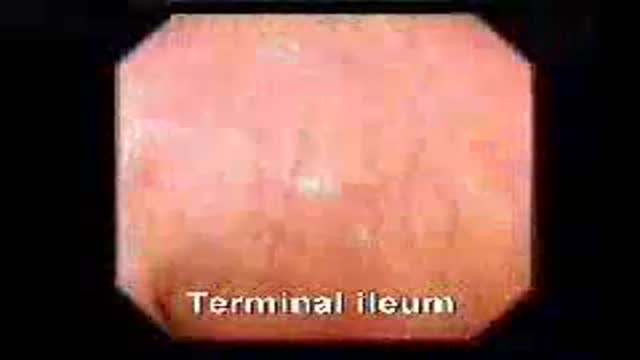
A video showing normal colonoscopy
This 40 yr male had upper abdominal pain for 3 months. A video-endoscopic examination of esophagus, stomach and duodenum was performed. A large 2.5x2.5 cm chronic ulcer was detected in the first part (bulb) of duodenum. A gastric biopsy was taken for diagnosis of Helicobacter infection and a rapid urease test done which was positive. He received triple therapy (2 antibiotics and acid suppressive drug for one week) to eradicate Helicobacter pylori infection. Ulcer disease showed rapid clinical and endoscopic healing. Eradication of Helicobacter pylori infection led to permanent ulcer cure.
Bleeding from Duodenal Ulcer
Educational video of male patient receiving an anoscopy.
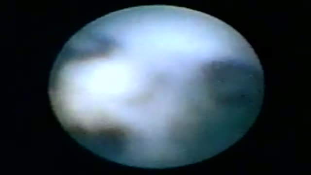
Endoscopy of Mammary Ducts with Micro-Endoscope called Mammary Ductoscopy. Indication:- Nipple Discharge. In this case Papilloma seen quite clearly. Biopsy can also be possible with Ductoscopy. Mammary Ductoscopy is very useful for diagnosis of Breast Cancer in early stage.
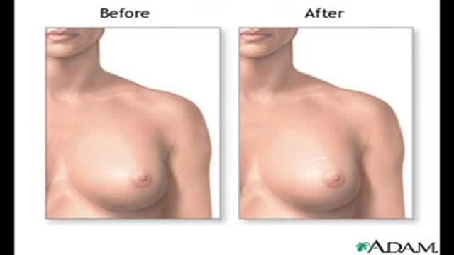
Symptoms of carcinoma of the breast
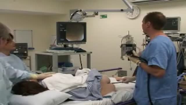
A video describing the procedure of colonoscopy or flexible fibre-optic examination of the colon.
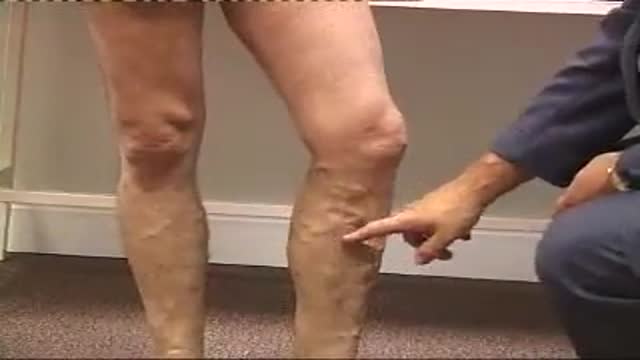
Examination of varicose veins
Examination of the shoulder
Examination of the Shoulder and Elbow
Examination of different gaits
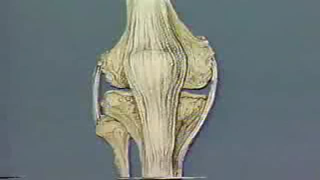
Full clinical and physical assessment of the knee and the knee joint
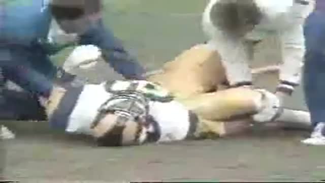
Physical Medical examination of the knee
