- Physical Examination
- Surgical Examination
- Ophthalmology
- Clinical Skills
- Orthopedics
- Surgery Videos
- Laparoscopy
- Pediatrics
- Funny Videos
- Cardiothoracic Surgery
- Nursing Videos
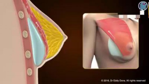
- Plastic Surgery
- Otorhinolaryngology
- Histology and Histopathology
- Neurosurgery
- Dermatology
- Pediatric Surgery
- Urology
- Dentistry
- Oncology and Cancers
- Anatomy Videos
- Health and Fitness
- Radiology
- Anaesthesia
- Physical Therapy
- Pharmacology
- Interventional Radiology
- Cardiology
- Endocrinology
- Gynecology
- Emergency Medicine
- Psychiatry and Psychology
- Childbirth Videos
- General Medical Videos
- Nephrology
- Physiology
- Diet and Food Health
- Diabetes Mellitus
- Neurology
- Women Health
- Osteoporosis
- Gastroenterology
- Pulmonology
- Hematology
- Rheumatology
- Toxicology
- Nuclear Medicine
- Infectious Diseases
- Vascular Disease
- Reproductive Health
- Burns and Wound Healing
- Other
Latest videos
Replacing PFM Crowns on Teeth 8 & 9 with BruxZir Solid Zirconia Crowns
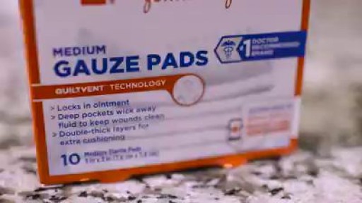
Minor burns can typically be treated at home -- but it's important to know when you need to seek treatment.
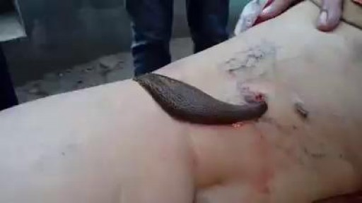
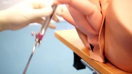
Leech therapy is the use of leeches in the treatment of disease conditions. Medicinal leeches are bloodsucking worms that they live in both aquatic and terrestrial environments.
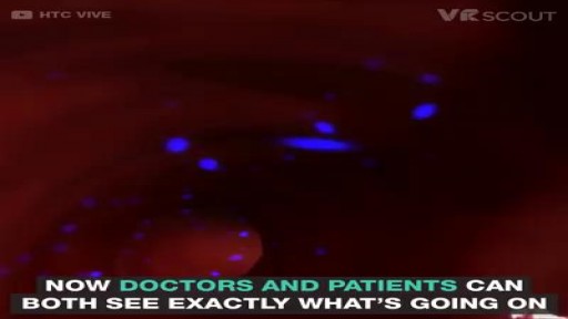
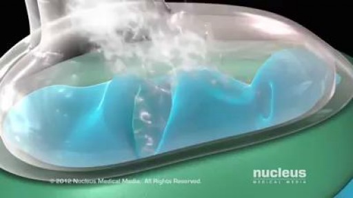
VR medical training takes you inside the human body.
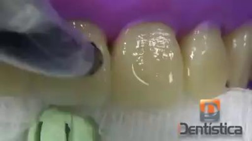
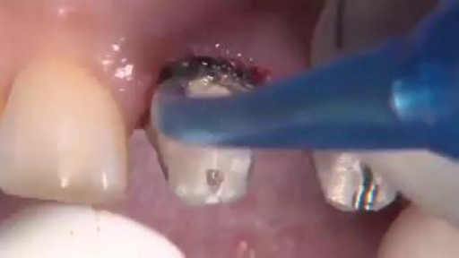
Tooth colored composite fillings are chemically bonded to teeth. For this reason, the placement of white fillings does not always require numbing the area being restored. Numbing (anesthetizing) the area is often required if tooth decay has progressed beneath the enamel layer and into the underlying dentin layer which surrounds the nerve of the tooth. Once decay is removed, the tooth is cleaned and a primer (weak acid) is applied to the area being restored. The primer opens pores in the enamel and dentin. A bonding agent is then flowed into the open pores and cured. Curing prepares the bonding agent to adhere to the tooth colored filling material. The filling material is then placed inside the tooth. After shaping the tooth colored filling material to resemble the natural anatomy of your tooth it is hardened by curing with a strong curing light. Once the white filling hardens, your bite will be checked to make sure your teeth fit together properly. If the tooth filling extends into the space between your teeth your dentist will also make sure you can floss between your teeth properly. Adjustments will be made if necessary followed by smoothing and polishing of your new filling..
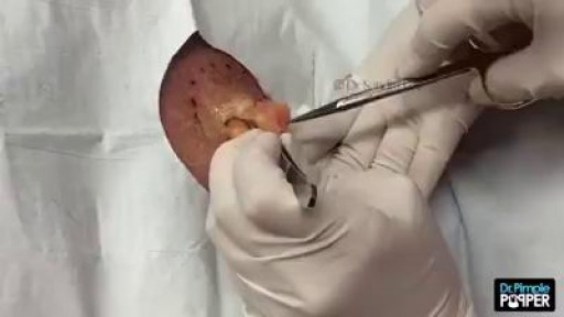
A lipoma is a growth of fat cells in a thin, fibrous capsule usually found just below the skin. Lipomas aren't cancer and don't turn into cancer. They are found most often on the torso, neck, upper thighs, upper arms, and armpits, but they can occur almost anywhere in the body. One or more lipomas may be present at the same time.
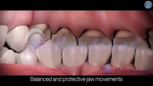
Your temporomandibular joint is a hinge that connects your jaw to the temporal bones of your skull, which are in front of each ear. It lets you move your jaw up and down and side to side, so you can talk, chew, and yawn. Problems with your jaw and the muscles in your face that control it are known as temporomandibular disorders (TMD). But you may hear it wrongly called TMJ, after the joint.
IMPLANT POCKETS - an educational animation explaining the different implant pockets
Skin Graft? Skin grafting is a surgical procedure that involves removing the skin from one area of the body and moving it, or transplanting it, to a different area of the body. This surgery may be done if a part of your body has lost its protective covering of skin due to burns, injury, or illness
Surgical procedure of Tonsillectomy and Adenoidectomy
Most babies will move into delivery position a few weeks prior to birth, with the head moving closer to the birth canal. When this fails to happen, the baby’s buttocks and/or feet will be positioned to be delivered first. This is referred to as “breech presentation.” Breech births occur in approximately 1 out of 25 full-term births.
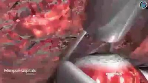
Beating Heart during surgery
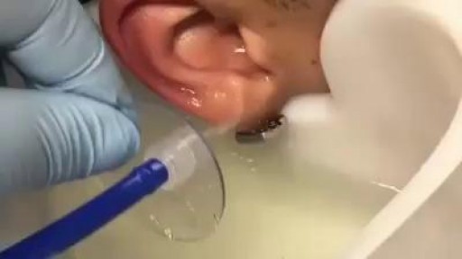
GIANT EAR WAX REMOVAL By using the elephant ear device.It's very useful video for medical students.Please share it!
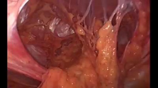
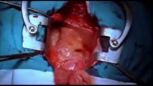
A giant abdominal wall hernia can develop from an existing ventral or incisional hernia, sometimes arising after one or more failed repair attempts. These hernias may also result from a traumatic injury where the abdomen was required to be left open and healing was delayed. In giant abdominal wall hernias, multiple loops of intestines and sometimes other abdominal organs reside within the hernia sac. The abdominal wall muscles then become conditioned to this and retract reducing the available space inside the abdomen.

Vediographic-Electric Beat-Pacemakers and the human heart
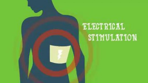
Nerve damage can start as numbness or tingling and progress to an intense feeling of burning or stabbing. What to know about treatment options:
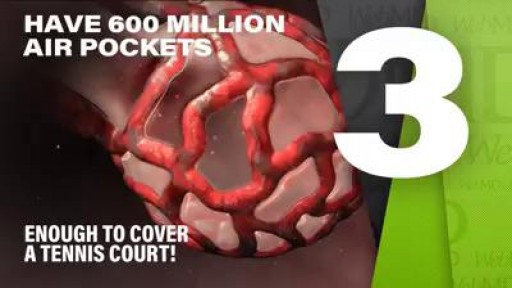
Your lungs are have 600 million air pockets -- enough to cover a tennis court.

Master perfect plank form and you .ll strengthen your core in no time.
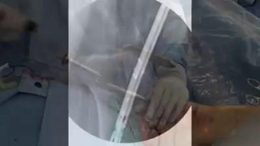
Intramedullary nailing of the tibia with suprapatellar entry and semi-extended positioning makes it technically easier to nail the proximal and distal fractures. The purpose of this article was to describe a simple method for suprapatellar nailing (SPN). A step-by-step run through of the surgical technique is described, including positioning of the patient. There are as yet only a few clinical studies that illustrate the complications with this method, and there has been no increased frequency of intraarticular damage. Within the body of the manuscript, information is included about intraarticular damage and comments with references about anterior knee pain.
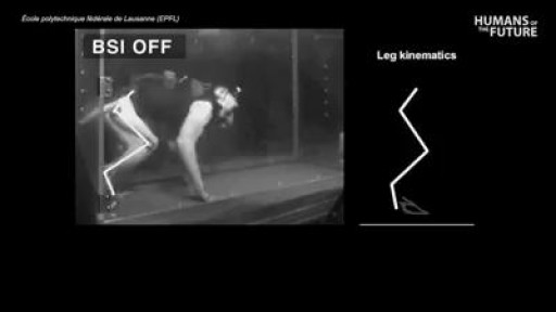
Watch as this wireless brain implant allows a paralyzed monkey to walk again
