- Physical Examination
- Surgical Examination
- Ophthalmology
- Clinical Skills
- Orthopedics
- Surgery Videos
- Laparoscopy
- Pediatrics
- Funny Videos
- Cardiothoracic Surgery
- Nursing Videos
- Plastic Surgery
- Otorhinolaryngology
- Histology and Histopathology
- Neurosurgery
- Dermatology
- Pediatric Surgery
- Urology
- Dentistry
- Oncology and Cancers
- Anatomy Videos
- Health and Fitness
- Radiology
- Anaesthesia
- Physical Therapy
- Pharmacology
- Interventional Radiology
- Cardiology
- Endocrinology
- Gynecology
- Emergency Medicine
- Psychiatry and Psychology
- Childbirth Videos
- General Medical Videos
- Nephrology
- Physiology
- Diet and Food Health
- Diabetes Mellitus
- Neurology
- Women Health
- Osteoporosis
- Gastroenterology
- Pulmonology
- Hematology
- Rheumatology
- Toxicology
- Nuclear Medicine
- Infectious Diseases
- Vascular Disease
- Reproductive Health
- Burns and Wound Healing
- Other
Latest videos
Zumba in Operation room
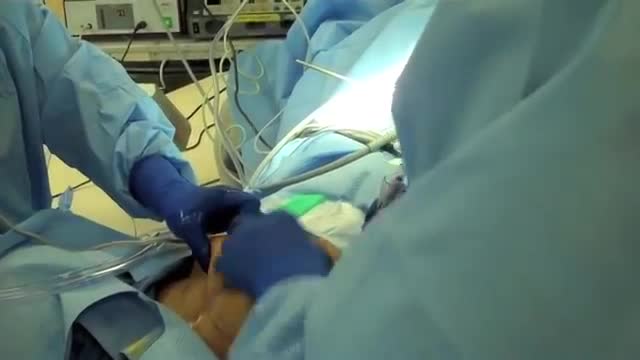
Laparoscopic use of Palmer's Point
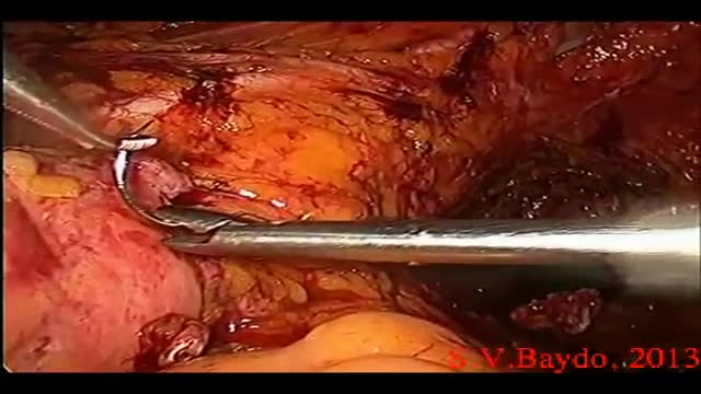
Iatrogenic injury to the ureter is a potentially devastating complication of modern surgery. The ureters are most often injured in gynecologic, colorectal, and vascular pelvic surgery. There is also potential for considerable ureteral injury during endoscopic procedures for ureteric pathology such as tumor or lithiasis. While maneuvers such as perioperative stenting have been touted as a means to avoid ureteral injury, these techniques have not been adopted universally, and the available literature does not make a case for their routine use. Distal ureteral injuries are best managed with ureteroneocystostomy with or without a vesico-psoas hitch. Mid-ureteral and proximal ureteral injuries can potentially be managed with ureteroureterostomy. If the distal segment is unsuitable for anastomosis then a number of techniques are available for repair including a Boari tubularized bladder flap, transureteroureterostomy, or renal autotransplantation. In rare cases renal autotransplantation or ureteral substitution with gastrointestinal segments may be warranted to re-establish urinary tract continuity. Laparoscopic and minimally invasive techniques have been employed to remedy iatrogenic ureteral injuries.
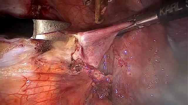
LAPAROSCOPIC END TO END URETERAL ANASTOMOSIS

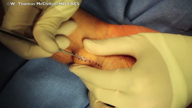
Rhinoplasty enhances facial harmony and the proportions of your nose. It can also correct impaired breathing caused by structural defects in the nose. Rhinoplasty surgery can change: Nose size in relation to facial balance Nose width at the bridge or in the size and position of the nostrils Nose profile with visible humps or depressions on the bridge Nasal tip that is enlarged or bulbous, drooping, upturned or hooked Nostrils that are large, wide, or upturned Nasal asymmetry If you desire a more symmetrical nose, keep in mind that everyone’s face is asymmetric to some degree. Results may not be completely symmetric, although the goal is to create facial balance and correct proportion.
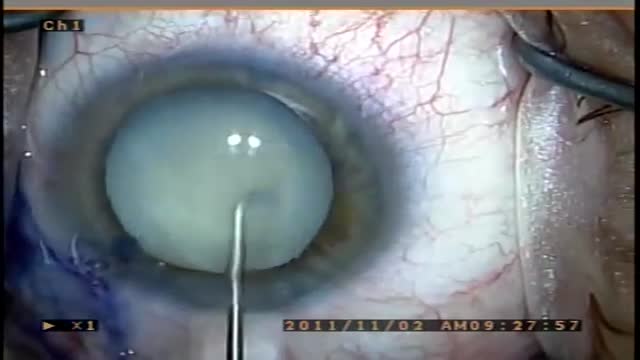
A cataract is a clouding of the lens in the eye that affects vision. Most cataracts are related to aging. Cataracts are very common in older people. By age 80, more than half of all Americans either have a cataract or have had cataract surgery. A cataract can occur in either or both eyes. It cannot spread from one eye to the other.
Ganglion Cyst Surgical Removal
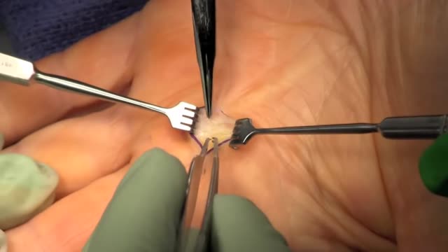
Trigger finger, also known as stenosing tenosynovitis (stuh-NO-sing ten-o-sin-o-VIE-tis), is a condition in which one of your fingers gets stuck in a bent position. Your finger may straighten with a snap — like a trigger being pulled and released. Trigger finger occurs when inflammation narrows the space within the sheath that surrounds the tendon in the affected finger. If trigger finger is severe, your finger may become locked in a bent position. People whose work or hobbies require repetitive gripping actions are at higher risk of developing trigger finger. The condition is also more common in women and in anyone with diabetes. Treatment of trigger finger varies depending on the severity.

These are a few common types of benign bone tumors: Osteochondroma is the most common benign bone tumor. ... Giant cell tumor is a benign tumor, typically affecting the leg (malignant types of this tumor are uncommon). Osteoid osteoma is a bone tumor, often occurring in long bones, that occurs commonly in the early 20s.

MRI of Bone Tumor

Enchondroma (Cartilage) benign tumor of the finger.
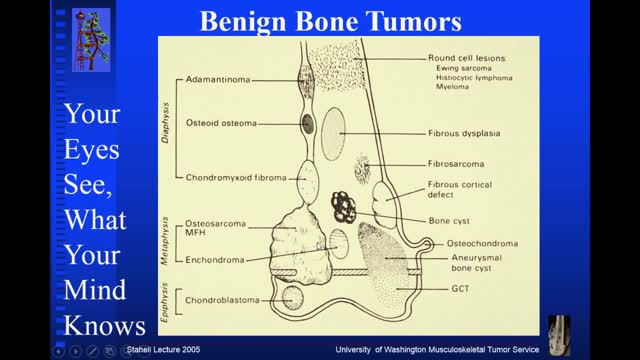
Osteochondroma. Osteochondromas (osteocartilaginous exostoses), the most common benign bone tumors, may arise from any bone but tend to occur near the ends of long bones. ... Enchondroma. ... Chondroblastoma. ... Chondromyxofibroma. ... Osteoid osteoma. ... Nonossifying fibroma (fibrous cortical defect) ... Benign giant cell tumor of bone.

Early Signs that Cancer is Growing in Your Body
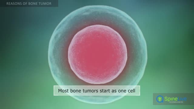
Bone tumors develop when cells in the bone divide without control, forming a mass of tissue. Most bone tumors are benign, which means they are not cancer and cannot spread. However, they may still weaken bone and lead to fractures or cause other problems. Bone cancer destroys normal bone tissue and may spread to other parts of the body (called metastasis). Benign Bone Tumors Benign tumors are more common than malignant tumors of the bones. These are a few common types of benign bone tumors: Osteochondroma is the most common benign bone tumor. It is more common in people under age 20. Giant cell tumor is a benign tumor, typically affecting the leg (malignant types of this tumor are uncommon). Osteoid osteoma is a bone tumor, often occurring in long bones, that occurs commonly in the early 20s. Osteoblastoma is a single tumor that occurs in the spine and long bones, mostly in young adults. Enchondroma usually appears in bones of the hand and feet. It often has no symptoms. It is the most common type of hand tumor.
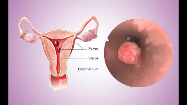
Uterine polyps are growths attached to the inner wall of the uterus that extend into the uterine cavity. Overgrowth of cells in the lining of the uterus (endometrium) leads to the formation of uterine polyps, also known as endometrial polyps. These polyps are usually noncancerous (benign), although some can be cancerous or can eventually turn into cancer (precancerous polyps). Uterine polyps range in size from a few millimeters — no larger than a sesame seed — to several centimeters — golf-ball-size or larger. They attach to the uterine wall by a large base or a thin stalk.
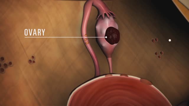
Endometriosis (en-doe-me-tree-O-sis) is an often painful disorder in which tissue that normally lines the inside of your uterus — the endometrium — grows outside your uterus. Endometriosis most commonly involves your ovaries, fallopian tubes and the tissue lining your pelvis. Rarely, endometrial tissue may spread beyond pelvic organs.

Colon polyp facts Colon polyps are growths on the inner lining of the colon and are very common. Colon polyps are important because they may be, or may become malignant (cancerous). They also are important because based on their size, number, and microscopic anatomy (histology); they can predict which patients are more likely to develop more polyps and colon cancer. Changes in the genetic material of cells lining the colon are the cause of polyps. There are different types of colon polyps with differing tendencies to become malignant and abilities to predict the development of more polyps and cancer. It is important to recognize families with members who have familial genetic conditions causing polyps because some of these conditions are associated with a very high incidence of colon cancer, and the cancer can be prevented or discovered early.
Digoxin is derived from the leaves of a digitalis plant. Digoxin helps make the heart beat stronger and with a more regular rhythm. Digoxin is also used to treat atrial fibrillation, a heart rhythm disorder of the atria (the upper chambers of the heart that allow blood to flow into the heart).
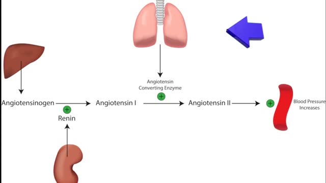
ACE inhibitors Email this page to a friend Print Facebook Twitter Google+ Angiotensin-converting enzyme (ACE) inhibitors are medicines. They treat heart, blood vessel, and kidney problems. How ACE inhibitors help ACE inhibitors are used to treat heart disease. These medicines make your heart work less hard by lowering your blood pressure. This keeps some kinds of heart disease from getting worse. Most people who have heart failure take these medicines. These medicines treat high blood pressure, strokes, or heart attacks. They may help lower your risk for stroke or heart attack. They are also used to treat diabetes and kidney problems. This can help keep your kidneys from getting worse. If you have these problems, ask your health care provider if you should be taking these medicines.

Renal artery stenosis is a narrowing of arteries that carry blood to one or both of the kidneys. Most often seen in older people with atherosclerosis (hardening of the arteries), renal artery stenosis can worsen over time and often leads to hypertension (high blood pressure) and kidney damage.
