
- Physical Examination
- Surgical Examination
- Ophthalmology
- Clinical Skills
- Orthopedics
- Surgery Videos
- Laparoscopy
- Pediatrics
- Funny Videos
- Cardiothoracic Surgery
- Nursing Videos
- Plastic Surgery
- Otorhinolaryngology
- Histology and Histopathology
- Neurosurgery
- Dermatology
- Pediatric Surgery
- Urology
- Dentistry
- Oncology and Cancers
- Anatomy Videos
- Health and Fitness
- Radiology
- Anaesthesia
- Physical Therapy
- Pharmacology
- Interventional Radiology
- Cardiology
- Endocrinology
- Gynecology
- Emergency Medicine
- Psychiatry and Psychology
- Childbirth Videos
- General Medical Videos
- Nephrology
- Physiology
- Diet and Food Health
- Diabetes Mellitus
- Neurology
- Women Health
- Osteoporosis
- Gastroenterology
- Pulmonology
- Hematology
- Rheumatology
- Toxicology
- Nuclear Medicine
- Infectious Diseases
- Vascular Disease
- Reproductive Health
- Burns and Wound Healing
- Kidney Zone Podcast
- Other
Latest videos
The newest additions to the MedicalVideos.com library — freshly curated medical and surgery videos, explainers, and lectures from educators and institutions worldwide.
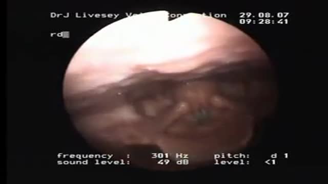

A check up at the Ear Nose & Throat doctor to make sure Genie's Opera singing vocal chords are working properly.
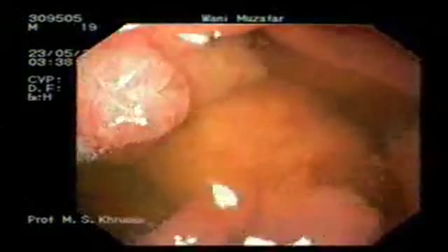
ENDOSCOPIC (NON-SURGICAL) REMOVAL OF MULTIPLE LARGE TUMORS FROM STOMACH IN A PATIENT WITH PEUTZ-JEGHERS SYNDROME
PEUTZ-JEGHERS SYNDROME: Peutz-Jeghers syndrome (PJS) is a familial syndrome consisting of mucocutaneous pigmentation, gastrointestinal polyposis and cancers of gut & other sites like breast, ovary, and testes. PJS has an autosomal dominant inheritance with variable and incomplete penetrance. Germline mutations of STK11/LKB1 gene on 19p cause this syndrome. Mucocutaneous pigmentation may be noted in early infancy. These deposits of melanin are most commonly found around the mouth, nose, lips, buccal mucosa, hands, and feet, and may also be present in perianal and genital areas. PJS polyps may be found in stomach, small intestine, or colon, but they tend to be prominent in the small intestine. These polyps may increase in size and cause small intestinal obstruction or intussusceptions that may occur in early infancy. Acute upper gastrointestinal bleeding and chronic faecal blood may complicate the disease.
PATIENT: The patient was a 25 yr male who had mucocutaneous pigmentation and multiple polyps in the stomach and duodenum. He presented with bleeding from gastric polyps. As the polyps in stomach were numerous, (more than 20 in number) and were large in size (some equal to small egg size), he had been advised to undergo surgery. Surgery planned was total gastrectomy.
PROCEDURE: The patient underwent video-endoscopy of the esophagus, stomach and duodenum. All polyps were examined for size and presence or absence of stalk. A plan to remove all the gastric polyps at endoscopy was made in the same sitting. He received light conscious sedation. Flat polyps were raised form the gastric wall by injection of saline in to polyp base to let these lesions have a stalk. This was done by needle injector. Each polyp was engaged in a snare and the polyp stalk was cut by coagulation cutting current. The cuts were clean without any bleeding. All polyps were recovered for histology. The histology revealed all polyps to be hamartomous lesions. None of the polyps were cancerous. Patient has been followed up for over one year and is doing fine without any further bleeding or pain.
Video shows the procedure of videoendoscpy and endoscopic removal of polyps.
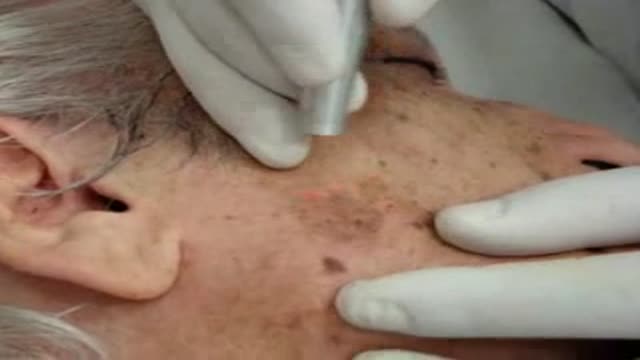
different aging and other skin spots can be treated succesfully with Co2 laser.
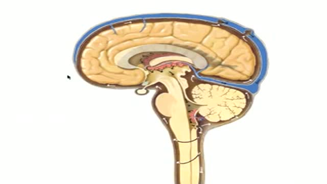
Neuroanatomy of CSF Flow

The complex circuitry interconnecting different areas in the brain, known collectively as white matter, is composed of millions of axons organized into fascicles and bundles. Upon macroscopic examination of sections of the brain, it is difficult to discern the orientation of the fibers. The same is true for conventional imaging modalities. However, recent advancements in magnetic resonance imaging (MRI) make such task possible in a live subject. By sensitizing an otherwise typical MRI sequence to the diffusion of water molecules it is possible to measure their diffusion coefficient in a given direction1. Normally, the axonal membrane and myelin sheaths pose barriers to the movement of water molecules and, thus, they diffuse preferentially along the axon2. Therefore, the direction of white matter bundles can be elucidated by determining the principal diffusivity of water. The three-dimensional representation of the diffusion coefficient can be given by a tensor and its mathematical decomposition provides the direction of the tracts3; this MRI technique is known as diffusion tensor imaging (DTI). By connecting the information acquired with DTI, three-dimensional depictions of white matter fascicles are obtained4. The virtual dissection of white matter bundles is rapidly becoming a valuable tool in clinical research.
Our journey begins with a transverse section of tightly packed axons as seen through light microscopy. Although represented as a two-dimensional "slice", we see that these axons in fact resemble tubes. A simulation of water molecules diffusing randomly inside the axons demonstrates how the membranes and myelin hinder their movement across them and shows the preferred diffusion direction --along the axons. The tracts depicted through DTI slowly blend in and we ride along with them. As we zoom out even more, we realize that it is a portion of the corpus callosum connecting the two sides of the brain we were traveling on and the great difference in relative scale of the individual axons becomes evident. The surface of the brain is then shown, as well as the rest of the white matter bundles--a big, apparently chaotic tangle of wires. Finally, the skin covers the brain.
With the exception of the simulated water molecules, all the data presented in the animation is obtained through microscopy and MRI. Computer algorithms for the extraction of the cerebral structures and a custom-built graphics engine make our journey through the brain's anatomy possible in a living person.
Micrograph courtesy of Dr. Christian Beaulieu, University of Alberta.
Music by Mario Mattioli.
References:
1. Stejskal, E.O., et al., J. Chem. Phys., 1965. 42:
2. Beaulieu, C., NMR Biomed., 2002. 15:435-55.
3. Basser, P.J., et al., J. Magn. Reson. B, 1994. 103:247-54.
4. Mori, S., et al., NMR Biomed., 2002. 15:468-80.
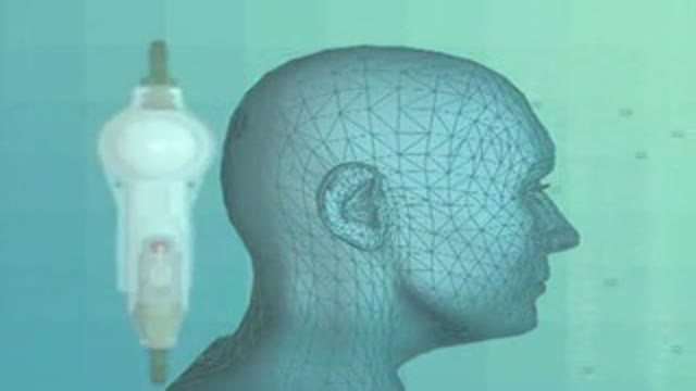
Insertion of a CSF shunt
Endoscopic fenestration of suprasellar cyst in a 4 years old girl
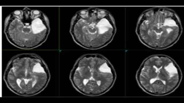
Endoscopic fenestration of arachnoid cyst in middle fossa
brain scans with arachnoid cyst, pre and post operative
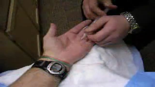
Ganglion Cyst Removal
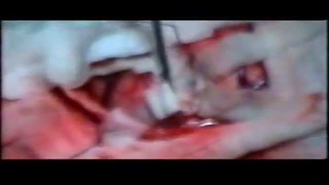
Hydatid Cyst Removal from the brain
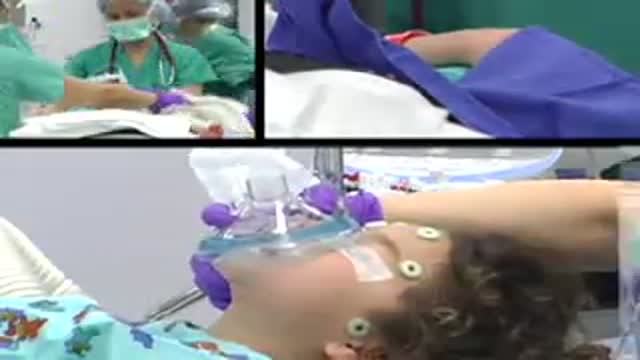
Watch as Dr. Benjamin Carson performs risky brain surgery on young Payton to remove a brain tumor. Dr. Carson, director of pediatric neurosurgery, is just one of the many reasons why Johns Hopkins Children's Center was recently ranked #1 in neurology and neurosurgery in America's Best Children's Hospitals 2008
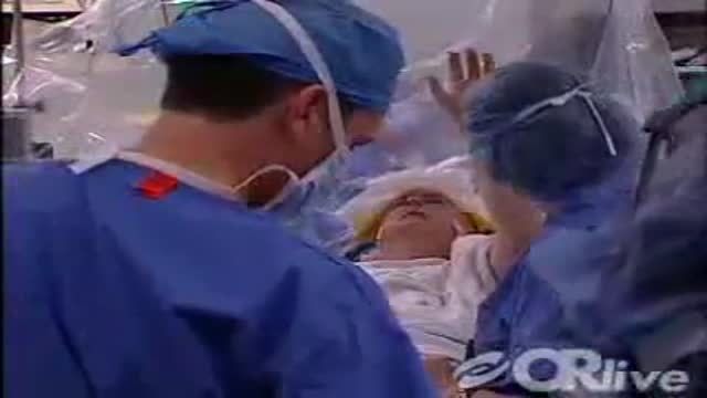
Vanderbilt Medical Center neurosurgeons and neurologists will be online demonstrating their 4-stage innovative technique used for Deep Brain Stimulation (DBS). Deep brain stimulation therapy utilizes an implantable neuro-stimulator to treat movement disorders such as Parkinson's disease, essential tremor, and dystonia.

M. Patrick Lowe, MD, renowned robotic surgeon and gynecologic oncologist at Northwestern Memorial Hospital, will demonstrate the use of robotic surgery to treat endometrial cancer.
Dr. Lowe, director of the robotics and minimally invasive surgical program for the Division of Gynecologic Oncology at Northwestern University's Feinberg School of Medicine, was among the early adopters of robotics to treat gynecologic malignancies, citing precision, improved dexterity and superior patient outcomes among the benefits.
"Women diagnosed with a gynecologic malignancy want the shortest route leading back to a degree of normalcy post treatment," says Lowe. "Robotic surgery offers the path of least resistance, combining shorter recovery times with superior outcomes."

Highlights of a fetal laser surgery for twin-to-twin transfusion syndrome (TTTS) will be shown from Tampa General Hospital
TTTS affects 10 to 15 percent of identical-twin pregnancies and is the result of abnormal blood exchange between identical twins through a common placenta. The larger of the twins, or recipient, is surrounded by too much amniotic fluid and in danger of heart failure as its body tries to pump the overwhelming volume of blood intended for both. The smaller, or donor twin, is encased in a shrinking amniotic sac deprived of blood. Without treatment, both will likely die.
Rubén Quintero, M.D., Medical Director of Maternal Fetal Medicine/Fetal Surgery at Tampa General Hospital and Professor and Director of the Division of Maternal Fetal Medicine, Department of Obstetrics and Gynecology, University of South Florida College of Medicine will narrate the procedure and answer e-mail questions live as taped highlights of the procedure are shown.

On Tuesday May 29th at 3:00pm EDT, University Hospitals Case Medical Center Cleveland, Ohio, will host a live webcast to demonstrate the removal of brain tumor and epileptic focus from an awake patient using intra-operative MRI and brain mapping. See this on OR-Live.com
The patient was a middle-aged gentleman with new onset seizures. An MRI showed what appeared to be a low grade glioma near the motor strip on the right. Studies have shown that complete removal can cure the seizures, improve quality of life and survival, but this is difficult to do with conventional technology without harming the surrounding normal brain because its difficult to determine where tumor ends and normal brain begins.

Cricothyroidotomy NEJM
Arthrocentesis of the Knee

The device has as great advantage that the accessibility increases in the hand, it maximizes the surgical area free of obstacles, it increases its functional versatility, for the material with the one that this made a maximum of durability is guaranteed, so that it can be sterilized in any team and it facilitates that the thumb is supported in relaxation state.
The considerable reduction of the surgical time of each intervention is inside the advantages that it provides this valuable instrument, also facilitating that they are executed with more security. For the stability that provide, it can also be used in bony fabrics of the hand. The instrument is both handle, of very easy use and great comfort in its handling. The standardization of most of its pieces makes it very simple. The solutions that are offered in this device for the subjection of the fingers and other parts of the hand are a novelty, but they also have the advantage that it commits very little surgical area and it guarantees a maximum of subjection staying the totally stable hand facilitating in great measure the surgeon's work. These pieces adapt to any diameter of fingers.
Endoscopic Carpal Tunnel Release Surgery
Medical Videos
Free medical and surgery videos for patients, students and professionals — trusted since 2007.
Founded by Dr. Mohamed Ibrahim, MD, PhD — physician-scientist.
Follow us on Facebook · Instagram