- Physical Examination
- Surgical Examination
- Ophthalmology
- Clinical Skills
- Orthopedics
- Surgery Videos
- Laparoscopy
- Pediatrics
- Funny Videos
- Cardiothoracic Surgery
- Nursing Videos
- Plastic Surgery
- Otorhinolaryngology
- Histology and Histopathology
- Neurosurgery
- Dermatology
- Pediatric Surgery
- Urology
- Dentistry
- Oncology and Cancers
- Anatomy Videos
- Health and Fitness
- Radiology
- Anaesthesia
- Physical Therapy
- Pharmacology
- Interventional Radiology
- Cardiology
- Endocrinology
- Gynecology
- Emergency Medicine
- Psychiatry and Psychology
- Childbirth Videos
- General Medical Videos
- Nephrology
- Physiology
- Diet and Food Health
- Diabetes Mellitus
- Neurology
- Women Health
- Osteoporosis
- Gastroenterology
- Pulmonology
- Hematology
- Rheumatology
- Toxicology
- Nuclear Medicine
- Infectious Diseases
- Vascular Disease
- Reproductive Health
- Burns and Wound Healing
- Other
Top videos

This animation shows you how a tummy tuck is performed at Boerhaave Medical Centre. Curious? Watch the video!
Boerhaave Medical Centre sets itself the goal of providing the highest quality care. Quality not just in terms of treatment, but also in terms of our staff and the before and after care given. By providing thorough information and clear advice in advance, carefully supporting our patients through the procedure and caring for them afterwards, we believe this quality is assured.
Although we are one of the largest clinics in the Netherlands and have built up many years of experience, we continuously strive to improve. After all, the Boerhaave Medical Centre intends to remain a pioneer in the healthcare sector, by working in accordance with the latest medical findings and techniques both now and in the future.
We offer the highest standard of plastic surgery in our cosmetic care department. For 365 days a year, you can also come to us for non-surgical treatments, such as injectables, permanent hair removal and gastric balloons.
We have been awarded the ZKN quality mark and are certified to ISO 9001-2008 for giving advice and carrying out plastic surgery, including after care.
Visit our website for more information: https://www.boerhaave.com/all-....treatments/upperbody
Follow us:
Facebook: https://www.facebook.com/boerhaavemc
Google+: https://plus.google.com/+BoerhaaveNl-Kliniek
Pinterest: https://nl.pinterest.com/BoerhaaveMC/
Instagram: https://www.instagram.com/BoerhaaveMC/

Access my FREE Online Membership today → https://www.thenotedanatomist.com
___
Unlock my Premium Tutoring Memberships → https://www.thenotedanatomist.com/premium-memberships
Lifetime Access to Online Anatomy Course
Foundational Q&A Cards Per Video
Notes and Key Takeaways
Downloadable Documents
Flashcards for Each Course
Weekly Group Tutoring Sessions
Direct Tutoring Sessions
___
Discover A Simplified Approach to Master the Complexity of Anatomy with me, Dr. David Morton ... The Noted Anatomist!
This video tutorial discusses an Introduction to Histology (study of tissues):
0:00. Intro
0:35. Hierarchical organization of living matter
1:56. H&E stains
3:00. Epithelium overview (characteristics and classifying scheme)
- 9:12. Simple squamous epithelium
- 11:05. Simple cuboidal epithelium
- 12:20. Simple columnar epithelium
- 13:36. Stratified squamous epithelium
- 15:51. Urinary epithelium (transitional epithelium)
- 16:45. Pseudo-stratified ciliated columnar epithelium (respiratory epithelium)
18:55. Connective tissue overview (characteristics and classifying scheme)
- 21.14. Connective tissue proper (loose CT, dense irregular CT, dense regular CT, adipose tissue)
- 24:50. Cartilage (hyaline cartilage, elastic cartilage, fibrocartilage)
- 26:04. Bone (osteoblasts, osteocytes, osteoclasts, calcium ...)
- 27:34. Blood (RBC, WBC, platelet, plasma)
28:54. Muscle tissue (skeletal muscle, cardiac muscle, smooth muscle)
32:54. Nervous tissue (neurons and glial cells)
36:58. In-a-Nutshell
37:07. Acknowledgements
For a more detailed study of histology go to The Histology Wizard: https://www.youtube.com/channe....l/UCAeLLruy9RkUWaW_r

A complete organized library of all my videos, digital slides, pics, & sample pathology reports is available here: https://kikoxp.com/posts/5084 (dermpath) & https://kikoxp.com/posts/5083 (bone/soft tissue sarcoma pathology)
Topics discussed:
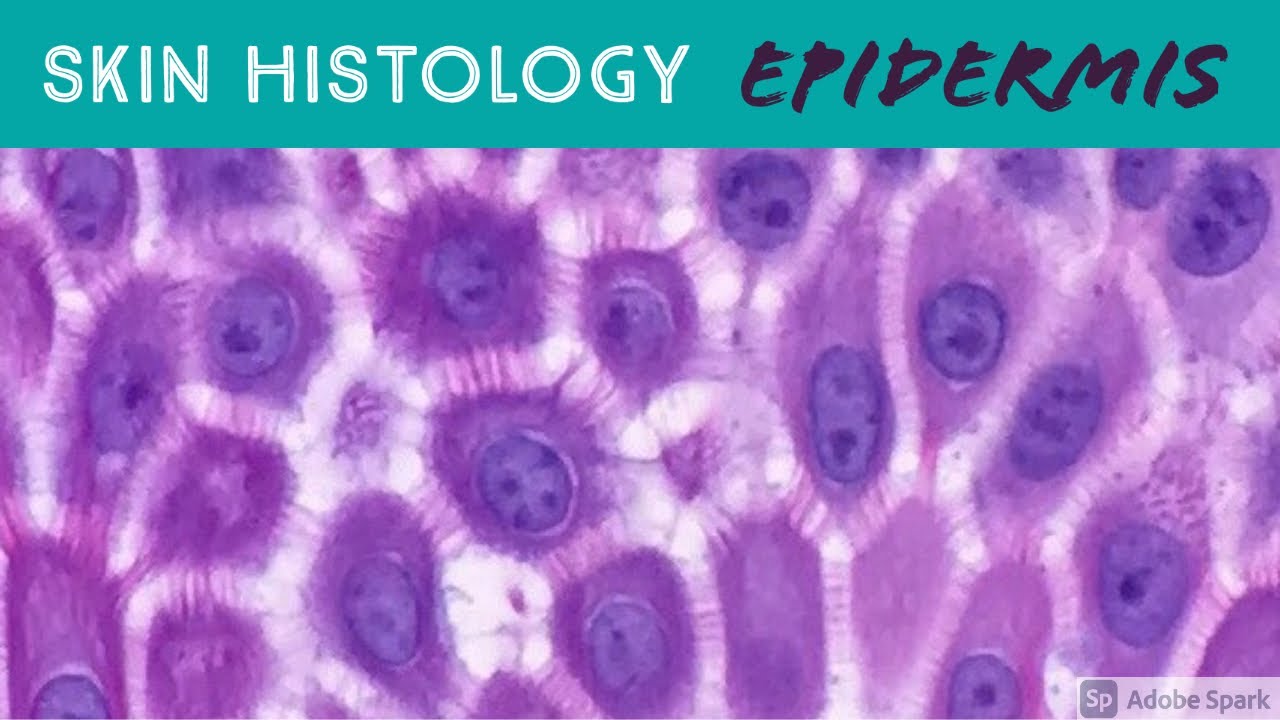
Epidermis:
Layers of epidermis: 0:10
Melanocytes vs Keratinocytes: 5:16
Langerhans cells: 10:10 & 33:30 & 57:30
Dermis:
Papillary and reticular dermis: 11:50
Three types of white empty spaces on a slide: vessels, glands/ducts/cysts, or artifact: 15:25
Blood vessels & nerves: 18:24 & 48:50 & 58:59
Arrector pili & other dermal smooth muscle: 20:00
Adnexal:
Sebaceous gland: 21:10
Hair follicle 23:14
Eccrine sweat glands and ducts 24:45 & 50:00
Gland/duct vs blood vessel 27:20 & 48:50
Apocrine glands: this video https://kikoxp.com/posts/7837 (at 12:30)
Acrosyringium: this video https://kikoxp.com/posts/7837 (at 10:00)
Three types of pink bundles: smooth muscle, nerve, dense connective tissue: 27:50
Acral skin (palm sole) with contact dermatitis 29:37
Parakeratosis 30:00
Perivascular lymphocytes 30:40
Eosinophils vs neutrophils 31:20
Spongiosis with desmosome keratinocyte spines 32:10
Spongiotic vesicles with Langerhans cells 33:30
Normal acral skin (palm & sole) with stratum lucidum 34:20
Normal glomus body/apparatus (canal of Sucquet-Hoyer) 35:40
Nerve 36:46 & 51:50
Adipose tissue (white fat cells) in subcutis with Lochkern 37:55
Normal scalp skin with large anagen hair follicles: 39:30
Hair follicle anatomy (bulb/matrix, inner root sheath, outer root sheath, hair shaft, isthmus, infundibulum): 40:55 (labeled images):
https://kikoxp.com/posts/3661 & https://kikoxp.com/posts/7899
Pacinian corpuscle 50:40
Meissner corpuscle 1:02:28
Dense regular connective tissue (Fascia/Tendon/Ligament) vs Smooth Muscle 53:00
Basic Normal Skin Immunohistochemistry:
-cytokeratin in epidermis: 55:33
-S100 in melanocytes and Langerhans cells and adipocytes: 57:30
-Desmin in smooth muscle (arrector pili and blood vessels): 58:59
-CD31 in endothelial cells of blood vessels: 59:33
-SOX-10 in melanocytes: 1:00:40
Digit/Finger/Toe histology (amputation for subungual acral melanoma) 1:04:10 & 1:08:30
-bone 1:05:40
-glomus body 1:05:15
-tendon/ligament 1:06:10
-artery 1:06:58
-fingernail/toenail 1:08:54
-acrosyringium 1:10:45
Solar elastosis (what wrinkles look like microscopically!) 1:11:50
Other videos you might like:
Tendon vs Nerve Histology Made Simple with the Ramen Noodle Sign (of Fulton) video: https://kikoxp.com/posts/4466
Melanocytes vs Keratinocytes made easy video: https://kikoxp.com/posts/3802
Blood Vessel vs Gland vs Artifact Made Easy video: https://kikoxp.com/posts/4808
The basic normal structures of the skin discussed and described by a dermatopathologist. This material is intended for use by medical students, junior pathology or dermatology residents, or for anyone else studying normal human histology. Special thanks to two of my medical students at UAMS for helping make this video possible. Miki Lindsey convinced me that I really needed to sit down and record this video. Akash Patel took time to edit the video and make it ready for YouTube. My sincere thanks to both of them for helping me overcome procrastination.
Huge thanks to Abigail Cline, a medical student at Medical College of Georgia, for volunteering to type a transcript of this ENTIRE video (over 14,000 words!) so that I could provide closed caption subtitles for those with hearing impairments and for those who may need assistance in understanding spoken English (particularly given how quickly I speak!). You can access a text version of her transcript of my video here: https://kikoxp.com/posts/5390
Correction - I made a mistake in the video. I said that sebaceous gland secretions are turned into smelly substances by bacteria and that this makes body odor. That is incorrect. That is actually true of APOCRINE gland secretions not sebaceous secretions.
Also, in the past I used "keratinocyte" and "squamous cell" interchangeably (this is because in dermatopathology, we see and talk about squamous cell carcinomas all the time, and those tumors are composed of keratinocytes). But technically, in normal skin histology, "squamous cell" refers only to the flattened keratinocytes in the superficial epidermis. Thankfully, a histology PhD colleague pointed this out to me and corrected my lazy nomenclature!
Please check out my Soft Tissue Pathology & Dermatopathology survival guide textbooks: http://bit.ly/2Te2haB
This video is geared towards medical students, pathology or dermatology residents, or practicing pathologists or dermatologists. Of course, this video is for educational purposes only and is not formal medical advice or consultation.
Presented by Jerad M. Gardner, MD. Please subscribe to my channel to be notified of new pathology teaching videos.
Follow me on:
Snapchat: JMGardnerMD
Twitter: @JMGardnerMD
Instagram: @JMGardnerMD
Facebook: https://www.facebook.com/JMGardnerMD/

#anatomy #histology #biology #bytesizemed
✨If you would like my help studying the structure of bones, check out my long-form video on it.
🔅Structure of Bone : https://youtu.be/MYInVEnnS_I
💫 For more videos like this, subscribe to my channel!
Byte Size Med: https://youtube.com/channel/UC....ZghvlgylH3r_CWfA18eF
📚Factual References & for Further Reading:
- DiFiore's Atlas of Histology
- Junqueira's Basic Histology
- Gartner's Concise Histology
- Openstax Anatomy and Physiology
https://openstax.org/details/b....ooks/anatomy-and-phy
- Openstax Biology
https://openstax.org/details/books/biology-2e
(The last two are links to open-source references. They are NOT affiliate links)
🌤 Note:
These are just a collection of my notes. So use them the way you would use borrowed notes from a friend. 📝
The images in this video are hand-drawn for illustration and explanation only.✍️ Hence, they may not be anatomically accurate. I am just one person making these videos. If there are any errors, that is unintentional. I try super hard to avoid them. Please let me know if you find any, so it gets clarified for other viewers. Science constantly evolves and changes. New discoveries are made everyday. So some of the information in these videos may become outdated. If you notice that, please let me know so I can update them.
⚡️Disclaimer:
These videos are NOT a substitute for a medical textbook. Textbooks are written by experts (which I do not claim to be), edited, proofread and referenced. Please use them.
The information has been sourced from multiple references as mentioned above. I draw all the pictures myself. But if I have inadvertently infringed on any copyright, that is completely unintentional. I only make these videos to impart education. If I have accidentally violated copyright in any way, do let me know so I can make the necessary changes or give credit to anyone who is owed the same.
These videos are NOT intended for patient education. They are NOT a substitute for diagnosis and treatment by a licensed medical professional. Always seek the advice of a qualified health care provider for any questions you may have regarding any medical condition, so that they can address your individual needs.
🔅They are ONLY meant to help students of medicine and health sciences with studying, and should be used for just that purpose and absolutely nothing else.
Byte Size Med. All Rights Reserved.
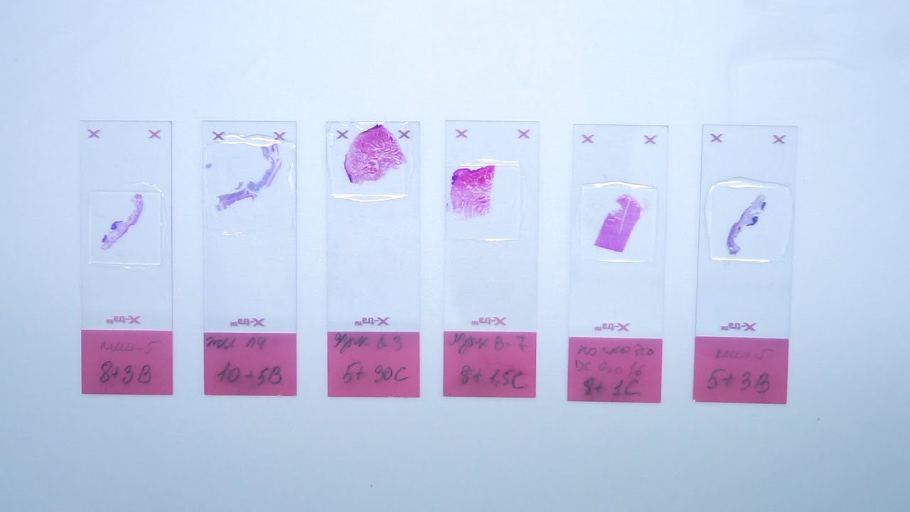
The most popular and one of the principal stains in histology is hematoxylin and eosin stain. It gives us an overview of the tissue and its structure. Hematoxylin binds with basophilic structures – for example DNA and RNA. So we can observe nuclei stained in blue or purple color. Eosin binds to acidophilic substances such as positively charged amino acid side chains. So as the result cytoplasm is pink or orange. All samples in laboratory are stained with H&E. There are several different types of hematoxylins and eosins used in histology which will give us different results.
In this video you will see, how we stain slides with different types of hematoxylins and eosins. Finally, we will compare the results.
• Subscribe to our channel: https://www.youtube.com/c/BioVitrumEN
• Watch other videos about histological process: https://www.youtube.com/playli....st?list=PLw4LQHit0MU
• Our website: http://en.biovitrum.ru/


The dentin is a hard tissue that forms the bulk of the tooth. It is similar to bone but is slightly harder, although softer than enamel. The dentin has numerous dentinal tubules that run across its length. Each dentinal tubule houses the cytoplasmic process of an odontoblast (odontoblastic process).
📄Notes for the video: https://www.hackdentistry.com/....bundles/revision-nin
💻Website: https://www.hackdentistry.com/
📰Blog: https://hackdentistry.substack.com/
Study resources on our website-
📖Oral pathology Revision Ninja (Notes, Videos & MCQs): https://www.hackdentistry.com/bundles/oral-pathology-revision-ninja
📖Oral Histology Revision Ninja (Notes, Videos & MCQs): https://www.hackdentistry.com/....bundles/revision-nin
📖Periodontics Revision Ninja (Notes & MCQs): https://www.hackdentistry.com/bundles/perio-rn
📖Question Bank: https://www.hackdentistry.com/bundles/question-bank
📖Access all content: https://www.hackdentistry.com/bundles/all-access-premium
References and further reading:
💡Berkovitz BKB, Hollan GR, Moxham BJ. Oral Anatomy, Histology and Embryology. 4th ed. Mosby Elsevier; 2009.
💡Nanci A. Tencate’s Oral Histology. Development, Structure and Function. 8th ed. Elsevier; 2013.
💡Kumar GS. Orban’s Oral Histology and Embryology.13th ed. Elsevier; 2011.
💡Avery JK. Oral development and Histology. 3rd ed. Thieme Medical Publishers; 2002.
Log in to https://www.hackdentistry.com and get access to:
I) Numerous Notes/Cheatsheets and Videos II) Thousands of quiz questions from our vast Question Bank!
HackDentistry is an edtech company that aims make learning dentistry fun,engaging and light hearted.
1) It focuses on helping students understand and retain core concepts in dentistry through highly visual sketch/whiteboard style video animations.
2) The platform helps improve exam performance by providing Revision Bundles and allowing students to test themselves using thousands of Practice Questions from a vast Question Bank. (multiple choice format).
3) It also provides for a community platform where students can come together, and engage with fellow dental students and dentists across the globe!
Facebook:
https://www.facebook.com/hackdentistry
Instagram:
https://www.instagram.com/hack.dentistry/
Twitter:
https://twitter.com/hckdentistry

Join the Amoeba Sisters a they explore different muscle tissues and then focus on the sliding filament theory in skeletal muscle! This video also briefly talks about muscle naming, some vocabulary (such as agonists and antagonists) before focusing on the sliding filament model. Video also mentions general roles of tropomyosin and troponin.
---------------------------------------------------------
Table of Contents:
00:00 Intro
0:39 Muscle Tissue Types
1:58 Muscle Characteristics
2:33 Skeletal Muscle Naming and Arrangement
3:26 Actin Myosin and Sarcomere
4:32 Sliding Filament Model
6:55 Tropomyosin an Troponin
---------------------------------------------------------
Factual References:
Betts, J. Gordon, et al. “10.3 Muscle Fiber Contraction and Relaxation - Anatomy and Physiology 2e | OpenStax.” Openstax.org, 20 Apr. 2022, openstax.org/books/anatomy-and-physiology-2e/pages/10-3-muscle-fiber-contraction-and-relaxation.
Urry, Lisa A, et al. Campbell Biology. 11th ed., New York, Ny, Pearson Education, Inc, 2017.
---------------------------------------------------------
Further Reading Recommendations:
What about I and A bands? What actually initiates the power stroke? How does calcium get released and from where? Remember, there is a lot more detail! We recommend this page from Openstax to learn more:
https://openstax.org/books/bio....logy-2e/pages/38-4-m
-----------------------------------------------
The Amoeba Sisters videos demystify science with humor and relevance. The videos center on Pinky's certification and experience in teaching biology at the high school level. Amoeba Sisters videos only cover concepts that Pinky is certified to teach, and they focus on her specialty: secondary life science. Learn more about our videos here: https://www.amoebasisters.com/our-videos
Support Us? https://www.amoebasisters.com/support-us
Our Resources and Handouts: https://www.amoebasisters.com/handouts
Biology Video Playlist: https://www.youtube.com/playli....st?list=PLwL0Myd7Dk1
GIFs: https://www.amoebasisters.com/gifs.html
Comics: https://www.amoebasisters.com/....parameciumparlorcomi
Unlectured Series: https://www.amoebasisters.com/unlectured
Connect with us!
Website: https://www.AmoebaSisters.com
Twitter: https://www.twitter.com/AmoebaSisters
Facebook: https://www.facebook.com/AmoebaSisters
Tumblr: https://www.amoebasisters.tumblr.com
Pinterest: https://www.pinterest.com/AmoebaSisters
Webtoon: https://www.webtoons.com/en/challenge/amoeba-sisters-sisterhood/list?title_no=289479&page=1
Instagram: https://www.instagram.com/amoebasistersofficial/
TikTok: https://www.tiktok.com/@amoebasistersofficial
Visit our Redbubble store at https://www.amoebasisters.com/store
TIPS FOR VIEWING EDU YOUTUBE VIDEOS:
Want to learn tips for viewing edu YouTube videos including changing the speed, language, viewing the transcript, etc? https://www.amoebasisters.com/....pinkys-ed-tech-favor
MUSIC:
Our intro music designed and performed by Jeremiah Cheshire.
End music in this video is listed free to use/no attribution required from the YouTube audio library.
COMMUNITY:
We take pride in our AWESOME community, and we welcome feedback and discussion. However, please remember that this is an education channel. See YouTube's community guidelines and how YouTube handles comments that are reported by the community. We also reserve the right to remove comments.
TRANSLATIONS:
Spanish Subtitles Translated by Jeremy García
Hindi Subtitles: Translated by Alisha Aggarwal
We gladly accept subtitle translations from our community. Learn more here: https://www.amoebasisters.com/....pinkys-ed-tech-favor We want to thank our amazing community for the generosity of their time in continuing to create translated subtitles.
We also have videos dubbed in Spanish and Portuguese using an artificial voice via https://aloud.area120.google.com to increase accessibility. See our Amoeba Sisters en Español channel https://www.youtube.com/channe....l/UC1Njo3LBy53cOPngz and Amoeba Sisters em Português https://www.youtube.com/channe....l/UCYTQPX2X_mXe0ZMPi

Excerpt from my Normal Skin Histology video: https://kikoxp.com/posts/3660.
A complete organized library of all my videos, digital slides, pics, & sample pathology reports is available here: https://kikoxp.com/posts/5084 (dermpath) & https://kikoxp.com/posts/5083 (bone/soft tissue sarcoma pathology).
Please check out my Soft Tissue Pathology & Dermatopathology survival guide textbooks: http://bit.ly/2Te2haB
Also, in the past I used "keratinocyte" and "squamous cell" interchangeably (this is because in dermatopathology, we see and talk about squamous cell carcinomas all the time, and those tumors are composed of keratinocytes). But technically, in normal skin histology, "squamous cell" refers only to the flattened keratinocytes in the superficial epidermis. Thankfully, a histology PhD colleague pointed this out to me and corrected my lazy nomenclature!
This video is geared towards medical students, pathology or dermatology residents, or practicing pathologists or dermatologists. Of course, this video is for educational purposes only and is not formal medical advice or consultation.
Presented by Jerad M. Gardner, MD. Please subscribe to my channel to be notified of new pathology teaching videos.
Follow me on:
Snapchat: JMGardnerMD
Twitter: @JMGardnerMD
Instagram: @JMGardnerMD
Kiko: https://kikoxp.com/profile/jer....ad_gardner1/content?
Facebook: https://www.facebook.com/JMGardnerMD/

© 2023 Elsevier. All rights reserved. Histologically the cervix is different from the rest of the uterus and also has a mucosa that doesn’t shed during menstruation.
Find our full video library only on Osmosis Prime: http://osms.it/more.
Join over 3 million current & future clinicians who learn by Osmosis, and over 130 universities around the world who partner with us to make medical and health education more engaging and efficient. We have unparalleled tools and materials to prepare you to succeed in school, on board exams, and as a future clinician. Sign up for a free trial at http://osms.it/more. If you're interested in exploring an institutional partnership, visit osmosis.org/educators to request a personalized demo.
Follow us on social:
Facebook: http://osms.it/facebook
Twitter: http://osms.it/twitter
Instagram for med: http://osms.it/instagram
Instagram for nursing: https://osms.it/ignursing
Linkedin: https://osms.it/linkedin
Our Vision: Everyone who cares for someone will learn by Osmosis.
Our Mission: To empower the world’s clinicians and caregivers with the best learning experience possible. Learn more here: http://osms.it/mission
Medical disclaimer: Knowledge Diffusion Inc (DBA Osmosis) does not provide medical advice. Osmosis and the content available on Osmosis's properties (Osmosis.org, YouTube, and other channels) do not provide a diagnosis or other recommendation for treatment and are not a substitute for the professional judgment of a healthcare professional in diagnosis and treatment of any person or animal. The determination of the need for medical services and the types of healthcare to be provided to a patient are decisions that should be made only by a physician or other licensed health care provider. Always seek the advice of a physician or other qualified healthcare provider with any questions you have regarding a medical condition. © 2023 Elsevier. All rights reserved.

An animated description of the composition of bones.
Visit www.orthofilms.com for more videos and info.
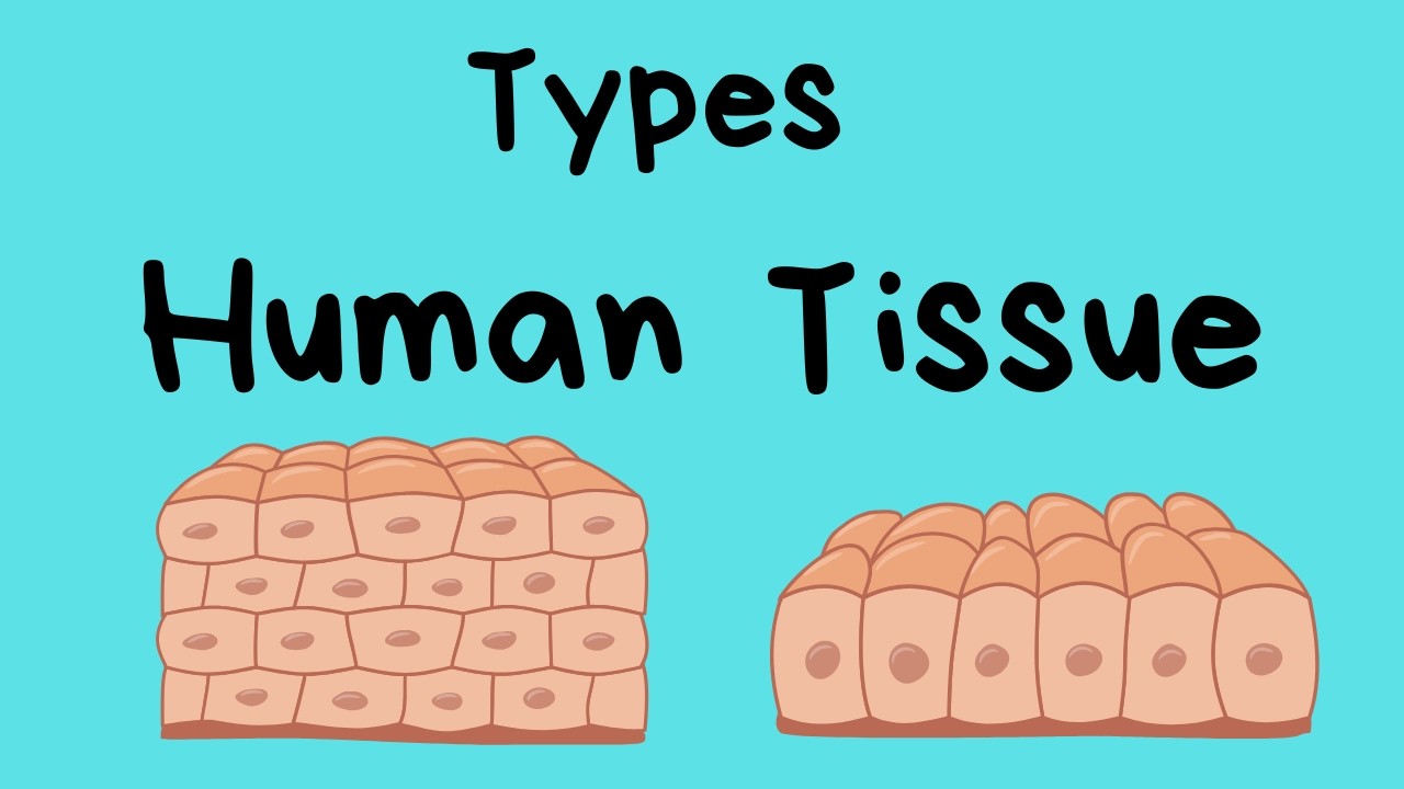
Types of Human Body Tissue
In this video, I review four types of tissue.
Connective tissue, epithelial tissue, muscle tissue, and nerve tissue.
Tissues are made up of cells working together.
*
*
For more Life Science videos and summaries see,
http://www.moomoomath.com/Midd....le-School-Science-an
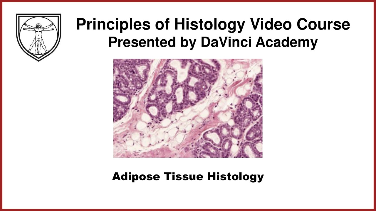
Covers the histological structure for adipose tissue and relevant cellular physiology for adipocytes. This video is a part of our Histology Video Course (https://youtube.com/playlist?l....ist=PLnr1l7WuQdDynxT
All Histology Videos: https://youtube.com/playlist?l....ist=PLnr1l7WuQdDynxT
Thank you to our sponsor Doc2Doc Lending, the Personal Lending platform designed for Doctors, by Doctors. Check out https://doc2doclending.com/davinci to learn more today.
DaVinci Academy Merch - Coffee mugs, T-shirts, hoodies and more: https://my-store-d90f46.creator-spring.com
Additional YouTube Content
Biochemistry videos: https://youtube.com/playlist?l....ist=PLnr1l7WuQdDzCUC
Anatomy Videos: https://youtube.com/playlist?l....ist=PLnr1l7WuQdDz2dK
DaVinci Cases Videos: https://youtube.com/playlist?l....ist=PLnr1l7WuQdDyJUl
The DaVinci Hour Podcast: https://youtube.com/playlist?l....ist=PLnr1l7WuQdDwSm9
DaVinci Academy Website: https://www.dviacademy.com/

#anatomy #histology #bytesizemed
✨If you would like my help studying about cartilage, you can check out my long-form video linked at the bottom of the screen.
💫 For more videos like this, subscribe to my channel, Byte Size Med.
📚Factual References & for Further Reading:
- DiFiore's Atlas of Histology
- Junqueira's Basic Histology
- Gartner's Concise Histology
- Openstax Anatomy and Physiology
https://openstax.org/details/b....ooks/anatomy-and-phy
- Openstax Biology
https://openstax.org/details/books/biology-2e
(The last two are links to open-source references. They are NOT affiliate links)
🌤 Note:
These are just a collection of my notes. So use them the way you would use borrowed notes from a friend. 📝
The images in this video are hand-drawn for illustration and explanation only.✍️ Hence, they may not be anatomically accurate. I am just one person making these videos. If there are any errors, that is unintentional. I try super hard to avoid them. Please let me know if you find any, so it gets clarified for other viewers. Science constantly evolves and changes. New discoveries are made everyday. So some of the information in these videos may become outdated. If you notice that, please let me know so I can update them.
⚡️Disclaimer:
These videos are NOT a substitute for a medical textbook. Textbooks are written by experts (which I do not claim to be), edited, proofread and referenced. Please use them.
The information has been sourced from multiple references as mentioned above. I draw all the pictures myself. But if I have inadvertently infringed on any copyright, that is completely unintentional. I only make these videos to impart education. If I have accidentally violated copyright in any way, do let me know so I can make the necessary changes or give credit to anyone who is owed the same.
These videos are NOT intended for patient education. They are NOT a substitute for diagnosis and treatment by a licensed medical professional. Always seek the advice of a qualified health care provider for any questions you may have regarding any medical condition, so that they can address your individual needs.
🔅They are ONLY meant to help students of medicine and health sciences with studying, and should be used for just that purpose and absolutely nothing else.
Byte Size Med. All Rights Reserved.

Classification of epithelium, discussion on lining epithelium of 3 major system (GIT, Urogenital and Respiratory system

Learn about the structural unit of compact bone (the osteon) and it's four basic parts: central canal, lamellae, lacunae, and canaliculi

✨This video is on the structure and functions of the three types of cartilage (Hyaline, Elastic and Fibrocartilage). I hope it helps! ☀️
🌟What's in this video?
0:00 - Intro
0:07 - Connective Tissue Recap
1:16 - Structure of Connective Tissue
1:57 - Structural Components of Cartilage
3:38 - Types of Cartilage
3:49 - Hyaline Cartilage
8:05 - Elastic Cartilage
8:55 - Fibrocartilage
✨ Other videos you may need:
🔅 Connective Tissue : https://youtu.be/xw_ALdt5n-A
🔅 Collagen : https://youtu.be/3e2JYMNS_W4
🔅 Ossification: https://youtu.be/86V9SNWD_No
🔅Histology: https://www.youtube.com/playli....st?list=PL1rG930trF2
💫 For more videos like this, subscribe to my channel!
Byte Size Med: https://youtube.com/channel/UC....ZghvlgylH3r_CWfA18eF
📚Factual References & for Further Reading:
- DiFiore's Atlas of Histology
- Junqueira's Basic Histology
- Gartner's Concise Histology
- Openstax Anatomy and Physiology
https://openstax.org/details/b....ooks/anatomy-and-phy
- Openstax Biology
https://openstax.org/details/books/biology-2e
(The last two are links to open-source references. They are NOT affiliate links)
🌤 Note:
These are just a collection of my notes. So use them the way you would use borrowed notes from a friend. 📝
The images in this video are hand-drawn for illustration and explanation only.✍️ Hence, they may not be anatomically accurate. I am just one person making these videos. If there are any errors, that is unintentional. I try super hard to avoid them. Please let me know if you find any, so it gets clarified for other viewers. Science constantly evolves and changes. New discoveries are made everyday. So some of the information in these videos may become outdated. If you notice that, please let me know so I can update them.
⚡️Disclaimer:
These videos are NOT a substitute for a medical textbook. Textbooks are written by experts (which I do not claim to be), edited, proofread and referenced. Please use them.
The information has been sourced from multiple references as mentioned above. I draw all the pictures myself. But if I have inadvertently infringed on any copyright, that is completely unintentional. I only make these videos to impart education. If I have accidentally violated copyright in any way, do let me know so I can make the necessary changes or give credit to anyone who is owed the same.
These videos are NOT intended for patient education. They are NOT a substitute for diagnosis and treatment by a licensed medical professional. Always seek the advice of a qualified health care provider for any questions you may have regarding any medical condition, so that they can address your individual needs.
🔅They are ONLY meant to help students of medicine and health sciences with studying, and should be used for just that purpose and absolutely nothing else.
Byte Size Med. All Rights Reserved.
![Histology of Exocrine Gland [Epithelium 7 of 7]](https://i.ytimg.com/vi/NkU7YJ7eYd0/maxresdefault.jpg)
Histological features and cellular biology of exocrine glands. This video is a part of our Histology Video Course (https://youtube.com/playlist?l....ist=PLnr1l7WuQdDynxT
Additional YouTube Content
Biochemistry videos: https://youtube.com/playlist?l....ist=PLnr1l7WuQdDzCUC
Anatomy Videos: https://youtube.com/playlist?l....ist=PLnr1l7WuQdDz2dK
DaVinci Cases Videos: https://youtube.com/playlist?l....ist=PLnr1l7WuQdDyJUl
The DaVinci Hour Podcast: https://youtube.com/playlist?l....ist=PLnr1l7WuQdDwSm9
DaVinci Academy Website: https://www.dviacademy.com/

Brain tumor survivor Robert Alvarez and neurosurgeon Sujit Prabhu, M.D., explain why and how Robert played the guitar during his surgery for a grade II astrocytoma. It was the first time a brain tumor patient played a musical instrument during an awake craniotomy at MD Anderson.
Read Robert Alvarez's story: https://www.mdanderson.org/pub....lications/cancerwise
Learn about awake craniotomy for brain tumors: https://www.mdanderson.org/pub....lications/cancerwise
Request an appointment at MD Anderson by calling 1-877-632-6789 or online at: https://my.mdanderson.org/Requ....estAppointment?cmpid
