- Physical Examination
- Surgical Examination
- Ophthalmology
- Clinical Skills
- Orthopedics
- Surgery Videos
- Laparoscopy
- Pediatrics
- Funny Videos
- Cardiothoracic Surgery
- Nursing Videos
- Plastic Surgery
- Otorhinolaryngology
- Histology and Histopathology
- Neurosurgery
- Dermatology
- Pediatric Surgery
- Urology
- Dentistry
- Oncology and Cancers
- Anatomy Videos
- Health and Fitness
- Radiology
- Anaesthesia
- Physical Therapy
- Pharmacology
- Interventional Radiology
- Cardiology
- Endocrinology
- Gynecology
- Emergency Medicine
- Psychiatry and Psychology
- Childbirth Videos
- General Medical Videos
- Nephrology
- Physiology
- Diet and Food Health
- Diabetes Mellitus
- Neurology
- Women Health
- Osteoporosis
- Gastroenterology
- Pulmonology
- Hematology
- Rheumatology
- Toxicology
- Nuclear Medicine
- Infectious Diseases
- Vascular Disease
- Reproductive Health
- Burns and Wound Healing
- Other
Top videos

G-Shot (G-Spot Amplification)


A young patient undergoes state of the art robotic surgery for Ovarian Cancer and Endometrial Cancer in Chicago, IL. The surgery is performed by noted gynecologic oncologist and expert robotic surgeon M. Patrick Lowe MD. Dr Lowe has been performing robotic surgery since 2006 and is one of a few gynecologic oncologist in the United States who utilizes robotics for ovarian cancer.
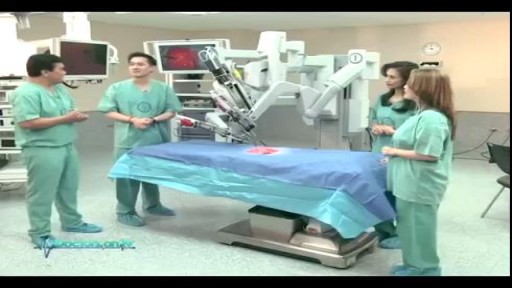
Understand how this world-class surgery platform operates a minimally invasive robotic surgery during a medical procedure for prostate cancer.

Dont worry sister!
Pharyngitis is caused by swelling in the back of the throat (pharynx) between the tonsils and the voice box (larynx). Most sore throats are caused by colds, the flu, coxsackie virus or mono (mononucleosis). Bacteria that can cause pharyngitis in some cases: Strep throat is caused by group A streptococcus.

The infection is generally transmitted by direct contact with the mucus or sores of someone else with strep. Common symptoms include sore throat, fever, and swollen lymph nodes in the neck. Rarely, complications can involve the heart or kidneys. Treatment is important to reduce complications. Oral antibiotics like penicillin, amoxicillin, cephalexin, or azithromycin are commonly used. Other medicines such as acetaminophen or ibuprofen can help with pain and fever.
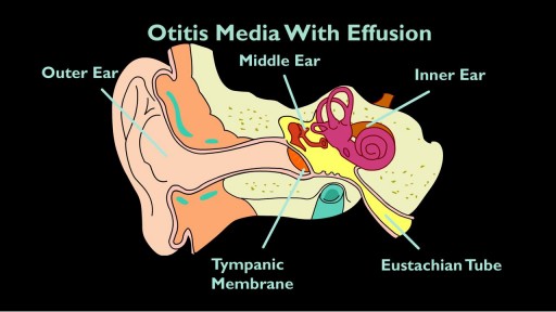
The eustachian tube drains fluid from your ears to the back of your throat. If it clogs, otitis media with effusion (OME) can occur. If you have OME, the middle part of your ear fills with fluid, which can increase the risk of ear infection. OME is very common. According to the Agency of Healthcare Research and Quality, about 90 percent of children will have OME at least once by the age of 10.

A myringotomy is a procedure in which your doctor creates a small hole in the eardrum so fluids such as water, blood, or pus can drain out. In many cases, your doctor will put in a tube so it won't get backed up again. The tube, which will usually fall out on its own in about six to 18 months, lets air flow through and keeps the middle ear dry. Tubes also: Reduce pain Improve hearing Cut down on the number of infections your child may have

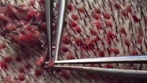
What Is a Hair Transplant? It's a type of surgery that moves hair you already have to fill an area with thin or no hair. Doctors have been doing these transplants in the U.S. since the 1950s, but techniques have changed a lot in recent years. You usually have the procedure in the doctor's office. First, the surgeon cleans your scalp and injects medicine to numb the back of your head. Your doctor will choose one of two methods for the transplant: follicular unit strip surgery (FUSS) or follicular unit extraction (FUE). With FUSS, the surgeon removes a 6- to 10-inch strip of skin from the back of your head. He sets it aside and sews the scalp closed. This area is immediately hidden by the hair around it. Next, the surgeon’s team divides the strip of removed scalp into 500 to 2,000 tiny grafts, each with an individual hair or just a few hairs. The number and type of graft you get depends on your hair type, quality, color, and the size of the area where you’re getting the transplant. If you’re getting the FUE procedure, the surgeon’s team will shave the back of your scalp. Then, the doctor will remove hair follicles one by one from there. The area heals with small dots, which your existing hair will cover. After that point, both procedures are the same. After he prepares the grafts, the surgeon cleans and numbs the area where the hair will go, creates holes or slits with a scalpel or needle, and delicately places each graft in one of the holes. He’ll probably get help from other team members to plant the grafts, too. Depending on the size of the transplant you’re getting, the process will take about 4 to 8 hours. You might need another procedure later on if you continue to lose hair or decide you want thicker hair. Expectations and Recovery After the surgery, your scalp may be very tender. You may need to take pain medications for several days. Your surgeon will have you wear bandages over your scalp for at least a day or two. He may also prescribe an antibiotic or an anti-inflammatory drug for you to take for several days. Most people are able to return to work 2 to 5 days after the operation. Within 2 to 3 weeks after surgery, the transplanted hair will fall out, but you should start to notice new growth within a few months. Most people will see 60% of new hair growth after 6 to 9 months. Some surgeons prescribe the hair-growing drug minoxidil (Rogaine) to improve hair growth after transplantation, but it’s not clear how well it works. Risks and Costs of Treatment The price of a hair transplant will depend largely on the amount of hair you’re moving, but it generally ranges from $4,000 to $15,000. Most insurance plans don’t cover it.
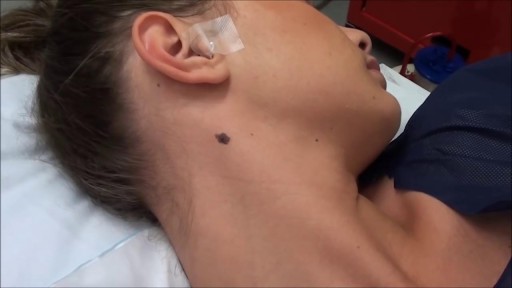
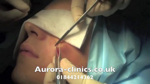
Most people develop several moles (nevi) throughout adulthood. Moles can be found anywhere on the body, usually in sun-exposed areas, and are usually brown, smooth, and slightly raised. In most cases, a nevus is benign and doesn't require treatment. Rarely, they turn into melanoma or other skin cancers. A nevus that changes shape, grows bigger, or darkens should be evaluated for removal.

The condition is caused by a blockage in the lymphatic system, part of the immune and circulatory systems. Lymphedema is most commonly caused by lymph node removal or damage due to cancer treatment. The main symptom is swelling in an arm or leg that may be accompanied by pain or discomfort. Exercise, wrapping, massage, and compression can help.

Dysmenorrhea, or chronic menstrual pain, is the most common gynecological pain condition, affecting from 45% to 95% of menstruating women. But because it is commonly considered a normal aspect of the menstrual cycle,

Do I Need to See My Doctor for Menstrual Cramps? || Common gynaecological problems in women It's perfectly normal to experience mild cramps during your period, and the good news is that these cramps can usually be eased with simple therapies like a heating pad or an over-the-counter pain reliever. However, some women's menstrual cramps may not feel better with these basic remedies. If this is the case for you, making an appointment with your doctor is important. This way you not only get the pain relief you deserve but also ensure there is nothing else going on.

Heavy period blood can be especially alarming if it contains clots. In most cases, though, red, brown, or even black menstrual blood clots are normal—just bits of the endometrium (the lining of the uterus) that are shed during menstruation.
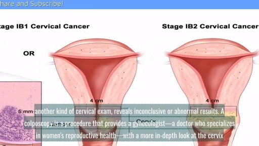
A cervical biopsy is a procedure that is sometimes done on women during an exam called a colposcopy to remove cervical tissue for examination. It is also called a punch biopsy. It is usually performed when a Pap smear result is either inconclusive or abnormal and a doctor wants to screen further for any cervical dysplasia or cervical cancer.
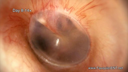
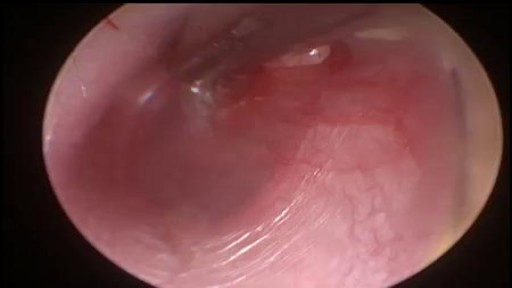
The is a time lapse video animation of a complicated ear infection with a ruptured eardrum causing drainage with eventual healing. The video also shows why a period of hearing loss and clogged/muffled ear sensation may occur.
