- Physical Examination
- Surgical Examination
- Ophthalmology
- Clinical Skills
- Orthopedics
- Surgery Videos
- Laparoscopy
- Pediatrics
- Funny Videos
- Cardiothoracic Surgery
- Nursing Videos
- Plastic Surgery
- Otorhinolaryngology
- Histology and Histopathology
- Neurosurgery
- Dermatology
- Pediatric Surgery
- Urology
- Dentistry
- Oncology and Cancers
- Anatomy Videos
- Health and Fitness
- Radiology
- Anaesthesia
- Physical Therapy
- Pharmacology
- Interventional Radiology
- Cardiology
- Endocrinology
- Gynecology
- Emergency Medicine
- Psychiatry and Psychology
- Childbirth Videos
- General Medical Videos
- Nephrology
- Physiology
- Diet and Food Health
- Diabetes Mellitus
- Neurology
- Women Health
- Osteoporosis
- Gastroenterology
- Pulmonology
- Hematology
- Rheumatology
- Toxicology
- Nuclear Medicine
- Infectious Diseases
- Vascular Disease
- Reproductive Health
- Burns and Wound Healing
- Other
Top videos
Worst Nail Infection: Paronychia

How to Remove Blackhead from the Face

Worms Inside Human Stomach

Laser Cystic Acne and Pimples Extraction

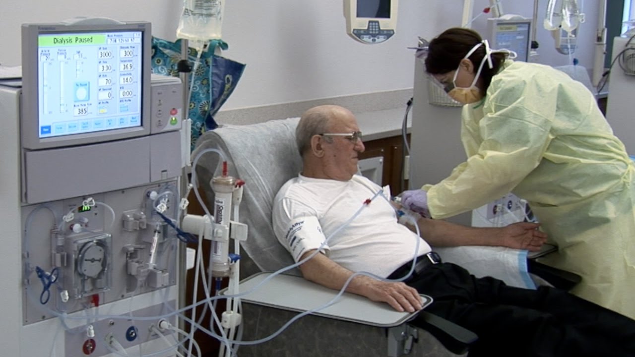
What is hemodialysis, and why would someone need it? How does hemodialysis work? Can people perform hemodialysis at home? John Kevin Tucker, M.D., Nephrologist at Brigham and Women's Hospital and Vice President for Education at Mass General Brigham, discusses hemodialysis and how it helps people who have lost their kidney function to maintain normal lives.
Subscribe Link: https://www.youtube.com/channe....l/UCYrLjATd88gPwIKnt
0:00 - Intro
0:26 - The Condition
2:06 - Hemodialysis: How It Works
4:37 - In-Center Hemodialysis Care Team
About Mass General Brigham:
Mass General Brigham combines the strength of two world-class academic medical centers, five nationally ranked specialty hospitals, 11 community hospitals, and dozens of health centers. Our doctors and researchers accelerate medical breakthroughs and drive innovations in patient care. They are leaders in medical education, serving as Harvard Medical School faculty and training the next generation of physicians. Mass General Brigham’s mission is to deliver the best, affordable health care to patients everywhere. Together, we transform the health of our communities and beyond.
#MassGeneralBrigham #MGB #Hemodialysis
Visit Mass General Brigham: https://www.massgeneralbrigham.org/
Find us on social:
Twitter: https://twitter.com/MassGenBrigham
Instagram: https://www.instagram.com/massgeneralbrigham/
Facebook: https://www.facebook.com/MassGeneralBrigham/
LinkedIn: https://www.linkedin.com/compa....ny/mass-general-brig
Mass General Brigham:
https://www.youtube.com/massgeneralbrigham
Kidney Failure: Signs, Dialysis Options, and Hemodialysis Explained | Mass General Brigham
https://youtu.be/azy7yc19QYQ

Hemodialysis is the process of cleaning the patient’s blood outside the body. Learn more about this renal replacement therapy option.
Read more: http://www.freseniusmedicalcar....e.com/en/patients-fa


Check out our new website http://www.evanshealthlab.com/
Follow Dr. Mike for new videos! http://twitter.com/docmikeevans
Dr. Mike Evans is founder of the Health Design Lab at the Li Ka Shing Knowledge Institute, an Associate Professor of Family Medicine and Public Health at the University of Toronto, and a staff physician at St. Michael's Hospital.
Written and Narrated by Dr. Mike Evans
Executive Producer, Dr. Mike Evans
Illustrations by Liisa Sorsa
Produced, Directed, and Photographed by Nick De Pencier
Editor, David Schmidt
Story/Graphic Facilitator, Disa Kauk
Production Assistant, Chris Niesing
Director of Operations, Mike Heinrich
©2014 Michael Evans and Reframe Health Films Inc.


The objectives of hemodialysis are to extract toxic nitrogenous substances from the blood and to remove excess water. In hemodialysis, the blood, laden with toxins and nitrogenous wastes, is diverted from the patient to a machine, a dialyzer, in which the blood is cleansed and then returned to the patient. Diffusion, osmosis, and ultrafiltration are the principles on which hemodialysis is based.
The toxins and wastes in the blood are removed by diffusion—that is, they move from an area of higher concentration in the blood to an area of lower concentration in the dialysate. The dialysate is a solution made up of all the important electrolytes in their ideal extracellular concentrations.
The electrolyte level in the patient’s blood can be brought
under control by properly adjusting the dialysate bath. The semipermeable membrane impedes the diffusion of large molecules,
such as red blood cells and proteins.
#hemodialysis #dialysis #viral #urinaryinfection #shorts #medical #animation

***SUBSCRIBE WITHIN THE NEXT 28 DAYS FOR A CHANCE TO WIN $1,000!***
Did you know only 20% of our video content is on YouTube? Try out our membership for FREE today! → https://bit.ly/3mWibYe
Try our NCLEX Prep FREE → https://bit.ly/3xYAOkT
Head to https://bit.ly/3mWibYe to get access to the other 80%, along with 800+ study guides, customizable quiz banks with 3,000+ test-prep questions, and answer rationales!
Popular Playlists:
NCLEX Fluid & Electrolytes: https://bit.ly/39BSHXs
Heart Failure (CHF): https://bit.ly/2u5zfDm
Myocardial Infarction (MI): https://bit.ly/3bN9AAk
Addison’s vs. Cushing: https://bit.ly/2STvute
Diabetes Mellitus & DKA vs HHNS: https://bit.ly/37D8nbs
Cardiomyopathy: https://bit.ly/38CwcSg
IV Fluids: Hypertonic, Hypotonic & Isotonic: https://bit.ly/2P45BWx
SIADH vs Diabetes Insipidus: https://bit.ly/2wq6Bhb
Follow us on social media for more EXCLUSIVE content 👋
More Videos: https://bit.ly/37CRttH
Instagram: https://www.instagram.com/simplenursing.com_
TikTok: https://www.tiktok.com/simplenursing
Thank you for the support & for tuning in!
Remember… don’t be scared, BE PREPARED!

Add Me on
Instagram-https://instagram.com/_dialysi....s_therapist?igshid=Y
Telegram-https://t.me/dialysistherapist
Dialysis
Dialysis technician
Dialysis nurse
Kidney dialysis
Hemolysis
Dialysis technology
Dialysis therapist
Dialysis technician vacancy
Dialysis technician salary
Dialysis scope
Dialysis course scope
B.sc dialysis scope
Salary of dialysis technician
Role of dialysis technician
Salary of dialysis technician in india
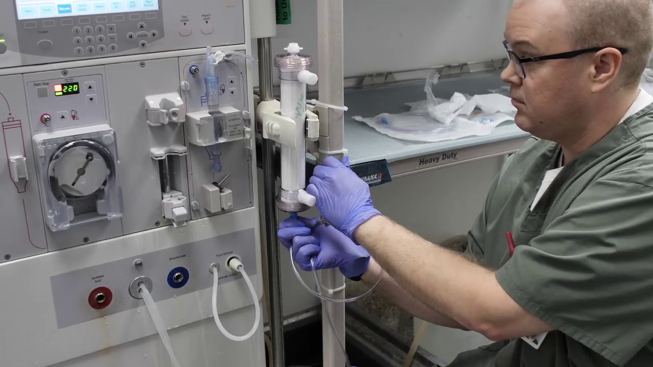
In this instructional video, Director of Critical Care Nephrology, Sevag Demirjian, MD goes over the steps for in-hospital production of ultra-pure continuous hemodialysis fluid.
By using the information in this video and/or any other materials made available by Cleveland Clinic related to the dialysate solution, you agree to comply with and be bound by the terms of the Permissive Use Agreement, a copy of which is available at https://bit.ly/3f9lN4j
Rhode Island Hospital's outpatient dialysis program cares for patients with chronic kidney disease. Learn more about the program, which includes a new, state of the art dialysis center in East Providence. http://www.rhodeislandhospital.....org/outpatient-dial
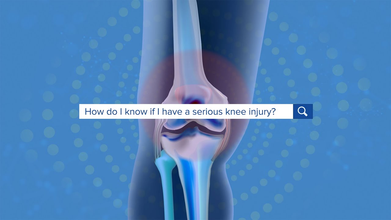
For more information please visit: https://www.yalemedicine.org/c....onditions/acl-injury
Serious injuries, by and large, cause a lot of swelling in the knee. Especially in younger patients. Now, someone could be arthritic and they overdo it going for a big long hike and they get some swelling the next day. But rapid onset of swelling, it's like hard to make out where your kneecap is, is a pretty big cardinal sign that there's something serious that's happened to your knee. Rapid onset swelling is usually due to blood in the joint. "A meniscus that really tears and flips in the front. You tear your quad or your patellar tendon, your kneecap dislocates, you tear a little blood vessel, your ACL tears, a piece of cartilage in bone gets knocked off and causes bleeding. So a lot of the really significant injuries, people get rapid onset swelling within three to four hours and they should seek attention There's always exceptions to rules, but if your knee looks like a grapefruit, you should go get it checked.

Demystify knee pain and discover nine of the most common causes of pain in this complex joint. Join Burke Selbst PT as we work through our simple screening for the most common types of problems.
Burke is the founder and clinical director of Focus Physical Therapy in Bend Oregon.
Find him:
https://focusptbend.com
https://facebook.com/focusphysio
Intro Song Credit
Adventures by A Himitsu https://www.youtube.com/channel/UCgFw...
Creative Commons — Attribution 3.0 Unported— CC BY 3.0
http://creativecommons.org/licenses/b...
Music released by Argofox https://youtu.be/8BXNwnxaVQE
Music provided by Audio Library https://youtu.be/MkNeIUgNPQ8

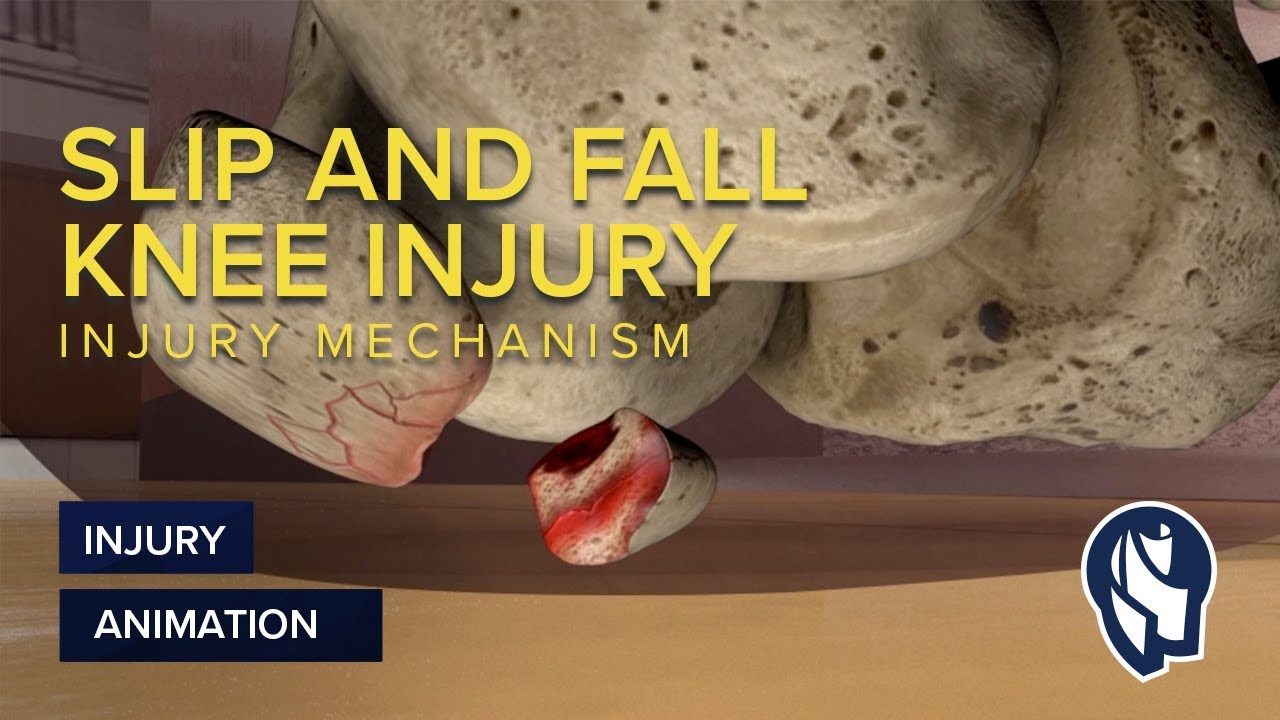
MotionLit can help you multiply the value of your case by portraying the mechanisms of injuries in a 3D Accident Reconstruction Animation. Call (855)850-0650 or visit motionlit.com to learn & earn more for your client!
MotionLit is a one-stop-shop for litigators, offering complete trial services from video production, animation, litigation support, exhibit design, trial technician, and video depositions. Our visuals have proven to help attorneys obtain record-breaking verdicts with our persuasive trial presentations, settlement documentaries, and demonstratives.
Contact Us At:
www.motionlit.com
(213) 291 9141
info@motionlit.com
Follow Us On:
Twitter: @motionlit
Instagram: https://www.instagram.com/motionlit/
Facebook: https://www.facebook.com/MotionLit/


If you have pain on the inside of your knee, it’s likely due to an injury or arthritis. The following exercises will help strengthen and stretch your muscles to prevent further damage and improve mobility.
#kneepain #arthritis #kneepainrelief #kneeosteoarthritis
- - - - - - - - - - - - - - - - - - - - - - - - - - - - - - - - - - - - -
DISCLAIMER: This video and any related comments are not medical advice. Check with your own healthcare professional before attempting anything in this video. This information is only intended to show you the correct technique for physical therapy exercises and should not be used to self-diagnose or self-treat any medical condition. If you experience any pain or difficulty while doing these exercises, stop immediately and see your healthcare professional.
