
- Physical Examination
- Surgical Examination
- Ophthalmology
- Clinical Skills
- Orthopedics
- Surgery Videos
- Laparoscopy
- Pediatrics
- Funny Videos
- Cardiothoracic Surgery
- Nursing Videos
- Plastic Surgery
- Otorhinolaryngology
- Histology and Histopathology
- Neurosurgery
- Dermatology
- Pediatric Surgery
- Urology
- Dentistry
- Oncology and Cancers
- Anatomy Videos
- Health and Fitness
- Radiology
- Anaesthesia
- Physical Therapy
- Pharmacology
- Interventional Radiology
- Cardiology
- Endocrinology
- Gynecology
- Emergency Medicine
- Psychiatry and Psychology
- Childbirth Videos
- General Medical Videos
- Nephrology
- Physiology
- Diet and Food Health
- Diabetes Mellitus
- Neurology
- Women Health
- Osteoporosis
- Gastroenterology
- Pulmonology
- Hematology
- Rheumatology
- Toxicology
- Nuclear Medicine
- Infectious Diseases
- Vascular Disease
- Reproductive Health
- Burns and Wound Healing
- Other
Top videos


Laparoscopy by Dr. Emadi in Qatar
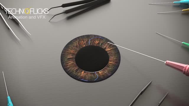
Cosmetic iris implants have not been evaluated by any U.S. regulatory agency or tested for safety in clinical trials. While the implants are not approved by the U.S. Food and Drug Administration, it has been reported in the media this month that the surgery is being performed overseas. During iris implant surgery, an artificial iris made of silicone is folded and inserted into a slit that has been cut into the cornea. Then the iris is unfolded and adjusted to cover the natural iris. Local anesthesia is used.
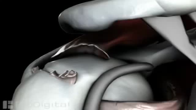
Recovery can take 4 to 6 months, depending on the size of the tear and other factors. You may have to wear a sling for 4 to 6 weeks after surgery. Pain is usually managed with medicines. Physical therapy can help you regain the motion and strength of your shoulder.
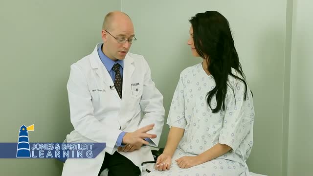
The examination room should be quiet, warm and well lit. After you have finished interviewing the patient, provide them with a gown (a.k.a. "Johnny") and leave the room (or draw a separating curtain) while they change. Instruct them to remove all of their clothing (except for briefs) and put on the gown so that the opening is in the rear. Occasionally, patient's will end up using them as ponchos, capes or in other creative ways. While this may make for a more attractive ensemble it will also, unfortunately, interfere with your ability to perform an examination! Prior to measuring vital signs, the patient should have had the opportunity to sit for approximately five minutes so that the values are not affected by the exertion required to walk to the exam room. All measurements are made while the patient is seated. Observation: Before diving in, take a minute or so to look at the patient in their entirety, making your observations, if possible, from an out-of-the way perch. Does the patient seem anxious, in pain, upset? What about their dress and hygiene? Remember, the exam begins as soon as you lay eyes on the patient. Temperature: This is generally obtained using an oral thermometer that provides a digital reading when the sensor is placed under the patient's tongue. As most exam rooms do not have thermometers, it is not necessary to repeat this measurement unless, of course, the recorded value seems discordant with the patient's clinical condition (e.g. they feel hot but reportedly have no fever or vice versa). Depending on the bias of a particular institution, temperature is measured in either Celcius or Farenheit, with a fever defined as greater than 38-38.5 C or 101-101.5 F. Rectal temperatures, which most closely reflect internal or core values, are approximately 1 degree F higher than those obtained orally. Respiratory Rate: Respirations are recorded as breaths per minute. They should be counted for at least 30 seconds as the total number of breaths in a 15 second period is rather small and any miscounting can result in rather large errors when multiplied by 4. Try to do this as surreptitiously as possible so that the patient does not consciously alter their rate of breathing. This can be done by observing the rise and fall of the patient's hospital gown while you appear to be taking their pulse. Normal is between 12 and 20. In general, this measurement offers no relevant information for the routine examination. However, particularly in the setting of cardio-pulmonary illness, it can be a very reliable marker of disease activity. Pulse: This can be measured at any place where there is a large artery (e.g. carotid, femoral, or simply by listening over the heart), though for the sake of convenience it is generally done by palpating the radial impulse. You may find it helpful to feel both radial arteries simultaneously, doubling the sensory input and helping to insure the accuracy of your measurements. Place the tips of your index and middle fingers just proximal to the patients wrist on the thumb side, orienting them so that they are both over the length of the vessel.

Carpal Tunnel Syndrome Ergonomics
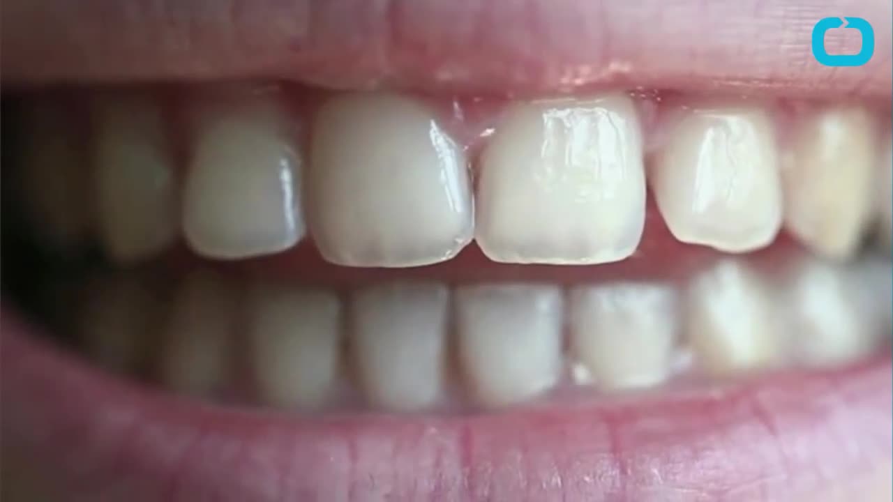
You May Be Able to Repair Cavities Without Getting a Filling
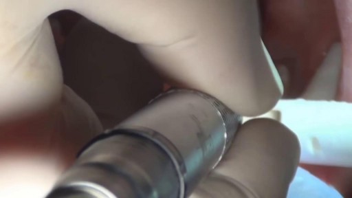
A filling is a way to restore a tooth damaged by decay back to its normal function and shape. When a dentist gives you a filling, he or she first removes the decayed tooth material, cleans the affected area, and then fills the cleaned out cavity with a filling material. By closing off spaces where bacteria can enter, a filling also helps prevent further decay. Materials used for fillings include gold, porcelain, a composite resin (tooth-colored fillings), and an amalgam (an alloy of mercury, silver, copper, tin and sometimes zinc).

Mode of action of NNRTIs

Probes, landmarks, and general windows to obtain transthoracic echo views
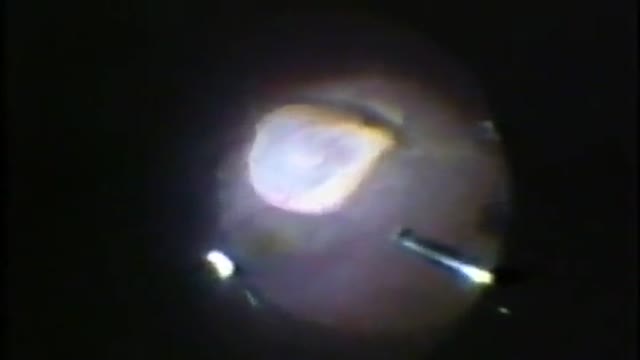
Eye Cyst Removal By Vitrectomy
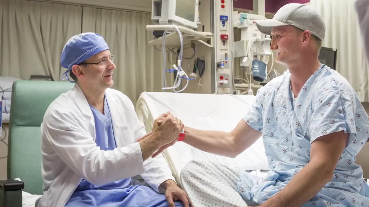
3D Printed Models Used in the Mayo Clinic's First Face Transplant
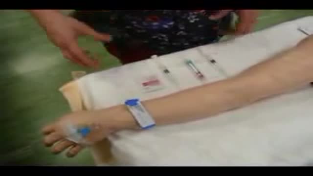
Drugs Through a Saline Lock
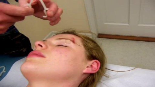
Demonstration of Burke-Baier wound closure forceps on simulated wound near eyebrow.

The E.coli bacteria has claimed 14 lives and infected more than 300 hundred in the country. It has now spread to other European states.
Facing an increasing number of cases, German health authorities warned people to avoid eating raw cucumber, tomatoes and lettuce.
Reinhard Burger, President of Robert Koch Institute, said, "As for the present situation there is no reason to give the all-clear yet and it is possible the original source of the infection is still active and could lead to further infections."
The first cases of the EHEC outbreak were noticed in Northern Germany, but infections are now spreading across the country.
Cases haves spiked compared to other years, and are still rising.
Daniel Bahr, German Health Minister, said, "The result is that we unfortunately still have to prepare for a rising number of cases. Exercising caution is still recommended and we ask our citizens to be particularly careful. "
But, the German government says it's working around the clock to stop the outbreak and clarify how it arose.
Ilse Aigner, German Agriculture Minister, said, "Together, we face a big challenge to piece together hundreds or thousands of mosaic pieces from Germany and abroad into an overall picture that gives us a clear answer on how this terrible infection arose. "
In the meantime, experts are advising not to eat pre-packaged or prepared salad, which may contain the bacteria.
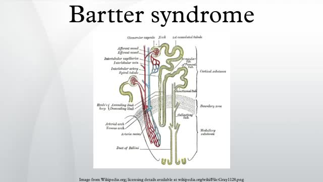
Bartter syndrome, originally described by Bartter and colleagues in 1962, [1] represents a set of closely related, autosomal recessive renal tubular disorders characterized by hypokalemia, hypochloremia, metabolic alkalosis, and hyperreninemia with normal blood pressure. The underlying renal abnormality results in excessive urinary losses of sodium, chloride, and potassium.
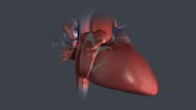
Every 10 minutes, someone is added to the national transplant waiting list, and every day, 22 people on average die waiting for a match, according to the United Network for Organ Sharing. But, thanks to innovations in bioengineering, all of that could change. Conceived nearly 60 years ago, the total artificial heart (TAH) has helped sustain the sickest biventricular failure patients waiting for a transplant. While the design of the primary TAH used today has mostly remained stagnant since the ’80s, when it was first implanted in a patient, new models and clinical trials may lead to a better device and, one day, a permanent solution. “We are still many years away from that,” Dr. Nader Moazami, director of the Cardiac Transplantation and Ventricular Assist Device Therapy Program at the Cleveland Clinic, told FoxNews.com of a permanent artificial heart. “Although tremendous strides have been made, biocompatibility will always remain a challenge.”
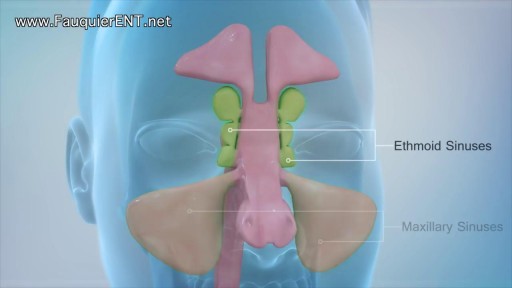
Explained (Balloon Sinuplasty and Endoscopic Sinus Surgery)

http://www.landging.com/accident-animation-workers-compensation-falling-stuff.html
This falling stuff accident animation demonstrates the injury covered by workers compensation program.

Dermatologist in New Orleans, Dr. Lupo, talks in a television interviews about Dermatology.

What is jaundice? Well, jaundice is a condition where the skin and eyes take on a yellowish color due to increased levels of bilirubin in the bloodstream
