- Physical Examination
- Surgical Examination
- Ophthalmology
- Clinical Skills
- Orthopedics
- Surgery Videos
- Laparoscopy
- Pediatrics
- Funny Videos
- Cardiothoracic Surgery
- Nursing Videos
- Plastic Surgery
- Otorhinolaryngology
- Histology and Histopathology
- Neurosurgery
- Dermatology
- Pediatric Surgery
- Urology
- Dentistry
- Oncology and Cancers
- Anatomy Videos
- Health and Fitness
- Radiology
- Anaesthesia
- Physical Therapy
- Pharmacology
- Interventional Radiology
- Cardiology
- Endocrinology
- Gynecology
- Emergency Medicine
- Psychiatry and Psychology
- Childbirth Videos
- General Medical Videos
- Nephrology
- Physiology
- Diet and Food Health
- Diabetes Mellitus
- Neurology
- Women Health
- Osteoporosis
- Gastroenterology
- Pulmonology
- Hematology
- Rheumatology
- Toxicology
- Nuclear Medicine
- Infectious Diseases
- Vascular Disease
- Reproductive Health
- Burns and Wound Healing
- Other
Top videos
Treating Hernia with Laparscopic Inguinal Hernia Repair
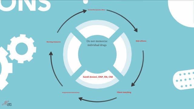
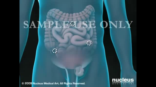
Remembering Medications & The Body Systems Affected
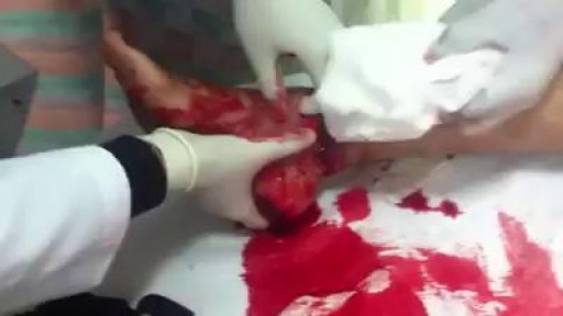
Broken or Dislocated Ankle Joint
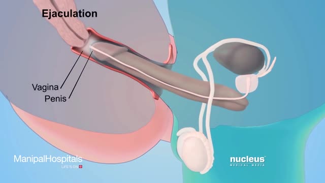
How To Use Male Condom Correctl
Diagnosis of HIV infection in infants is aided by HIV culture or DNA/RNA polymerase chain reaction (PCR); positive results are confirmed by repeating the test. In suspected cases, HIV testing should occur in the newborn period (ie, before the infant is 48 h old), at age 1-2 months, and again at age 3-6 months.

Because the continuous supply of glucose is stopped after birth, the neonate develops hypoglycemia because of insufficient substrate. Stimulation of fetal insulin release by maternal hyperglycemia during labor significantly increases the risk of early hypoglycemia in these infants.
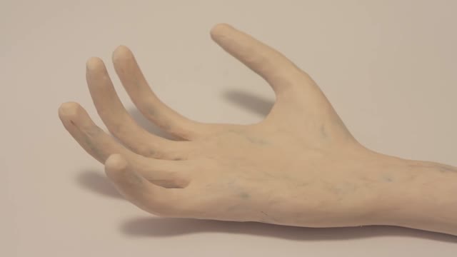
What happens to our bodies after we die?

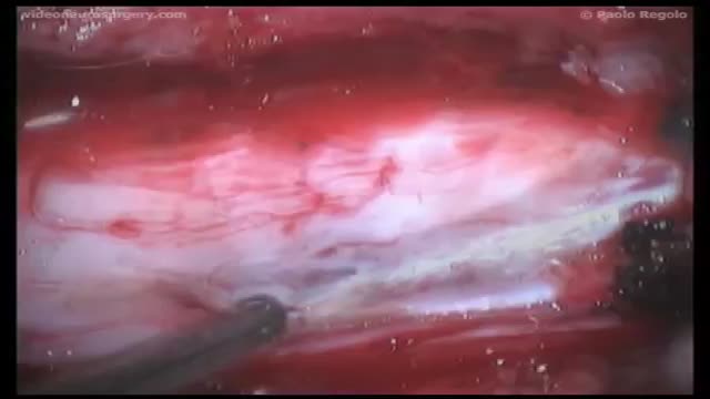
UChicago Medicine organ transplant surgeon Dr. Rolf Barth explains a how the laparoscopic donor nephrectomy – also known as the single-port nephrectomy – procedure works to remove an organ donor’s kidney from their body to be transplanted into a recipient. This minimally invasive kidney donor transplant surgery allows living organ donors the get back to their lives more quickly than the traditional approach and leaves them with a nearly invisible scar in the belly button.
Learn more about living kidney donation: https://www.uchicagomedicine.o....rg/conditions-servic
Syringomyelia is a cystic cavitation of the spinal cord associated with Chiari I malformation (70%) or basilar invagination (10%) or tumor. It may be a post-traumatic condition. There are 2 main forms: communicating with the central canal or subarachnoid spaces (Chiari I malformation); non communicating (trauma, tumors).

Johns Hopkins orthopaedic hip and knee surgeon, Savyasachi "Savya" Thakkar, explains how to prepare for knee replacement surgery, and what to expect before and after surgery. To learn more about our hip and knee replacement division, visit https://www.hopkinsmedicine.org/ortho. #KneeReplacement #JohnsHopkins
Q&A's
0:15 What causes someone to need a knee replacement?
0:29 What should patients do in advance of surgery?
1:10 Do you recommend physical therapy BEFORE surgery?
1:43 Will joint implants set off metal detectors at airports?
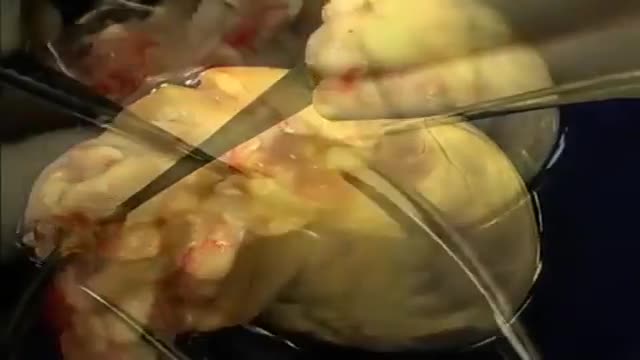
The first operation is harvesting the heart from the donor. The donor is usually an unfortunate person who has suffered irreversible brain injury, called "brain death". Very often these are patients who have had major trauma to the head, for example, in an automobile accident. The victim's organs, other than the brain, are working well with the help of medications and other "life support" that may include a respirator or other devices. A team of physicians, nurses, and technicians goes to the hospital of the donor to remove donated organs once brain death of the donor has been determined. The removed organs are transported on ice to keep them alive until they can be implanted. For the heart, this is optimally less than six hours. So, the organs are often flown by airplane or helicopter to the recipient's hospital.
Researchers have received approval to bring 20 brain-dead humans back to life.

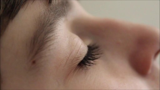
Tourette syndrome (also called Tourette's disorder or simply, "Tourette's") is an abnormal neurological condition characterized by motor and vocal tics. Tics are involuntary, rapid, sudden repetitive movements or sounds. Tics can be classified in a variety of ways. Motor tics can affect any part of the body including the head, neck, face, arms, shoulders, hands, feet, or legs. Facial tics, especially eye blinking, are usually the first symptoms of TS. Vocal tics are sounds that are made involuntarily. Vocal tics can include clearing the throat, coughing, sniffing, grunting, yelping, or shouting. In a few cases, vocal tics can include strange, inappropriate, or obscene words and phrases (called coprolalia). Vocal tics can also appear as constantly repeating the words of others (echolalia).

Thousands of Whiteheads! Why Do I Get Them?
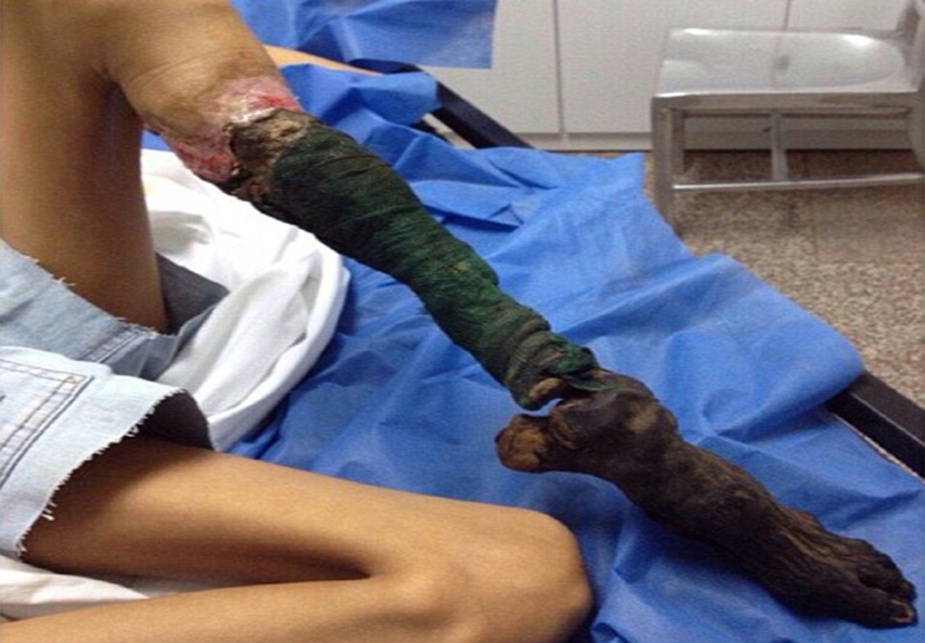
Watch that video of a Snake bite causes girl’s leg to rot away
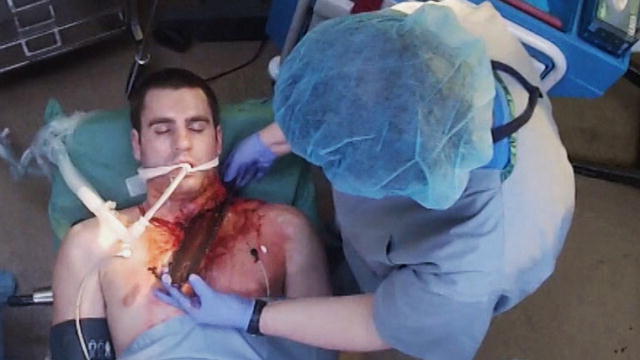
Watch that video of Surgery to Remove Chainsaw Blade From Man's Neck
Retinitis pigmentosa is a rare, inherited degenerative eye disease that causes severe vision impairment. Symptoms often begin in childhood. They include decreased vision at night or in low light and loss of side vision (tunnel vision).
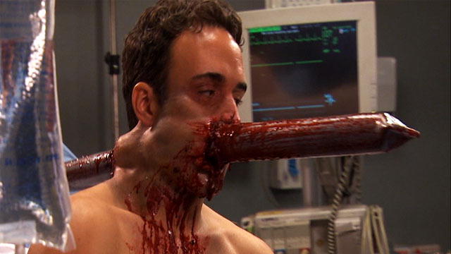
Watch that video for Removing Steel Pipe Penetrated Man's Head
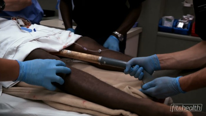
Watch that video of A Man Impaled by Shovel Inside His Butt - ER Stories
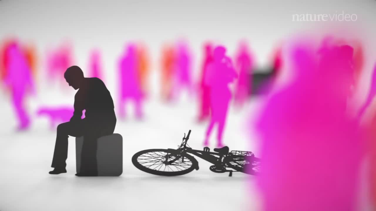
When you’re depressed, it can feel like you’ll never get out from under a dark shadow. However, even the most severe depression is treatable. So, if your depression is keeping you from living the life you want to, don’t hesitate to seek help. Learning about your depression treatment options will help you decide what approach is right for you. From therapy to medication to healthy lifestyle changes, there are many effective treatments that can help you overcome depression and reclaim your life.
