
- Drug Guide
- Lab Values
- Vital Signs
- Muscles A–Z
- Clinical Signs
- Medical Abbreviations
- Medical Terminology
- Physical Examination
- Surgical Examination
- Ophthalmology
- Clinical Skills
- Orthopedics
- Surgery Videos
- Laparoscopy
- Pediatrics
- Funny Videos
- Cardiothoracic Surgery
- Nursing Videos
- Plastic Surgery
- Otorhinolaryngology
- Histology and Histopathology
- Neurosurgery
- Dermatology
- Pediatric Surgery
- Urology
- Dentistry
- Oncology and Cancers
- Anatomy Videos
- Health and Fitness
- Radiology
- Anaesthesia
- Physical Therapy
- Pharmacology
- Interventional Radiology
- Cardiology
- Endocrinology
- Gynecology
- Emergency Medicine
- Psychiatry and Psychology
- Childbirth Videos
- General Medical Videos
- Nephrology
- Physiology
- Diet and Food Health
- Diabetes Mellitus
- Neurology
- Women Health
- Osteoporosis
- Gastroenterology
- Pulmonology
- Hematology
- Rheumatology
- Toxicology
- Nuclear Medicine
- Infectious Diseases
- Vascular Disease
- Reproductive Health
- Burns and Wound Healing
- Kidney Zone Podcast
- Other
Top videos
The most-watched medical videos on MedicalVideos.com — ranked by views from our community of medical students, healthcare professionals, and patients learning how the body works.
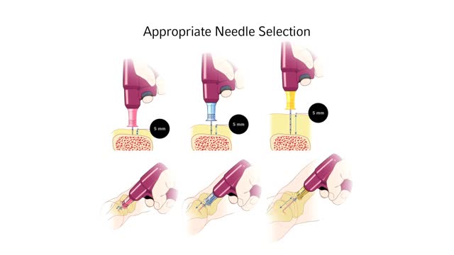

Infant Child Needle Selection and Insertion Technique Animation Video
PKU is inherited in families in an autosomal recessive pattern. Autosomal recessive inheritance means that a person has two copies of the gene that is altered. Usually, each parent of an individual who has PKU carries one copy of the altered gene. ... Gene alterations (mutations) in the PAH gene cause PKU.
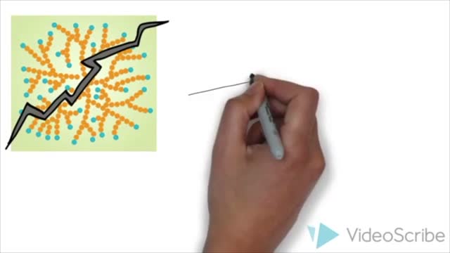
Pompe disease is a rare multisystem genetic disorder that is characterized by absence or deficiency of the lysosomal enzyme alpha-glucosidase (GAA). This enzyme is required to breakdown (metabolize) the complex carbohydrate glycogen and convert it into the simple sugar glucose.
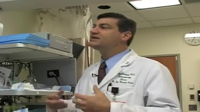
When is endoscopy used? Endoscopes were first developed to look at parts of the body that couldn’t be seen any other way. This is still a common reason to use them, but endoscopy now has many other uses too. It’s often used in the prevention, early detection, diagnosis, staging, and treatment of cancer. To prevent and screen for cancer Some types of endoscopes are used to look for cancer in people who have no symptoms. For example, colonoscopy (KO-lun-AH-skuh-pee) and sigmoidoscopy (SIG-moid-AH-skuh-pee) are used to screen for colon and rectal cancer. These procedures can also help prevent cancer because they let doctors find and remove polyps (growths) that might become cancer if left alone. To find cancer early Endoscopy can sometimes be used to find cancer early, before it has had a chance to grow or spread. Looking for causes of symptoms When people go to the doctor with certain symptoms, endoscopy can sometimes be used to help find a cause. For instance: Laryngoscopy to look at the vocal cords in people with long-term hoarseness Upper endoscopy in people having trouble swallowing Colonoscopy in people with anemia (low red blood cell counts) with an unknown cause Colonoscopy in people with blood in their stool Looking at problems found on imaging tests Imaging tests such as x-rays and CT scans can sometimes show physical changes within the body. But these tests may only give information about the size, shape, and location of the problem. Doctors use endoscopes to see more details, like color and surface texture, when trying to find out what’s going on. Newer methods of endoscopy that include high magnification are being tested to find out whether they are more useful in detecting cancer and other abnormal cells on the inner surfaces of the body. To diagnose and find out the stage (extent) of cancer To get a tissue sample Going one step further, most types of endoscopes have tools on the end that the doctor can use to take out small tissue samples. This procedure is called a biopsy (BY-op-see). Samples can be taken from suspicious areas and then looked at under a microscope or tested in other ways to see if cancer is there. A biopsy is usually the best way to find out if a growth or change is cancer or something else. Getting a closer look In some cases endoscopes are used to help find out how far a cancer has spread. Thoracoscopy (THOR-uh-KAHS -kuh-pee) and laparoscopy (LAP-uh-RAHS-kuh-pee) can be very useful in finding out if cancer has spread into the thorax (chest) or abdomen (belly). The surgeon can look into these places making only a small incision (cut) in the skin.
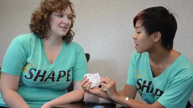
Female Condom Demonstration
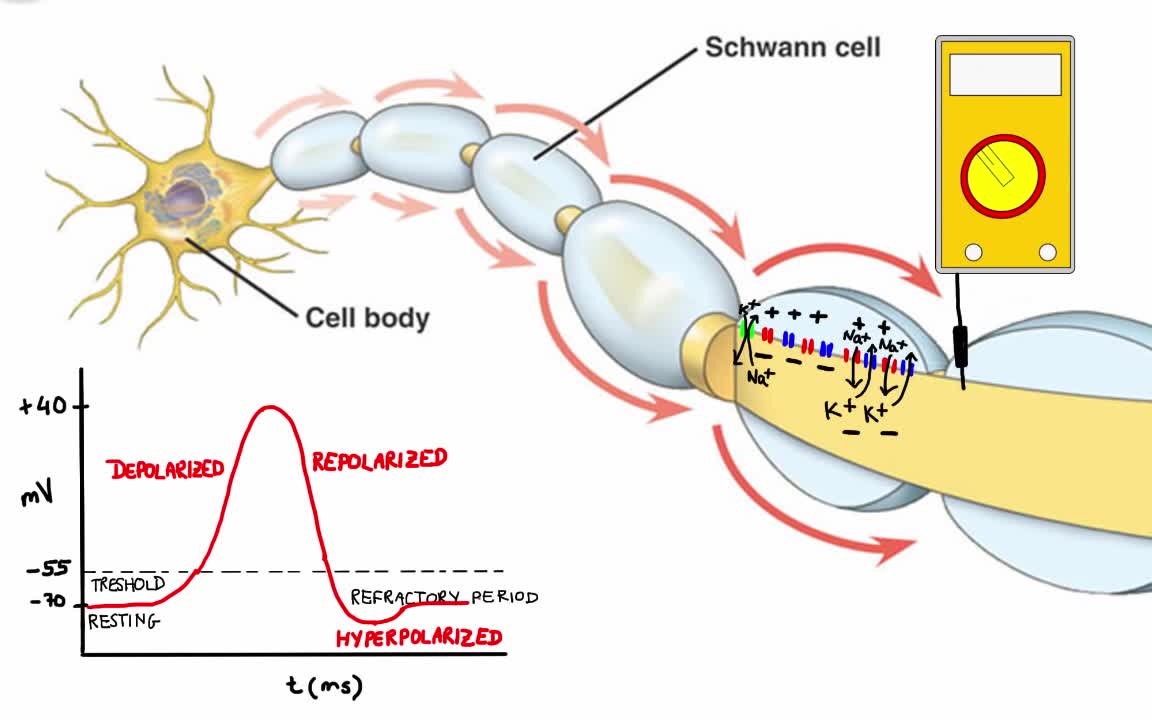
Your body has nerves that connect your brain to the rest of your organs and muscles, just like telephone wires connect homes all around the world. When you want your hand to move, your brain sends signals through your nerves to your hand telling the muscles to contract. But your nerves don’t just say “hand, move.” Instead your nerves send lots of electrical impulses (called action potentials) to different muscles in your hand, allowing you to move your hand with extreme precision.
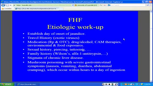
Fulminant hepatic failure (FHF) or acute liver failure (ALF) is defined as the rapid development of acute liver injury with severe impairment of the synthetic function and hepatic encephalopathy in a patient without obvious, previous liver disease.

Nearly 300 million people experience the world without certain colors every day. ‘Color For the Colorblind’ is a short documentary about what happened when we partnered with EnChroma, maker of color blindness-correcting glasses, to help people experience colors for the first time.
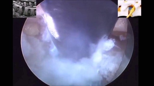
If you have been diagnosed with a bulging disc, you are not alone. Bulging discs, also known as a disc protrusion, are a very common occurrence. They usually remain asymptomatic; however, they can cause discomfort and disability in various parts of the body if the disc compresses an adjacent nerve root or the spinal cord. As we age, the outer fibrous portion of our discs can weaken. Pressure from the central core of the disc can then stretch to the outer rim, causing the disc to bulge. If left untreated, the disc can continue to bulge until it tears, which is classified as a herniated disc.

A semen analysis, also called "seminogram" evaluates certain characteristics of a male's semen and the sperm contained therein. It is done to help evaluate male fertility, whether for those seeking pregnancy or verifying the success of vasectomy
Parathyroid cancer is a rare disease in which malignant (cancer) cells form in the tissues of a parathyroid gland. The parathyroid glands are four pea-sized organs found in the neck near the thyroid gland. The parathyroid glands make parathyroid hormone (PTH or parathormone). PTH helps the body use and store calcium to keep the calcium in the blood at normal levels.
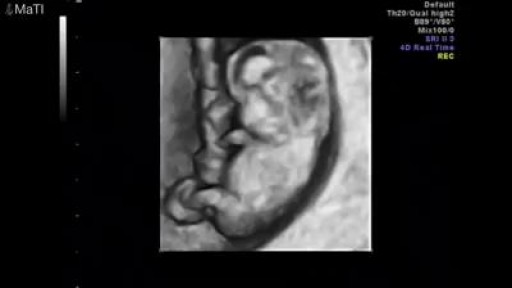
Pregnancy ultrasounds are performed mainly using transabdominal ultrasound. For many women, especially after 8 weeks gestation, sufficient information about the baby may be obtained with transabdominal ultrasound only. However, in the early pregnancy, the developing embryo is very small (at 6 weeks gestation, the baby is only 5-9mm long) and a transvaginal ultrasound may be required to get a better image of the baby. Transvaginal ultrasound is safe and commonly performed during all stages of pregnancy, including the first trimester. It will not harm you or your baby.
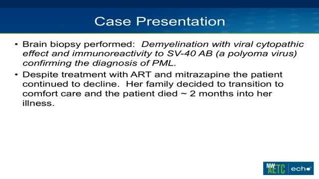
Progressive multifocalleukoencephalopathy is a demyelinating illness of the central nervous system that typically occurs in immunosuppressed patients, especially those with AIDS. It is caused by reactivation of the polyomavirus JC (JC virus) and presents with neurologic deficits including hemiparesis, gait ataxia, visual symptoms, and altered mental status. It is not seen in non-immunosuppressed patients, and fever is not typical
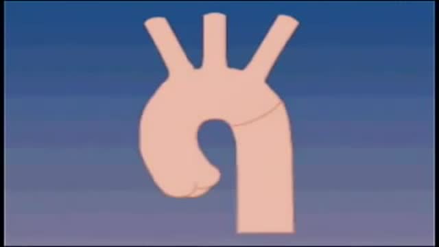
Although the exact cause of abdominal aortic aneurysms is unknown, a number of factors may play a role, including: Tobacco use. ... Hardening of the arteries (atherosclerosis). ... High blood pressure. ... Blood vessel diseases in the aorta. ... Infection in the aorta. ... Trauma. ... Heredity.
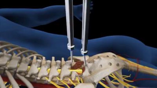
At each level of the spine, there is a disc space in the front and paired facet joints in the back. Working together, these structures define a motion segment (Fig. 1A). Back pain may result when injury or degenerative changes allow abnormal movement of the vertebrae to rub against one another, known as an unstable motion segment (Fig. 1B). Two vertebrae need to be fused to stop the motion at one segment. For example, an L4-L5 fusion is a one-level spinal fusion (Fig. 1C). A two-level fusion joins three vertebrae together and so on.
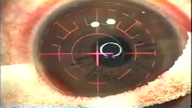
LASIK, which stands for laser in-situ keratomileusis, is a popular surgery used to correct vision in people who are nearsighted, farsighted, or have astigmatism. All laser vision correction surgeries work by reshaping the cornea, the clear front part of the eye, so that light traveling through it is properly focused onto the retina located in the back of the eye. LASIK is one of a number of different surgical techniques used to reshape the cornea.
t’s the brain, after all, that devises experiments and interprets their results. How the brain perceives, how it makes decisions and judgments, and how those judgments can go awry are at least as important to science as knowing the intricacies of nonbiotic experimental machinery. And as any brain scientist will tell you, there’s still a long way to go before understanding the brain will get crossed off science’s to-do list. But there has been progress. A recent special issue of the journal Neuron offers a convenient set of “perspective” papers exploring the current state of understanding of the brain’s inner workings. Those papers show that a lot is known. But at the same time they emphasize that there’s a lot we don’t know.
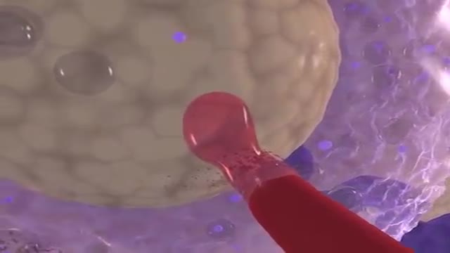
What does the placenta do? The placenta is an organ that develops in your uterus during pregnancy. This structure provides oxygen and nutrients to your growing baby and removes waste products from your baby's blood. The placenta attaches to the wall of your uterus, and your baby's umbilical cord arises from it. In most pregnancies, the placenta attaches at the top or side of the uterus.
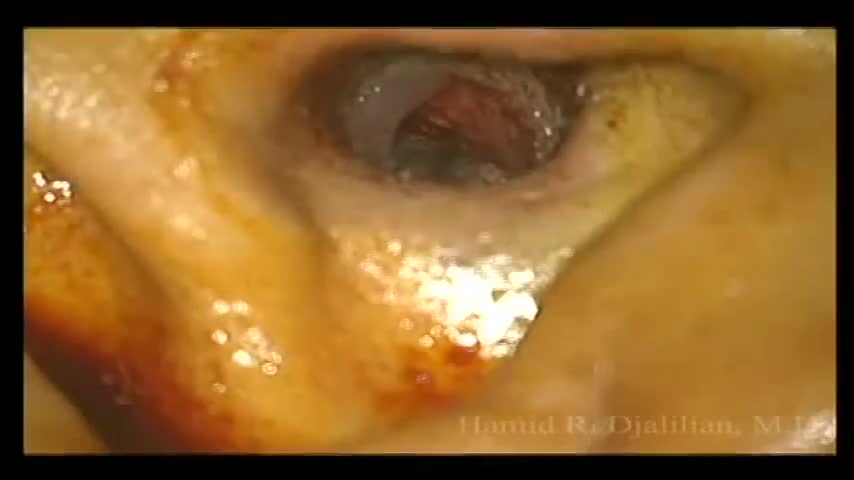
This is a surgery showing the removal of a large exostosis. Exostoses are bony growths in the ear canal from chronic exposure to cold water/air, most commonly from surfing. This patient had growths in both ears, which were completely obstructing the ear canals. This patient had a single exostosis that was blocking this side (the right side).
Medical Videos
Free medical and surgery videos for patients, students and professionals — trusted since 2007.
Founded by Dr. Mohamed Ibrahim, MD, PhD — physician-scientist.
Follow us on Facebook · Instagram