- Physical Examination
- Surgical Examination
- Ophthalmology
- Clinical Skills
- Orthopedics
- Surgery Videos
- Laparoscopy
- Pediatrics
- Funny Videos
- Cardiothoracic Surgery
- Nursing Videos
- Plastic Surgery
- Otorhinolaryngology
- Histology and Histopathology
- Neurosurgery
- Dermatology
- Pediatric Surgery
- Urology
- Dentistry
- Oncology and Cancers
- Anatomy Videos
- Health and Fitness
- Radiology
- Anaesthesia
- Physical Therapy
- Pharmacology
- Interventional Radiology
- Cardiology
- Endocrinology
- Gynecology
- Emergency Medicine
- Psychiatry and Psychology
- Childbirth Videos
- General Medical Videos
- Nephrology
- Physiology
- Diet and Food Health
- Diabetes Mellitus
- Neurology
- Women Health
- Osteoporosis
- Gastroenterology
- Pulmonology
- Hematology
- Rheumatology
- Toxicology
- Nuclear Medicine
- Infectious Diseases
- Vascular Disease
- Reproductive Health
- Burns and Wound Healing
- Other
Top videos

Watch that Knife Fully Stabbed Inside Chest Removal Surgery

New-born baby having a bath

karnim herbal capsules


Buy your new Medical Marijuana Card for $59 & renewals for $45. Chat with our licensed doctors from your place and obtain your card by email in less than 10 minutes. Visit https://www.onlinemmjlosangeles.com/

Watch that video of an Ingrown Hair Causes Huge Tumor in a Man's Stomach


http://permanently-cure-your-ulcer.info-pro.co/ Symptoms Of An Ulcer, H Pylori Natural Treatment, H Pylori Treatment Natural, Diet For H Pylori. Are You Sure You Have An Ulcer? There are many symptoms that are associated with ulcers. Some ulcer sufferers only experience mild symptoms while others experience more severe. The more common symptoms of an ulcer are listed below. Abdominal discomfort is the most common symptom of an ulcer. This discomfort usually: is a dull, gnawing ache. • comes and goes for several days or weeks. • occurs 2 to 3 hours after a meal. • occurs in the middle of the night (when the stomach is empty). • is relieved by eating. • is relieved by antacid medications. Other symptoms include: • weight loss • poor appetite • bloating • burping • nausea • vomiting If you have some or all of these symptoms, it’s a good indicator that you may have an ulcer or be developing an ulcer. Discover my 100% natural cure for ulcers. click here. http://permanently-cure-your-ulcer.info-pro.co/

A 28 years old man lost his right arm with a conveyor device in 2014. The video is taken 2 years after replantation. You can see another videos in my site: https://drliaghatclinic.com, https://instagram.com/liaghatclinic, https://t.me/liaghatclinic

Arm Replantation of a Child By Dr. Omid Liaghat : https:drliaghatclinic.com

As a curious child, you might remember staring at an older relative's thick stockings at the blue, gnarled veins lying under the skin like bumpy snakes. Known as varicose veins, these blood vessels, which return blood from the legs to the heart, are actually a more superficial system. The real, working venous system for the legs lies deeper, says to Robert A. Weiss, MD, assistant professor of dermatology at the Johns Hopkins School of Medicine in Baltimore. This is good news, because it means that if the surface veins begin to clump up and bulge, they can be removed or destroyed without ruining circulation to the leg. The National Institutes of Health estimates that 60% of all men and women suffer from some form of vein disorder. A quarter of varicose vein sufferers are men, although Weiss notes that it is almost always women who seek help for spider veins.

http://cfs-cure.plus101.com ----- Chronic Fatigue Syndrome Diet , Cures For Fatigue, Cure For Chronic Fatigue Syndrome. Chronic Fatigue Syndrome Treatment Chronic Fatigue Syndrome (CFS) is variable and unpredictable, and the condition takes its toll on the patient physically, mentally and emotionally. A number of studies have been performed on CFS, with one particular study determining poor early management of the disorder as a primary risk factor for severe CFS. Among the medical community, there is still no consensus on the best course of action for CFS. Most doctors feel that there is no cure for this condition, and limit their treatment to managing the symptoms. There is controversy over different approaches, and main ones being: • Prescription medications • Lifestyle changes • Diet • Nutritional supplements • Graded exercise therapy • Cognitive behavioral therapy • Other alternative/complementary treatments As CFS affects the patients not only physically but also mentally and emotionally, a holistic approach needs to be taken. It is also important that the people around CFS patients understand the condition, and realize that the patient is not just "being lazy" or "constantly feeling down" - chronic fatigue syndrome IS a serious illness and has severe symptoms. Cognitive Behavioral Therapy Cognitive behavioral therapy helps individuals to interpret their symptoms, which in turn helps the patient to shape their behavior in a way to better react to the symptoms. Graded Exercise Therapy A physical therapist can help determine the best exercises for the individual. Programs will start with low levels of exercising, increasing the intensity as the individual gradually builds strength and endurance. Lifestyle Changes Lifestyle changes will also be necessary, including individuals pacing themselves, lowering stress levels, eating a well-balanced diet, engaging in regular moderate exercise, and improving sleep habits. The individual’s work schedule may also need to be modified, as many individuals with CFS find maintaining their regular work schedule too draining. Diet and Chronic Fatigue Syndrome Treatment Diet is crucial in CFS, and dietary supplements may be needed. Certain foods may need to be restricted from the diet, as these may trigger or exacerbate CFS symptoms. A diet-symptom journal can help individuals to identify problem foods. In addition, a significant number of CFS cases may be caused or worsened by un-diagnosed food allergies and intolerances. Therefore, it should be a priority for every patient to check for these using a food-symptom diary and elimination diet, especially if in addition to fatigue you experience gastrointestinal symptoms such as stomach cramps, constipation, or diarrhea. Prescriptions and Medications Depression is often associated with CFS. Antidepressants may be prescribed to treat depression, which in turn will help individuals to cope with CFS-related problems. Studies also show antidepressants administered in low doses may help to relieve pain and improve sleep. Prescription sleep aids may also be prescribed to help individuals improve their sleep. Other drugs that may be prescribed include antiviral drugs, ADD/ADHD medications and anti-anxiety drugs. Alternative/Alternative Chronic Fatigue Syndrome Treatment While the usefulness of alternative/complementary therapy may still be controversial in the scientific community, many patients experience tremendous benefits from these. Main ones include:

Actual Footage of Cell Division (Kidney Cells) see more http://www.kidneymy.com/

wearable dialysis - and we expect to begin clinical trials in 2018 see more http://www.kidneymy.com/

Ewing's sarcoma typically occurs in children and young adults. It often begins in the legs, bones of the pelvis, and arms. Bone pain, localized swelling, and tenderness are symptoms. In rare cases bone fractures may also be found. Treatments include chemotherapy, surgery, and radiation.
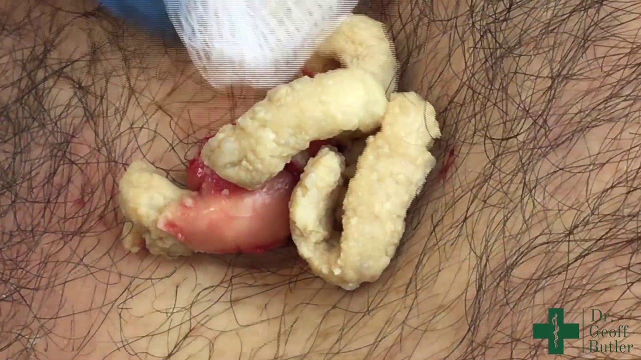
Watch that video of Removal of a large epidermal cyst in the chest

Watch that video of Removing Hand Caught Inside Meat Grinder

Watch that video of Unbelievable Mutations and Medical Condition Photos