Video hàng đầu

Watch that video of Unreal Mutations and Medical Condition
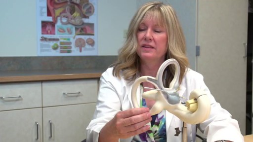
Vertigo is a sensation of feeling off balance. If you have these dizzy spells, you might feel like you are spinning or that the world around you is spinning.
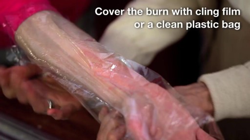
A burn is tissue damage that results from scalding, overexposure to the sun or other radiation, contact with flames, chemicals or electricity, or smoke inhalation. Is it a major or minor burn? Call 911 or seek immediate care for major burns, which: Are deep Cause the skin to be dry and leathery May appear charred or have patches of white, brown or black Are larger than 3 inches (about 8 centimeters) in diameter or cover the hands, feet, face, groin, buttocks or a major joint A minor burn that doesn't require emergency care may involve: Superficial redness similar to a sunburn Pain Blisters An area no larger than 3 inches (about 8 centimeters) in diameter Treating major burns Until emergency help arrives: Protect the burned person from further harm. If you can do so safely, make sure the person you're helping is not in contact with the source of the burn. For electrical burns, make sure the power source is off before you approach the burned person. Make certain that the person burned is breathing. If needed, begin rescue breathing if you know how. Remove jewelry, belts and other restrictive items, especially from around burned areas and the neck. Burned areas swell rapidly. Cover the area of the burn. Use a cool, moist bandage or a clean cloth. Don't immerse large severe burns in water. Doing so could cause a serious loss of body heat (hypothermia). Elevate the burned area. Raise the wound above heart level, if possible. Watch for signs of shock. Signs and symptoms include fainting, pale complexion or breathing in a notably shallow fashion. Treating minor burns For minor burns: Cool the burn. Hold the burned area under cool (not cold) running water or apply a cool, wet compress until the pain eases. Remove rings or other tight items from the burned area. Try to do this quickly and gently, before the area swells. Don't break blisters. Fluid-filled blisters protect against infection. If a blister breaks, clean the area with water (mild soap is optional). Apply an antibiotic ointment. But if a rash appears, stop using the ointment. Apply lotion. Once a burn is completely cooled, apply a lotion, such as one that contains aloe vera or a moisturizer. This helps prevent drying and provides relief. Bandage the burn. Cover the burn with a sterile gauze bandage (not fluffy cotton). Wrap it loosely to avoid putting pressure on burned skin. Bandaging keeps air off the area, reduces pain and protects blistered skin. If needed, take an over-the-counter pain reliever, such as ibuprofen (Advil, Motrin IB, others), naproxen sodium (Aleve) or acetaminophen (Tylenol, others).

For that matter, every healthcare professional undergoes this emotional hardship..
Never looked away once great video
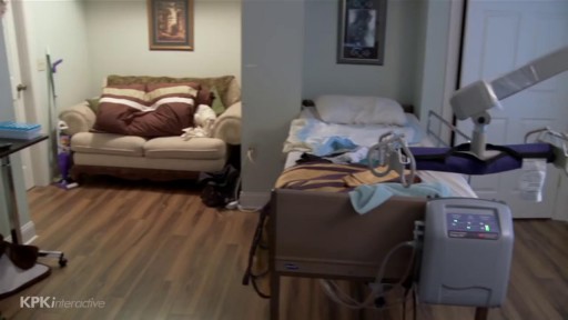

In this video I discuss sexual function for people with a spinal cord injury. The amount of feeling and function can vary drastically depending on the level and severity of the injury to the spinal cord.

A new study from Mayo Clinic finds the use of the drug therapy etanercept ineffective in treating alcoholic hepatitis, an acute inflammation of the liver caused by excessive consumption of alcohol. Alcoholic hepatitis is a major cause of morbidity and mortality worldwide. Severe alcohol-related liver disease carries a poor prognosis. Several research studies have worked to find a successful treatment for alcoholic hepatitis, but no consensus has been reached on the most effective treatment regimen.
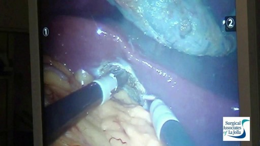
Blepharoplasty

Like a fine whiskey barrel and wine cellar, cannabis also comes at its best when aged in a dark, cool place. Though there is no steadfast expiration date for cannabis, the method you use for preserving the cannabis makes a big difference in maintaining the buds’ freshness and potency. The question is, how do you store cannabis in a way that could extend its longevity while maintaining the vigor and freshness? Experts have described different methods. However, here are some time-proven methods that are easy and inexpensive and require very less equipment. Use air-tight glass containers to store the weed Use clean air-tight glass containers or jars to store cannabis. You can buy glass containers from any ordinary supermarket or hardware store. The tricky part is to make sure you do leave some air in the container while the air stored with cannabis isn’t in detrimental extent. Always leave 1/4 space at the top of the canister or container. Do not fill the containers to the brim with the buds. If you leave no air, then the buds will dry out. If you have too much air, the buds will get damp and moldy. Freeze your cannabis in a convenient temperature The best way to store your buds is in air-tight glass jars, in a cool and dark place under an ideal temperature between 60 and 70 degrees Fahrenheit. If you need to store a high volume of cannabis, you can freeze them after keeping them completely dry for a period of 4 weeks. On this note, you should know that you must not handle frozen buds until it becomes normal in room temperature as trichomes become brittle and can easily break off in freezing temperature. Refrigerate your cannabis (Not Recommended) Even if you use airtight jars, cannabis can grow mold in the fridge. So, you should avoid storing cannabis in the fridge. If you can’t help but doing it, make sure the weed is completely dry and put them in the back where the humidity and temperature don’t fluctuate. Plastic Baggies (Worst method!) Albeit this is very common among people who aren’t expert in handling cannabis, this is the worst of all storage methods. Cannabis gets brittle and dries out in plastic bags. It also loses its natural smell, and the potency deteriorates sharply. So, it should be avoided entirely or can be used for a short-term if there is no better alternative. Here are some things you should know while storing cannabis - Make sure cannabis has been cured for at least 4 weeks before putting them into long-term storage. Without proper curing before storage, the buds can lose their strength and smoothness. - Sunlight can stop the medicinal qualities of cannabis. Your cannabis, if stored correctly, can maintain its medicinal qualities for a few years. Exposure to Sun will turn your cannabis brown, no matter how you have stored it away. - Air-tight, nonporous glass jar are the best way for storing the buds for long term. You can use metal or plastic box/bag, but that could reduce the smell and taste after a while. - Avoid heat and middling temperature in the place where you store your buds. The ideal temperature is 60-70°F (15-21°C) or under 32°F (0°C). Extra heat, cold or middling temperature cause the cannabis potency to decrease. - Keep your cannabis away from any electronic devices or appliances that will expose the cannabis to heat. Keeping cannabis on top of a microwave, or near a laptop or mobile charge is a bad idea. Now, as you know that how to store cannabis properly and make it last for years, enjoy the best form of your weeds even it comes from the previous year. Do write to us in the comments section if you have any questions. Also, don’t forget to hit the subscribe button below. Visit OnlineMedicalCard.com now to get an MMJ recommendation online in less than 10 minutes.
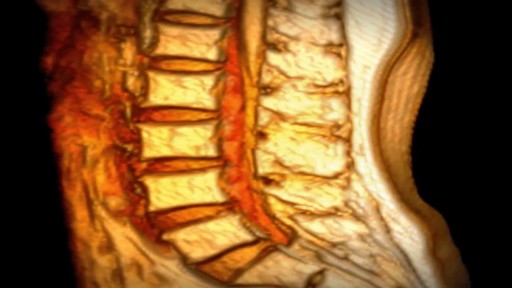
If you have been diagnosed with a bulging disc, you are not alone. Bulging discs, also known as a disc protrusion, are a very common occurrence. They usually remain asymptomatic; however, they can cause discomfort and disability in various parts of the body if the disc compresses an adjacent nerve root or the spinal cord. As we age, the outer fibrous portion of our discs can weaken. Pressure from the central core of the disc can then stretch to the outer rim, causing the disc to bulge. If left untreated, the disc can continue to bulge until it tears, which is classified as a herniated disc. Because a bulging disc does not always show symptoms, many people have bulging discs without realizing it. As long as the bulging area does not press against a nearby nerve, no symptoms occur. When the bulging disc does cause a pinched nerve, however, you may begin to experience symptoms. In the lower back, the damaged disc can cause pain to travel to the hips, buttocks, legs and feet. In the cervical spine, pain can radiate from the neck, down the arm and to the fingers.


Systemic lupus erythematous is an autoimmune condition characterised by damage to organ systems due to autoantibodies and immune complex deposition. Genes, epigenetic changes and environment play a role in its pathogenesis. SLE is a truly multi system disease causing widespread clinical manifestations in almost all organ systems. Autoantibodies in SLE are numerous and mainly include ANA, dsDNA, Sm and others.
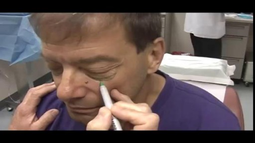
Birthmark Removal with Laser

URBN Dental is at your service to provide professional dental tips and quality service. Do you ever wonder how often you should be changing your toothbrush (or toothbrush head if you are using an electric one)? Switching your electric head or tossing your toothbrush is recommended every three months. If you are sick or have a cold sore or canker sore, it’s highly suggested to also switch your brushes. The toothbrush bristles can contain a lot of harmful bacteria and need to be replaced to decrease your risk of potential systemic illnesses. Also the bristles themselves can get worn and frayed and will decrease the efficiency of the toothbrush. Without replacing the toothbrush bristles, you suffer from a greater risk of encountering gum disease and cavities, so be sure you swap those bristles! Schedule a dental appointment now to learn more! Click on our website to book today: https://www.urbndental.com/

How to Imporve Sexual Health or Stamina Part 2 https://youtu.be/S17bCnwCLuI Dr. Aslam Naveed is a well known sexologist in Pakistan. He has treated more than 1 Lac patients since last 30 years of clinical Practice in sexology, he knows how to help the people facing sexual disorders. Contact: 021-34595050, 03432821919 sexologistpakistan.com facebook.com/menssexcareclinic/ Address: Men's Care Clinic, 2nd floor, The Modern Hospital Opposite Safari Park, University Road. Karachi.


