- Physical Examination
- Surgical Examination
- Ophthalmology
- Clinical Skills
- Orthopedics
- Surgery Videos
- Laparoscopy
- Pediatrics
- Funny Videos
- Cardiothoracic Surgery
- Nursing Videos
- Plastic Surgery
- Otorhinolaryngology
- Histology and Histopathology
- Neurosurgery
- Dermatology
- Pediatric Surgery
- Urology
- Dentistry
- Oncology and Cancers
- Anatomy Videos
- Health and Fitness
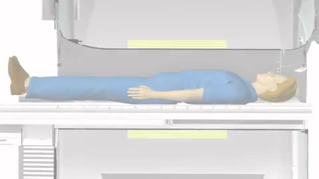
- Radiology
- Anaesthesia
- Physical Therapy
- Pharmacology
- Interventional Radiology
- Cardiology
- Endocrinology
- Gynecology
- Emergency Medicine
- Psychiatry and Psychology
- Childbirth Videos
- General Medical Videos
- Nephrology
- Physiology
- Diet and Food Health
- Diabetes Mellitus
- Neurology
- Women Health
- Osteoporosis
- Gastroenterology
- Pulmonology
- Hematology
- Rheumatology
- Toxicology
- Nuclear Medicine
- Infectious Diseases
- Vascular Disease
- Reproductive Health
- Burns and Wound Healing
- Other
Top videos
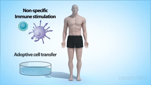
Cancer immunology is a branch of immunology that studies interactions between the immune system and cancer cells (also called tumors or malignancies). It is a growing field of research that aims to discover innovative cancer immunotherapies to treat and retard progression of the disease.
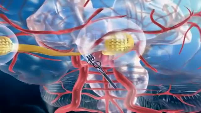
A brain (cerebral) aneurysm is a bulging, weak area in the wall of an artery that supplies blood to the brain. In most cases, a brain aneurysm causes no symptoms and goes unnoticed. In rare cases, the brain aneurysm ruptures, releasing blood into the skull and causing a stroke. When a brain aneurysm ruptures, the result is called a subarachnoid hemorrhage. Depending on the severity of the hemorrhage, brain damage or death may result. The most common location for brain aneurysms is in the network of blood vessels at the base of the brain called the circle of Willis. What causes a brain aneurysm? A person may inherit the tendency to form aneurysms, or aneurysms may develop because of hardening of the arteries (atherosclerosis) and aging. Some risk factors that can lead to brain aneurysms can be controlled, and others can't. The following risk factors may increase your risk for an aneurysm or, if you already have an aneurysm, may increase your risk of it rupturing: Family history. People who have a family history of brain aneurysms are more likely to have an aneurysm than those who don't. Previous aneurysm. People who have had a brain aneurysm are more likely to have another. Gender. Women are more likely to develop a brain aneurysm or to suffer a subarachnoid hemorrhage. Race. African Americans are more likely than whites to have a subarachnoid hemorrhage. High blood pressure. The risk of subarachnoid hemorrhage is greater in people who have a history of high blood pressure. Smoking. In addition to being a cause of high blood pressure, the use of cigarettes may greatly increase the chances of a brain aneurysm rupturing.
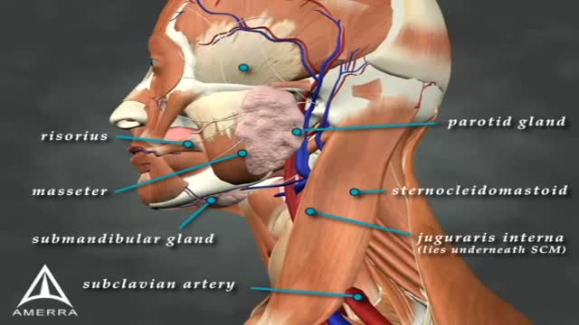
Sialorrhea or excessive drooling is a major issue in children with cerebral palsy and adults with neurodegenerative disorders. In this review, we describe the clinical features, anatomy and physiology of sialorrhea, as well as a review of the world literature on medical treatment using Yale University’s search engine; including but not limited to Medline and Erasmus. Level of drug efficacy is defined according to the guidelines of American Academy of Neurology. Current medical management is unsatisfactory. Topical agents (scopolamine and tropicamide) and oral agents (glyccopyrolate) combined render a level B evidence (probably effective); however, this treatment is associated with troublesome side effects. Double-blind and placebo-controlled studies of botulinum toxin (BoNT) provide a level A evidence for type B (two class I studies; effective and established) and both overall and individual B level of evidence for OnabotulinumtoxinA (A/Ona) and AbobotulinumtoxinA (A/Abo); these are probably effective. For IncobotulinumtoxinA (A/Inco), the level of evidence is U (insufficient) due to lack of blinded studies. Side effects are uncommon; transient and comparable between the two types of toxin. A clinical note at the end of this review comments on fine clinical points. Administration of BoNTs into salivary glands is currently the most effective way of treating sialorrhea.
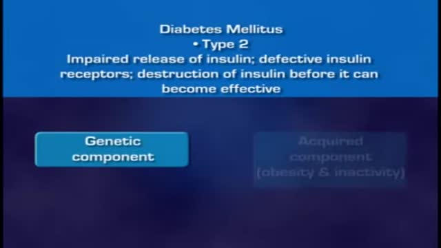
When food is taken, it is broken down into smaller components. Sugars and carbohydrates are thus broken down into glucose for the body to utilize them as an energy source. The liver is also able to manufacture glucose. In normal persons the hormone insulin, which is made by the beta cells of the pancreas, regulates how much glucose is in the blood. When there is excess of glucose in blood, insulin stimulates cells to absorb enough glucose from the blood for the energy that they need. Insulin also stimulates the liver to absorb and store any excess glucose that is in the blood. Insulin release is triggered after a meal when there is a rise in blood glucose. When blood glucose levels fall, during exercise for example, insulin levels fall too. High insulin will promote glucose uptake, glycolysis (break down of glucose), and glycogenesis (formation of storage form of glucose called glycogen), as well as uptake and synthesis of amino acids, proteins, and fat. Low insulin will promote gluconeogenesis (breakdown of various substrates to release glucose), glycogenolysis (breakdown of glycogen to release gluose), lipolysis (breakdown of lipids to release glucose), and proteolysis (breakdown of proteins to release glucose). Insulin acts via insulin receptors.
6 987 24 MORE How Does Anesthesia Work? Credit: itsmejust | Shutterstock If you’ve ever had surgery, unless you are super tough, you’ve gone through it with the benefit of anesthetics. But, how do these body-numbing elixirs work? Prior to the invention of anesthesia in the mid-1800s, surgeons had to hack off limbs, sew up wounds and remove mysterious growths with nothing to dull the patient's pain but opium or booze. While these drugs may have numbed the patient, they didn’t always completely block the pain, or erase the memory of it. Since then, doctors have gotten much better at putting us out with drug combinations that ease pain, relax muscles and, in some cases, put us in a deep state of hypnosis that gives us temporary amnesia. Today, there are two primary types of anesthesia drugs: those that knockout the whole body (general) and those that only numb things up locally.

Cosmetic Eye and Eyelid Surgery
Most people have general anesthesia right before surgery. This means you will be asleep and pain-free. Other kinds of anesthesia, like regional anesthesia or a block, may also be used for this surgery. The tissue to replace your damaged ACL will come from your own body or from a donor. A donor is a person who has died and chose to give all or part of his or her body to help others. Tissue taken from your own body is called an autograft. The two most common places to take tissue from are the knee cap tendon or the hamstring tendon. Your hamstring is the muscle behind your knee. Tissue taken from a donor is called an allograft. The procedure is usually performed with the help of knee arthroscopy. With arthroscopy, a tiny camera is inserted into the knee through a small surgical cut. The camera is connected to a video monitor in the operating room. Your surgeon will use the camera to check the ligaments and other tissues of your knee. Your surgeon will make other small cuts around your knee and insert other medical instruments. Your surgeon will fix any other damage found, and then will replace your ACL by following these steps: The torn ligament will be removed with a shaver or other instruments. If your own tissue is being used to make your new ACL, your surgeon will make a larger cut. Then, the autograft will be removed through this cut. Your surgeon will make tunnels in your bone to bring the new tissue through. This new tissue will be in the same place as your old ACL. Your surgeon will attach the new ligament to the bone with screws or other devices to hold it in place. As it heals, the bone tunnels fill in. This holds the new ligament in place. At the end of the surgery, your surgeon will close your cuts with sutures (stitches) and cover the area with a dressing. You may be able to view pictures after the procedure of what the doctor saw and what was done during the surgery.

Such foods include carrots, eggplant, cauliflower, green beans, broccoli, peppers, onions, lettuce, zucchini, tomatoes, peanuts and walnuts. These foods are generally safe for you to eat at each meal without spiking your blood sugar.

Here we’ll explain the symptoms of pancreatitis, how alcohol causes the condition and the other health problems it can lead to. You probably don’t pay much attention to your pancreas. But that small, tadpole-shaped organ behind your stomach and below your ribcage is pretty important. It produces two essential substances: digestive juices which your intestines use to break down food, and hormones that are involved in digestion, such as insulin, which regulates your blood sugar levels. Pancreatitis is when your pancreas becomes inflamed and its cells are damaged. Heavy drinking can cause pancreatitis. But if you drink within the government’s low risk unit guidelines, you should avoid upsetting this important organ.

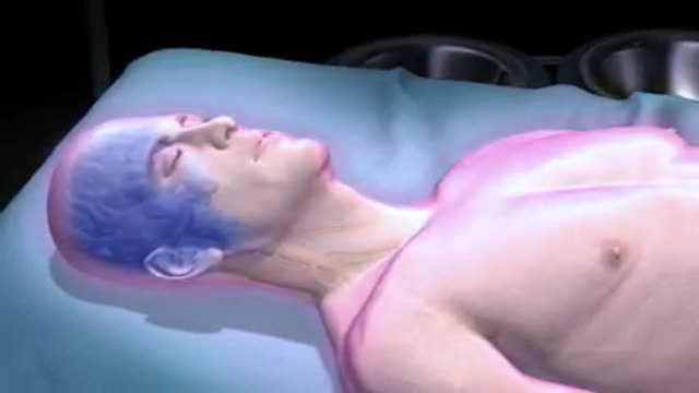
Preeclampsia is a pregnancy complication characterized by high blood pressure and signs of damage to another organ system, often the kidneys. Preeclampsia usually begins after 20 weeks of pregnancy in a woman whose blood pressure had been normal. Even a slight rise in blood pressure may be a sign of preeclampsia. Left untreated, preeclampsia can lead to serious — even fatal — complications for both you and your baby. If you have preeclampsia, the only cure is delivery of your baby. If you're diagnosed with preeclampsia too early in your pregnancy to deliver your baby, you and your doctor face a challenging task. Your baby needs more time to mature, but you need to avoid putting yourself or your baby at risk of serious complications.
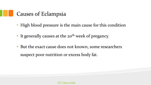
Ten percent of all pregnancies are complicated by hypertension. Eclampsia and preeclampsia account for about half of these cases worldwide, and these conditions have been recognized and described for years despite the general lack of understanding of the disease. [1] In the fifth century, Hippocrates noted that headaches, convulsions, and drowsiness were ominous signs associated with pregnancy. In 1619, Varandaeus coined the term eclampsia in a treatise on gynecology. [2, 3]
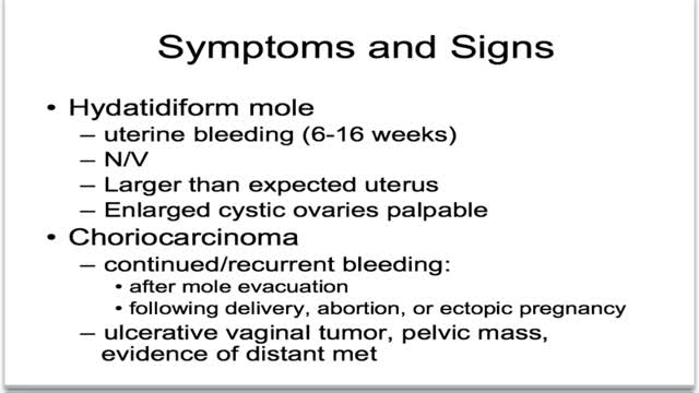
What is gestational trophoblastic disease? Cancer starts when cells in the body begin to grow out of control. Cells in nearly any part of the body can become cancer, and can spread to other areas of the body. To learn more about how cancers start and spread, see What Is Cancer? Gestational trophoblastic (jeh-STAY-shuh-nul troh-fuh-BLAS-tik) disease (GTD) is a group of rare tumors that involve abnormal growth of cells inside a woman's uterus. GTD does not develop from cells of the uterus like cervical cancer or endometrial (uterine lining) cancer do. Instead, these tumors start in the cells that would normally develop into the placenta during pregnancy. (The term gestational refers to pregnancy.) GTD begins in the layer of cells called the trophoblast (troh-fuh-BLAST) that normally surrounds an embryo. (Tropho- means nutrition, and -blast means bud or early developmental cell.) Early in normal development, the cells of the trophoblast form tiny, finger-like projections known as villi. The villi grow into the lining of the uterus. In time, the trophoblast layer develops into the placenta, the organ that protects and nourishes the growing fetus.

Researchers believe that the infectious agent that causes mad cow disease is an abnormal version of a protein normally found on cell surfaces, called a prion. For reasons still unknown, this protein becomes altered and destroys nervous system tissue -- the brain and spinal cord.
Diabetes is a growing global health concern, as is obesity. Diabetes and obesity are intrinsically linked: obesity increases the risk of diabetes and also contributes to disease progression and cardiovascular disease. Although the benefits of weight loss in the prevention of diabetes and as a critical component of managing the condition are well established, weight reduction remains challenging for individuals with type 2 diabetes due to a host of metabolic and psychological factors. For many patients, lifestyle intervention is not enough to achieve weight loss, and alternative options, such as pharmacotherapy, need to be considered. However, many traditional glucose-lowering medications may lead to weight gain. This article focuses on the potential of currently available pharmacological strategies and on emerging approaches in development to support the glycemic and weight-loss goals of individuals with type 2 diabetes. Two pharmacotherapy types are considered: those developed primarily for blood glucose control that have a favorable effect on body weight and those developed primarily to induce weight loss that have a favorable effect on blood glucose control. Finally, the potential of combination therapies for the management of obese patients with type 2 diabetes is discussed.
Initial treatment of a deviated septum may be directed at managing the symptoms of the tissues lining the nose, which may then contribute to symptoms of nasal obstruction and drainage. Your doctor may prescribe: Decongestants. Decongestants are medications that reduce nasal tissue swelling, helping to keep the airways on both sides of your nose open. Decongestants are available as a pill or as a nasal spray. Use nasal sprays with caution, however. Frequent and continued use can create dependency and cause symptoms to be worse (rebound) after you stop using them. Decongestants have a stimulant effect and may cause you to be jittery as well as elevate your blood pressure and heart rate. Antihistamines. Antihistamines are medications that help prevent allergy symptoms, including obstruction and runny nose. They can also sometimes help nonallergic conditions such as those occurring with a cold. Some antihistamines cause drowsiness and can affect your ability to perform tasks that require physical coordination, such as driving. Nasal steroid sprays. Prescription nasal corticosteroid sprays can reduce inflammation in your nasal passage and help with obstruction or drainage. It usually takes from one to three weeks for steroid sprays to reach their maximal effect, so it is important to follow your doctor's directions in using them. Medications only treat the swollen mucus membranes and won't correct a deviated septum.
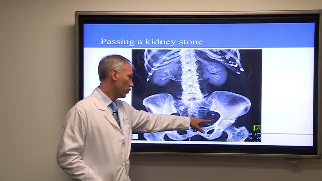
Treatment for kidney stones varies, depending on the type of stone and the cause. Small stones with minimal symptoms Most kidney stones won't require invasive treatment. You may be able to pass a small stone by: Drinking water. Drinking as much as 2 to 3 quarts (1.9 to 2.8 liters) a day may help flush out your urinary system. Unless your doctor tells you otherwise, drink enough fluid — mostly water — to produce clear or nearly clear urine. Pain relievers. Passing a small stone can cause some discomfort. To relieve mild pain, your doctor may recommend pain relievers such as ibuprofen (Advil, Motrin IB, others), acetaminophen (Tylenol, others) or naproxen sodium (Aleve). Medical therapy. Your doctor may give you a medication to help pass your kidney stone. This type of medication, known as an alpha blocker, relaxes the muscles in your ureter, helping you pass the kidney stone more quickly and with less pain. Large stones and those that cause symptoms Kidney stones that can't be treated with conservative measures — either because they're too large to pass on their own or because they cause bleeding, kidney damage or ongoing urinary tract infections — may require more extensive treatment. Procedures may include: Using sound waves to break up stones. For certain kidney stones — depending on size and location — your doctor may recommend a procedure called extracorporeal shock wave lithotripsy (ESWL). ESWL uses sound waves to create strong vibrations (shock waves) that break the stones into tiny pieces that can be passed in your urine. The procedure lasts about 45 to 60 minutes and can cause moderate pain, so you may be under sedation or light anesthesia to make you comfortable. ESWL can cause blood in the urine, bruising on the back or abdomen, bleeding around the kidney and other adjacent organs, and discomfort as the stone fragments pass through the urinary tract. Surgery to remove very large stones in the kidney. A procedure called percutaneous nephrolithotomy (nef-row-lih-THOT-uh-me) involves surgically removing a kidney stone using small telescopes and instruments inserted through a small incision in your back. You will receive general anesthesia during the surgery and be in the hospital for one to two days while you recover. Your doctor may recommend this surgery if ESWL was unsuccessful. Using a scope to remove stones. To remove a smaller stone in your ureter or kidney, your doctor may pass a thin lighted tube (ureteroscope) equipped with a camera through your urethra and bladder to your ureter. Once the stone is located, special tools can snare the stone or break it into pieces that will pass in your urine. Your doctor may then place a small tube (stent) in the ureter to relieve swelling and promote healing. You may need general or local anesthesia during this procedure. Parathyroid gland surgery. Some calcium phosphate stones are caused by overactive parathyroid glands, which are located on the four corners of your thyroid gland, just below your Adam's apple. When these glands produce too much parathyroid hormone (hyperparathyroidism), your calcium levels can become too high and kidney stones may form as a result. Hyperparathyroidism sometimes occurs when a small, benign tumor forms in one of your parathyroid glands or you develop another condition that leads these glands to produce more parathyroid hormone. Removing the growth from the gland stops the formation of kidney stones. Or your doctor may recommend treatment of the condition that's causing your parathyroid gland to overproduce the hormone.

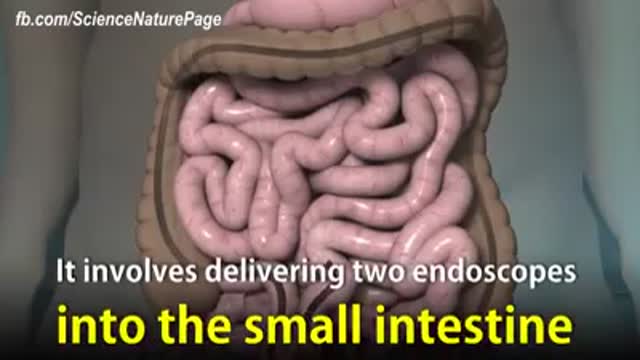
Endoscopic retrograde cholangiopancreatography, or ERCP, is a specialized technique used to study the bile ducts, pancreatic duct and gallbladder. Ducts are drainage routes; the drainage channels from the liver are called bile or biliary ducts. The pancreatic duct is the drainage channel from the pancreas.
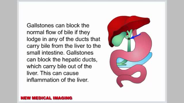
Gallstones are hardened deposits of digestive fluid that can form in your gallbladder. Your gallbladder is a small, pear-shaped organ on the right side of your abdomen, just beneath your liver. The gallbladder holds a digestive fluid called bile that's released into your small intestine. Gallstones range in size from as small as a grain of sand to as large as a golf ball. Some people develop just one gallstone, while others develop many gallstones at the same time. Gallstones are common in the United States. People who experience symptoms from their gallstones usually require gallbladder removal surgery. Gallstones that don't cause any signs and symptoms typically don't need treatment.
Magnetic resonance imaging (MRI) can be an important tool in the diagnosis of multiple sclerosis (MS). MRI can also be used to monitor the progression of the disease in people living with MS. How does it work? MRI uses very strong magnets, radio signals, and computer software to take 3-dimensional pictures of the inside of the body. Will I need contrast material? Maybe. Contrast material is a substance that temporarily changes the way imaging tools interact with the body. They are often used to visualize certain types of MS disease activity on the MRI. If your doctor thinks your scan requires this contrast material, you may get an injection before you get in the MRI machine. How long will it take? The time may vary based on the type of MRI. Be sure to discuss with your doctor in advance so he or she can provide you with exact timing. But don’t worry, you won’t have to stay still the whole time. The technician will let you know when they’re starting a new image.
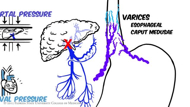
Portal hypertension is an increase in the blood pressure within a system of veins called the portal venous system. Veins coming from the stomach, intestine, spleen, and pancreas merge into the portal vein, which then branches into smaller vessels and travels through the liver.
