- Physical Examination
- Surgical Examination
- Ophthalmology
- Clinical Skills
- Orthopedics
- Surgery Videos
- Laparoscopy
- Pediatrics
- Funny Videos
- Cardiothoracic Surgery
- Nursing Videos
- Plastic Surgery
- Otorhinolaryngology
- Histology and Histopathology
- Neurosurgery
- Dermatology
- Pediatric Surgery
- Urology
- Dentistry
- Oncology and Cancers
- Anatomy Videos
- Health and Fitness
- Radiology
- Anaesthesia
- Physical Therapy
- Pharmacology
- Interventional Radiology
- Cardiology
- Endocrinology
- Gynecology
- Emergency Medicine
- Psychiatry and Psychology
- Childbirth Videos
- General Medical Videos
- Nephrology
- Physiology
- Diet and Food Health
- Diabetes Mellitus
- Neurology
- Women Health
- Osteoporosis
- Gastroenterology
- Pulmonology
- Hematology
- Rheumatology
- Toxicology
- Nuclear Medicine
- Infectious Diseases
- Vascular Disease
- Reproductive Health
- Burns and Wound Healing
- Other
Top videos
Fibromialgia Remedios Naturales, Como Curar La Fibromialgia, Medicamento Para Fibromialgia. http://fibromialgia-cura.info-pro.co/ -- Medicina Natural Para La Fibromialgia. Se estima que 5 millones de estadounidenses sufren de fibromialgia. Los dolores profundos y crónicos pueden tener un enorme impacto en la salud física y emocional. Desafortunadamente, los tratamientos son pocos y distantes entre sí, y los que existen a menudo vienen con la posibilidad de efectos secundarios desagradables. La medicina natural para la fibromialgia puede ser una bendición para los enfermos que la padecen. Estas terapias complementarias, suelen ser efectivas y pueden mejorar la calidad de vida y rejuvenecer un cuerpo atormentado por el dolor crónico. El masaje es a menudo uno de los métodos más eficaces para reducir los síntomas de fibromialgia. Alivia la rigidez, mejora el rango de movimiento, reduce el dolor y ayuda a controlar el estrés. Una técnica llamada liberación miofascial es especialmente adecuado para la fibromialgia el dolor calmante. La fascia es un tejido conectivo delgada que cubre y se extiende a lo largo del músculo. Los pacientes con fibromialgia sufren comúnmente de apriete de la fascia que contribuye al dolor y la fatiga muscular. La liberación miofascial es una técnica suave que relaja la fascia y reduce el dolor asociado. Las terapias naturales pueden ayudar desde dentro también. La investigación ha encontrado que muchos enfermos de fibromialgia tienen niveles bajos de vitamina D y magnesio. 100% natural aliviar el dolor y mejorar tu calidad de vida solo haciendo click aqui. http://fibromialgia-cura.info-pro.co
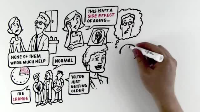
Hypothyroidism is a condition in which the body lacks sufficient thyroid hormone. Since the main purpose of thyroid hormone is to "run the body's metabolism," it is understandable that people with this condition will have symptoms associated with a slow metabolism. The estimates vary, but approximately 10 million Americans have this common medical condition. In fact, as many as 10% of women may have some degree of thyroid hormone deficiency. Hypothyroidism is more common than you would believe, and millions of people are currently hypothyroid and don't know it.
this animated surgery showing management of bone defects with the Precice Lengthening-Compression IM nail

Home Remedies For Acid Reflux, Ginger For Acid Reflux, Heartburn After Gallbladder Removal --- http://heartburn-acid-reflux.info-pro.co --- Stop using Pepto Bismol until you read the following… There is BREAKING scientific news reporting that many of America’s most popular antacids – both the ones you buy at the drug store and the ones you need prescriptions for… Are linked to more than a dozen forms of potentially DEADLY cancers. Click this link now to get the full story and see if you’re at risk. You’ll find out about a “just discovered” alternative to antacids…. Something that can permanently cure even the worst cases of acid reflux in as little few days, and that doesn’t require any pills or medications. Click Here: http://heartburn-acid-reflux.info-pro.co
It sounds like you're questioning whether or not your water may have broken, and this can actually be a hard thing for a lot of women to tell. Usually if your water breaks, it's just a trickle of fluid, and you're afraid to admit it to anyone because you think you peed your pants. And it is normal to pee your pants when you're pregnant because the bladder is right below the uterus, and if the baby moves just right, it might kick out a little bit of urine. So if you feel a trickle or a little tiny gush of fluid, what you want to do is put a pad or a pantie-liner on after going to the bathroom and emptying your bladder, and wait an hour and see if fluid continues to come out. And if it does, then you're not having bladder leakage issues - your water is probably broken.
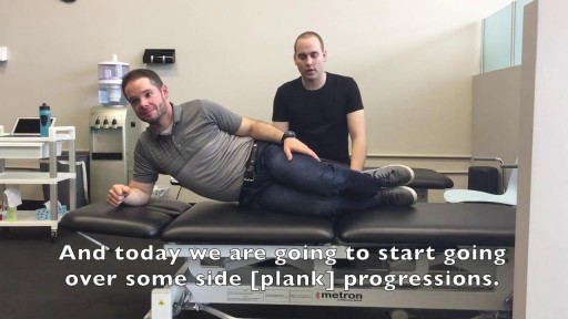
Wouldn’t it be great if there was an exercise that would work multiple muscles all at once? Well, today is your day! Mike and Tyler take you through the progressions of an exercise that targets not only your abs but your entire core (back muscles, transverses abdomens, rectus abdomens, obliques) AND your glute muscles. Get ready for the side plank and stay tuned for those progressions! Check us out on Social Media! Facebook: https://www.facebook.com/striveptandperformance/ Instagram: https://www.instagram.com/striveptandperf/ Twitter: https://twitter.com/StrivePTandPerf Blog: http://www.strivept.ca/blog
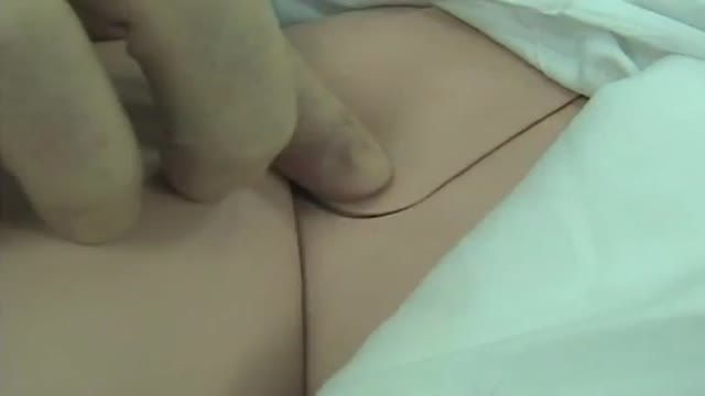
Femoral Venous Line Placement

Live in Caregiver Toronto - https://medwayhealthcare.com/ Foot Care Nurse - https://medwayhealthcare.com/foot-care/ Respite Care - https://medwayhealthcare.com/respite-care/
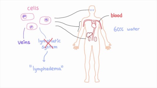
The lymphatic system is a network of specialized vessels (lymph vessels) throughout the body whose purpose is to collect excess lymph fluid with proteins, lipids, and waste products from the tissues. This fluid is then carried to the lymph nodes, which filter waste products and contain infection-fighting cells called lymphocytes. The excess fluid in the lymph vessels is eventually returned to the bloodstream. When the lymph vessels are blocked or unable to carry lymph fluid away from the tissues, localized swelling (lymphedema) is the result.
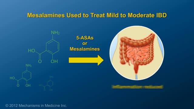
This animation describes risks of inflammatory bowel disease (IBD) and risks/benefits of medication (5-ASAs, steroids, immunomodulators, & biologics).
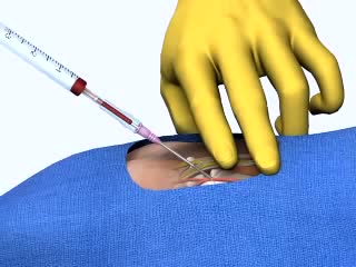
Arterial Cannulation
Acute hemothorax due to aortic rupture in aortic dissection with lung collapse and mediastinal shift.

UCLA Hand Transplant Procedure

Vaginal prolapse (also called vaginal vault prolapse) is quite common after a hysterectomy (surgery to remove the uterus), but not everyone who has a hysterectomy experiences POP. Without the uterine attachments to hold it up, the top of the vagina can drop into the vaginal canal.
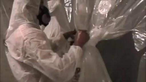
Every owner of a building where asbestos abatement activity occurs is responsible for the performance of the asbestos abatement activities by his/her agent, contractor, employee, or other representative. Each building owner is responsible for determining the amount of asbestos-containing material that may be disturbed during the course of work. The size and scope of the overall project, with particular reference to the total amount of asbestos-containing material that will be disturbed determines the reporting or filing requirements established in the Asbestos Control Program Rules. An asbestos project is defined as any form of work that will disturb more than 25 linear feet or more than 10 square feet of asbestos-containing material.

The hepatitis A virus, which causes the infection, usually is spread when a person ingests even tiny amounts of contaminated fecal matter. The hepatitis A virus infects liver cells and causes inflammation. The inflammation can impair liver function and cause other signs and symptoms of hepatitis A. Hepatitis A virus can be transmitted several ways, such as: Eating food handled by someone with the virus who doesn't thoroughly wash his or her hands after using the toilet Drinking contaminated water Eating raw shellfish from water polluted with sewage Being in close contact with a person who's infected — even if that person has no signs or symptoms Having sex with someone who has the virus
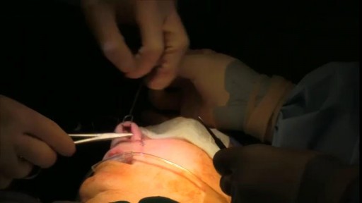
Third stage nasal econstuction: Nasolabial flap thinning, caudal septoplasty
Arthrocentesis involves both the puncture of a joint and the aspiration of its synovial fluid. It is typically used to make an accurate diagnosis of a painful, warm, swollen joint. Removal of excess fluid can be therapeutic. Analysis of the removed fluid helps to decipher its etiology. [1]
This 35 years old man lost his right wrist in metal lathe cut machine. the video is taken about 2 years after replantation. You can see another videos in my site: https://drliaghatclinic.com, https://instagram.com/liaghatclinic, https://t.me/liaghatclinic
Amanda walks Chelsea through how to do the basics of a one leg squat, as she tries not to fall over. Do it at home, at work, or at the gym! No equipment needed! Check us out on Social Media! Facebook: https://www.facebook.com/striveptandperformance/ Instagram: https://www.instagram.com/striveptandperf/ Twitter: https://twitter.com/StrivePTandPerf Blog: http://www.strivept.ca/blog
