- Physical Examination
- Surgical Examination
- Ophthalmology
- Clinical Skills
- Orthopedics
- Surgery Videos
- Laparoscopy
- Pediatrics
- Funny Videos
- Cardiothoracic Surgery
- Nursing Videos
- Plastic Surgery
- Otorhinolaryngology
- Histology and Histopathology
- Neurosurgery
- Dermatology
- Pediatric Surgery
- Urology
- Dentistry
- Oncology and Cancers
- Anatomy Videos
- Health and Fitness
- Radiology
- Anaesthesia
- Physical Therapy
- Pharmacology
- Interventional Radiology
- Cardiology
- Endocrinology
- Gynecology
- Emergency Medicine
- Psychiatry and Psychology
- Childbirth Videos
- General Medical Videos
- Nephrology
- Physiology
- Diet and Food Health
- Diabetes Mellitus
- Neurology
- Women Health
- Osteoporosis
- Gastroenterology
- Pulmonology
- Hematology
- Rheumatology
- Toxicology
- Nuclear Medicine
- Infectious Diseases
- Vascular Disease
- Reproductive Health
- Burns and Wound Healing
- Other
Top videos
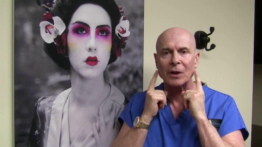
Mandibular Angle Augmentation with Injectable Filler
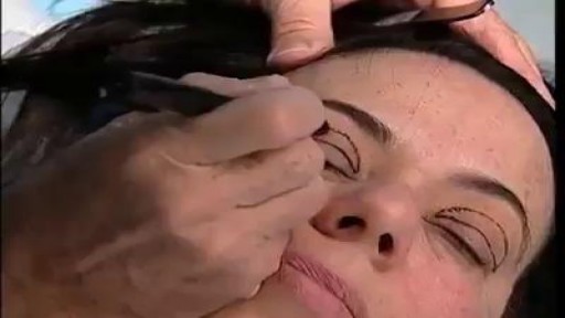
Plastic Eyelid Beauty surgery

Johns Hopkins orthopaedic hip and knee surgeon, Savyasachi "Savya" Thakkar, explains who is a candidate for knee replacement surgery, and what to expect during and after surgery. To learn more about our hip and knee replacement division, visit https://www.hopkinsmedicine.org/ortho #KneeReplacementSurgery #JohnsHopkins
Q&A's
0:15 What causes someone to need a knee replacement?
0:54 Describe the surgery.
1:36 What types of implants are used?
2:24 How is the recovery after surgery?
2:48 Describe the post-surgery physical therapy.
3:24 Do you perform revision surgery?

Roux-en-Y Gastric Bypass Surgery
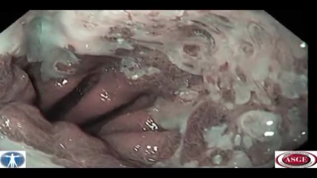
En Bloc Esophageal Mucosectomy, an experimental technique for the endolumenal management of Barrett's related dysplasia and neoplasia. High grade dysplasia is in indication for esophagectomy; however esophagectomy has a mortality rate up to 12 percent, and up to 56 percent of patients may develop s...erious post-operative complications. Multiple ablated lesions can progress under the neo-squamous layer, leading to buried Barrett's mucosa. With conventional piecemeal EMR, cautery effect limits evaluation in areas of interest, Barrett's epithelium is left behind, tissue is not evaluated in situ and invasive lesions may be missed due to incomplete sampling. A new technique, en bloc esophageal mucosectomy, or EEM, was developed. The technique begins with conventional EMR in the proximal esophagus to access the submucosal space. Conventional EMR is being performed here. The mucosa is resected using an electrothermal snare. The mucosal defect from the first EMR is seen here. EMR is then repeated on the opposing wall. Sequential EMR creates a complete concentric mucosal defect. In the following sequence the completely detached column of mucosa can be seen, bounded by submucosa and muscularis propria layers. Here in the stomach, the endoscope is retroflexed and is covered by a sleeve of esophageal mucosa which has been freed to the GE junction and inverted. This sequence demonstrates a double snare technique. This snare is alongside the endoscope. The snare has been passed through the working channel. The working channel snare is pulled back, and the snare alongside the scope is used to grasp the mucosal column. With tension on the column the working channel snare can be threatened and advanced. This sequence shows the snare as it is being passed down to the GE junction. At the GE junction, the snare is tightened and cautery is applied. This frees the column of mucosal tissue from the remaining attachment. The endoscope is then withdrawn. Then detached mucosal column can be grasped with a snare and retrieved. In the following sequence, the long column of mucosa is being withdrawn via the overtube. Here, endoscopic forceps have been passed through the column to demonstrates the concentric nature of the specimen. The length of mucosa can be seen here alongside 2 conventional EMR specimens. Approximately 15cm of tissues was removed in this case. On endoscopy immediately following the resection, there is no bleeding or evidence of perforation in the area of resection. The endoscope is advanced and the exposed submucosa can be appreciated down to the GE junction. This is the low power view of the histologic specimen generated by EEM. Metaplastic tissue adjacent to a dysplastic focus would be completely removed. With a high power view, the layers of the esophagus can be appreciated. The epithelium, lamina propria, muscularis mucosa and submucosa are visible, with no cautery artifact in the area of interest. The technique would remove metplasia, low grade dysplasia, high grade dysplasia, and intramucosal carcinoma, as well a T 1 a lesions. All the animals in this series tolerated the procedure well. A total of five non-survival procedures and 4 survival procedures were performed. In the survival procedures, all four swine thrived in the post-operative period. Two swine were then survived for 9 days following the procedure. On post —op day nine, after passing into the upper esophagus, the proximal margin of the mucosectomy is seen here. Healing appears to be occurring. There is no evidence of leak, and no stricting is seen at 9 days down to the GE junction Passing into the stomach, some residual feed can be seen. Two swine were then survived for 13 days. On this follow-up endoscopy, the area of the mucosectomy is again healing. There was a loose stricture in both animals and both were easily traversed with a 9.8 mm gastroscope. There was a gross appearance of re-epitheliazation in some areas. It is notable that the stricture was present in the proximal esophagus with no narrowing distally. At necropsy there was not eviden
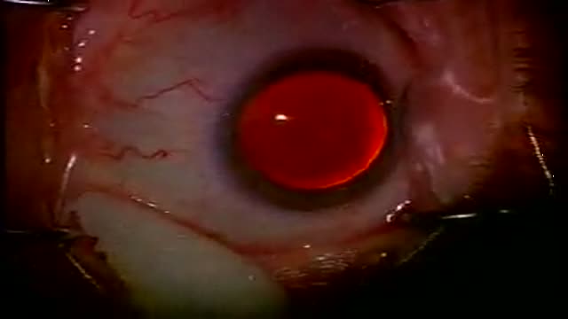
Cataract Surgery is a brief outpatient surgery. Done with modern advancements it reqires no stitches, no patches or no needles. It is done on an outpatient basis at http://redrocksurgerycenter.com by Drs. David Malitz and Dr. Surjeet Singh in Las Vegas, Nevada. Althouth the procedure is brief, there are risks, alternatives benefits and potential complications. To minimize these adverse effects, pick an experienced Surgeon. Just call 702-509-1733 for a FREE screening or visit http://sweyeinstitute.com. With our modern implants you can even reduce or eliminate your need for glasses after surgery! It is your eyes, it is worth the trip, most insurance accepted and uninsured patients are no problem!

Daca esti solist de muzica etno, populara sau usoara si ai un videoclip pe care vrei sa-l difuzezi pe Televiziunea Favorit, suna la 0722.410.597 Eugen Ungureanu. Pentru alte detalii urmareste emisiunea Ora Veseliei in fiecare vineri de la ora 18.00, sau viziteaza pagina de internet http://www.tvbucuresti.com
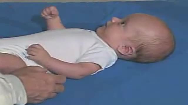
Pediatric Neurological Examination

Causas De La Diabetes, Signos De La Diabetes, Complicaciones Agudas De La Diabetes, Diabetico
http://todo-sobre-la-diabetes.good-info.co
Remedios Naturales Para Controlar La Diabetes
No es un hecho desconocido que en la actualidad los casos de diabetes se hayan incrementado considerablemente debido a la alimentación de la vida moderna y al estilo de vida que llevan las personas.
La diabetes tipo 2 es una enfermedad que se relaciona profundamente con la alimentación y se caracteriza por un elevado nivel de azúcar en sangre.
Este tipo de diabetes se puede controlar perfectamente llevando un estilo de vida saludable y una alimentación apropiada.
Existen muchos remedios naturales por los que puedes optar para luchar contra la diabetes:
Se trata de un remedio natural muy sencillo de realizar y que te resultará de gran utilidad para combatir la diabetes.
como eliminar la diabetes en pocos dias de manera natural y para siempre haciendo click aqui:
http://todo-sobre-la-diabetes.good-info.co
Haga Clic En El Enlace De Abajo Para Comprobar Que Funciona
http://todo-sobre-la-diabetes.good-info.co
Suscríbete A Nuestro Canal
https://www.youtube.com/user/VivirConSalud1
https://www.youtube.com/watch?v=i89z59Oi7Bg
Causas De La Diabetes, Signos De La Diabetes, Complicaciones Agudas De La Diabetes, Diabetico,
tipos de diabetes que existen,
Causa De La Diabetes,
que provoca la diabetes,
que ocasiona la diabetes,
historia natural de la diabetes mellitus,
fisiopatologia del pie diabetico,
cuales son las causas de la diabetes,
como se detecta la diabetes,
como detectar la diabetes,
clasificacion de la diabetes,
federacion internacional de diabetes
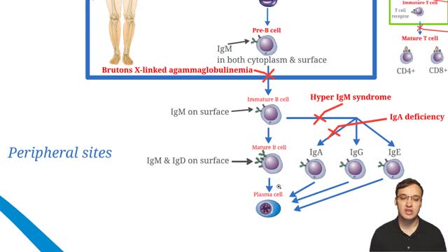
X-linked agammaglobulinemia (XLA), or Bruton agammaglobulinemia, is an inherited immunodeficiency disease caused by mutations in the gene coding for Bruton tyrosine kinase (BTK). The disease was first elucidated by Bruton in 1952, for whom the gene is named. BTK is critical to the maturation of pre–B cells to differentiating mature B cells. The BTK gene defect has been mapped to the long arm of the X chromosome at band Xq21.3 to Xq22, spanning 37.5kb with 19 exons forming 659 amino acids to complete the BTK cytosolic tyrosine kinase. A database of BTK mutations (BTKbase: Mutation registry for X-linked agammaglobulinemia) lists 544 mutation entries from 471 unrelated families showing 341 unique molecular events. No single mutation accounts for more than 3% of mutations in patients. In addition to mutations, a number of variants or polymorphisms have been found.
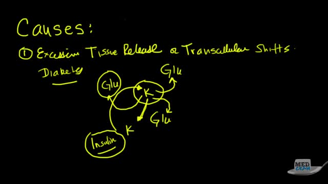
Hyperkalemia is defined as a serum potassium concentration higher than the upper limit of the normal range; the range in infants and children is age-dependent, whereas the range for adults is approximately 3.5-5.5 mEq/L. The upper limit may be considerably higher in young or premature infants, as high as 6.5 mEq/L.[5] Degrees of hyperkalemia are defined as follows[6] : 5.5-6.0 mEq/L – Mild 6.1-7.0 mEq/L – Moderate ≥7.0 mEq/L – Severe levels higher than 7 mEq/L can lead to significant hemodynamic and neurologic consequences. levels exceeding 8.5 mEq/L can cause respiratory paralysis or cardiac arrest and can quickly be fatal. Because of a paucity of distinctive signs and symptoms, hyperkalemia can be difficult to diagnose. Indeed, it is frequently discovered as an incidental laboratory finding. The physician must be quick to consider hyperkalemia in patients who are at risk for this disease process. (See Etiology.) However, any single laboratory study demonstrating hyperkalemia must be repeated to confirm the diagnosis, especially if the patient has no changes on electrocardiography (ECG). Because hyperkalemia can lead to sudden death from cardiac arrhythmias, any suggestion of hyperkalemia requires an immediate ECG to ascertain whether ECG signs of electrolyte imbalance are present (see Workup). Continuous ECG monitoring is essential if hyperkalemia is confirmed. Other testing is directed toward uncovering the condition or conditions that led to the hyperkalemia (see Workup). The aggressiveness of therapy for hyperkalemia is directly related to the rapidity with which the condition has developed, the absolute level of serum potassium, and the evidence of toxicity. The faster the rise of the potassium level, the higher it has reached, and the greater the evidence of cardiotoxicity, the more aggressive therapy should be. In severe cases, treatment focuses on immediate stabilization of the myocardial cell membrane, rapid shifting of potassium to the intracellular space, and total body potassium elimination. In addition, all sources of exogenous potassium should be immediately discontinued. (See Treatment.)
An increased prevalence of cardiovascular disease (CVD) has been found in women of childbearing age,[1] with the presence of CVD in pregnant women posing a difficult clinical scenario in which the responsibility of the treating physician extends to the unborn fetus. Profound changes occur in the maternal circulation that have the potential to adversely affect maternal and fetal health, especially in the presence of underlying heart conditions. Up to 4% of pregnancies may have cardiovascular complications despite no known prior disease. The European Society of Cardiology has published guidelines on the management of cardiovascular disease during pregnancy.[
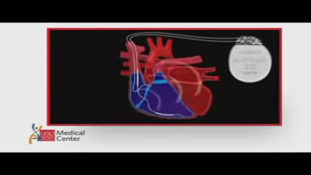
ICDs are useful in preventing sudden death in patients with known, sustained ventricular tachycardia or fibrillation. Studies have shown ICDs to have a role in preventing cardiac arrest in high-risk patients who haven't had, but are at risk for, life-threatening ventricular arrhythmias. View an animation of an ICD. Newer-generation ICDs may have a dual function which includes the ability to serve as a pacemaker. The pacemaker feature would stimulate the heart to beat if the heart rate is detected to be too slow. What is an Implantable Cardioverter Defibrillator (ICD)? An ICD is a battery-powered device placed under the skin that keeps track of your heart rate. Thin wires connect the ICD to your heart. If an abnormal heart rhythm is detected the device will deliver an electric shock to restore a normal heartbeat if your heart is beating chaotically and much too fast. ICDs have been very useful in preventing sudden death in patients with known, sustained ventricular tachycardia or fibrillation. Studies have shown that they may have a role in preventing cardiac arrest in high-risk patients who haven't had, but are at risk for, life-threatening ventricular arrhythmias.
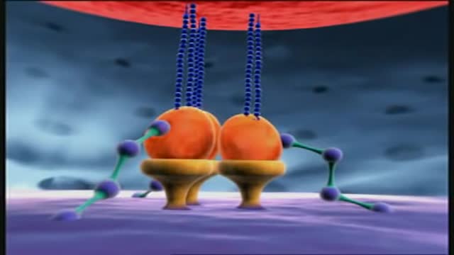
HIV gradually destroys the immune system by attacking and killing a type of white blood cell called a CD4 cell. CD4 cells play a major role in protecting the body from infection. HIV uses the machinery of the CD4 cells to multiply (make copies of itself) and spread throughout the body. This process, which is carried out in seven steps or stages, is called the HIV life cycle. HIV medicines protect the immune system by blocking HIV at different stages of the HIV life cycle. Antiretroviral therapy or ART is the use of HIV medicines to treat HIV infection. People on ART take a combination of HIV medicines from at least two different HIV drug classes every day. Because each class of drugs is designed to target a specific step in the HIV life cycle, ART is very effective at preventing HIV from multiplying. ART also reduces the risk of HIV drug resistance. ART can’t cure HIV, but HIV medicines help people with HIV live longer, healthier lives. ART also reduces the risk of HIV transmission (the spread of HIV to others).
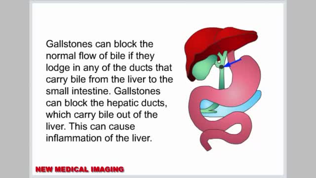
Gallstones are hardened deposits of digestive fluid that can form in your gallbladder. Your gallbladder is a small, pear-shaped organ on the right side of your abdomen, just beneath your liver. The gallbladder holds a digestive fluid called bile that's released into your small intestine. Gallstones range in size from as small as a grain of sand to as large as a golf ball. Some people develop just one gallstone, while others develop many gallstones at the same time. Gallstones are common in the United States. People who experience symptoms from their gallstones usually require gallbladder removal surgery. Gallstones that don't cause any signs and symptoms typically don't need treatment.
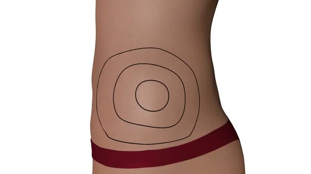
Questo Video 3D illustra la tecnica della Microlipocavitazione: sistema chirurgico ad ultrasuoni per ottenere l'emulsione del grasso in eccesso da eliminare. La Microlipocavitazione è una tecnica di chirurgia ambulatoriale, che richiede una modesta anestesia locale con un recupero delle proprie attività pressoché immediato.
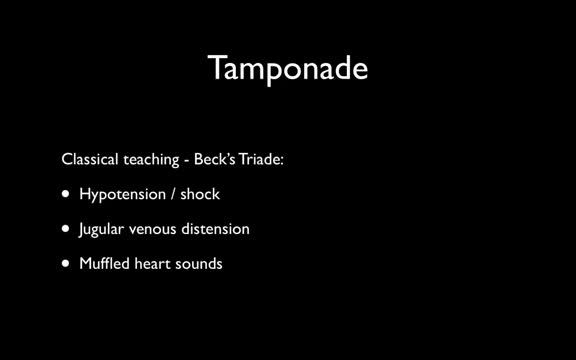
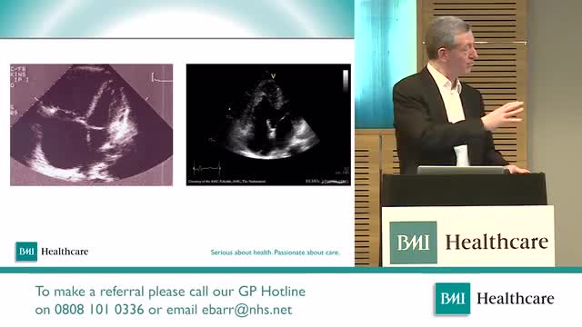
Cardiac tamponade is a medical emergency that requires urgent drainage of the pericardial fluid. Preferably, patients should be monitored in an intensive care unit. All patients should receive the following: Oxygen Volume expansion with blood, plasma, dextran, or isotonic sodium chloride solution, as necessary, to maintain adequate intravascular volume - Sagristà-Sauleda et al noted significant increase in cardiac output after volume expansion [24] (see the Cardiac Output calculator) Bed rest with leg elevation - This may help increase venous return Positive-pressure mechanical ventilation should be avoided because it may decrease venous return and aggravate signs and symptoms of tamponade. Inpatient care After pericardiocentesis, leave the intrapericardial catheter in place after securing it to the skin using sterile procedure and attaching it to a closed drainage system via a 3-way stopcock. Periodically check for reaccumulation of fluid, and drain as needed. The catheter can be left in place for 1-2 days and can be used for pericardiocentesis. Serial fluid cell counts can be useful for helping to discover an impending bacterial catheter infection, which could be catastrophic. If the white blood cell (WBC) count rises significantly, the pericardial catheter must be removed immediately. A Swan-Ganz catheter can be left in place for continuous monitoring of hemodynamics and to assess the effect of reaccumulation of pericardial fluid. A repeat echocardiogram and a repeat chest radiograph should be performed within 24 hours.
Chronic mesenteric ischemia (CMI) usually results from long-standing atherosclerotic disease of 2 or more mesenteric vessels. [1] Other nonatheromatous causes of CMI include the vasculitides, such as Takayasu arteritis. Symptoms are caused by the gradual reduction in blood flow to the intestine. [2] (See Presentation.) In 1958, Shaw and Maynard described the first thromboendarterectomy of the superior mesenteric artery (SMA) for the treatment of both acute mesenteric ischemia (AMI) and CMI. Several other surgical procedures have since been attempted, ranging from reimplantation of the visceral branch into the adjacent aorta to using an autogenous vein graft. In 1972, Stoney and Wylie introduced transaortic visceral thromboendarterectomy and aortovisceral bypass, which have proved to be highly effective techniques.
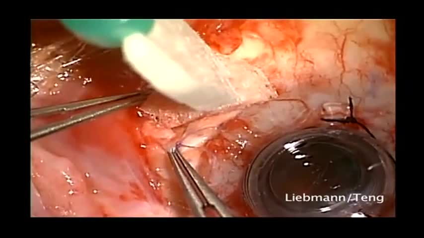
This patient had a history of herpes and had a previous corneal transplant that subsequently became opacified. There was also a previous Baerveldt implant placed into the anterior chamber. This surgery consists of Baerveldt tube being moved from the anterior chamber to posterior chamber, removal of failed graft, placement of temporary keratoprosthesis, pars plana vitectomy and corneal transplant. Jeffrey Liebmann, MD. Mark Speaker, MD. Uri Shabto, MD. Christopher Teng, MD.
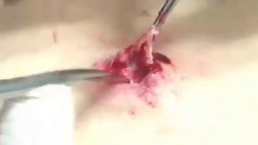
What is inside A Cyst? Watch it now
