- Physical Examination
- Surgical Examination
- Ophthalmology
- Clinical Skills
- Orthopedics
- Surgery Videos
- Laparoscopy
- Pediatrics
- Funny Videos
- Cardiothoracic Surgery
- Nursing Videos
- Plastic Surgery
- Otorhinolaryngology
- Histology and Histopathology
- Neurosurgery
- Dermatology
- Pediatric Surgery
- Urology
- Dentistry
- Oncology and Cancers
- Anatomy Videos
- Health and Fitness
- Radiology
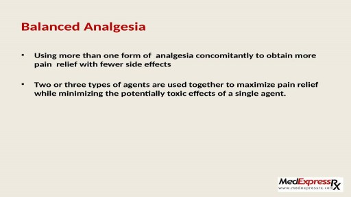
- Anaesthesia
- Physical Therapy
- Pharmacology
- Interventional Radiology
- Cardiology
- Endocrinology
- Gynecology
- Emergency Medicine
- Psychiatry and Psychology
- Childbirth Videos
- General Medical Videos
- Nephrology
- Physiology
- Diet and Food Health
- Diabetes Mellitus
- Neurology
- Women Health
- Osteoporosis
- Gastroenterology
- Pulmonology
- Hematology
- Rheumatology
- Toxicology
- Nuclear Medicine
- Infectious Diseases
- Vascular Disease
- Reproductive Health
- Burns and Wound Healing
- Other
Top videos
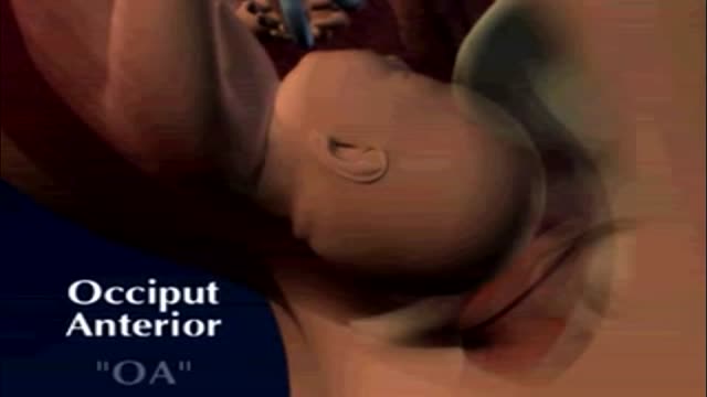
fetal position in womb at 34 weeks fetal position in womb week by week fetal position in womb at 19 weeksUnborn babies toss and turn and hold many different positions within the womb during the gestation period; pregnant women everywhere will attest to the fact that their children always start up the gymnastics at bedtime.
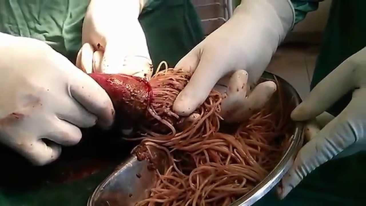
Watch that video of Bodybuilder's Colon Contains 10 lbs of Meat Worms
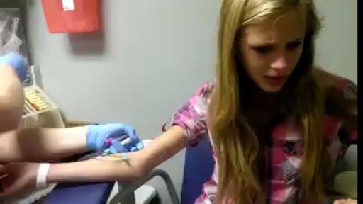
Panic attack from Injection:'(

Histology lab video reviewing the structure and cells of thin skin, thick skin, and skin sensory structures on digital histology slides. This video is a part of our Histology Video Course (https://youtube.com/playlist?l....ist=PLnr1l7WuQdDynxT
All Histology Videos: https://youtube.com/playlist?l....ist=PLnr1l7WuQdDynxT
Thank you to our sponsor Doc2Doc Lending, the Personal Lending platform designed for Doctors, by Doctors. Check out https://doc2doclending.com/davinci to learn more today.
DaVinci Academy Merch - Coffee mugs, T-shirts, hoodies and more: https://my-store-d90f46.creator-spring.com
Additional YouTube Content
Biochemistry videos: https://youtube.com/playlist?l....ist=PLnr1l7WuQdDzCUC
Anatomy Videos: https://youtube.com/playlist?l....ist=PLnr1l7WuQdDz2dK
DaVinci Cases Videos: https://youtube.com/playlist?l....ist=PLnr1l7WuQdDyJUl
The DaVinci Hour Podcast: https://youtube.com/playlist?l....ist=PLnr1l7WuQdDwSm9
DaVinci Academy Website: https://www.dviacademy.com/
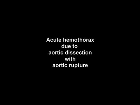
Acute hemothorax due to aortic rupture in aortic dissection with lung collapse and mediastinal shift.
DHI India - patient Ravish speaks about his For Alopecia Diagnosis, call 1800 103 9300 (Toll Free) Book your consultation - https://goo.gl/wBJh1o
Pain in joints or any part of body is very unpleasant and annoying experience. It is very common in people those suffering from arthritis. To get an end to all such pains, one can start using Generic Celebrex ( https://www.medexpressrx.com/celebrex-generic.aspx ). Here is a brief detail about this wonderful painkiller.
Are most spiders poisonous? The majority of the 3,000 spiders in the United States aren’t poisonous. Even if most spiders did bite, their fangs are too small or weak to puncture human skin. Their bites may leave itchy, red wounds that heal within a week or so. The spiders that do manage to bite through our skin and insert toxic venom can cause serious health complications. Read on to learn what spider bites look like, what spider varieties leave certain bites, and how to treat spider bites. What do spider bites look like? Identifying a spider bite is easier if you saw the spider that bit you, but it’s possible that you won’t notice the wound until hours later. Look for things like: swelling a red welt skin damage any troubling symptoms that accompany the bite Other possible symptoms that may accompany a spider bite include: itching or rash pain around the area of the bite muscle pain or cramping blister that’s red or purple in color sweating difficulty breathing headache nausea and vomiting fever chills anxiety or restlessness rashes swollen lymph glands high blood pressure Spider bites often take longer to heal than other insect bites, and they may affect skin tissues. It’s important to keep the bite clean to reduce the risk of infection. How to treat a spider bite at home In some cases, you can treat spider bites at home. For nonvenomous spider bites, follow these steps: Apply an ice pack on and off the bite for 10 minutes at a time. Elevate the area to reduce swelling. Take an antihistamine, such as diphenhydramine (Benadryl), to help with itching. Clean the area with soap and water to prevent infection. Apply antibiotic ointment to the area if blisters develop. Seek medical attention if you’re showing symptoms of a spider bite or if the symptoms don’t go away over time. Always seek medical attention if you suspect you’ve been bitten by one of the following species: brown recluse black widow hobo spider tarantula Brazilian wandering spider
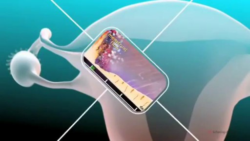
The menstrual cycle is the regular natural change that occurs in the female reproductive system that makes pregnancy possible. The cycle is required for the production of oocytes, and for the preparation of the uterus for pregnancy.
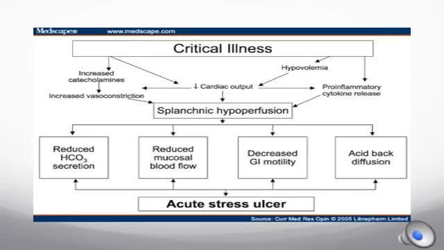
Stress-related mucosal disease (SRMD) is an acute, erosive gastritis representing conditions ranging from stress-related injury to stress ulcers (1, 2). Stress-related injury is superficial mucosal damage that presents primarily as erosions, whereas stress ulcers are deep, focal mucosal damage penetrating the submucosa with high risk for gastrointestinal bleeding (2, 3). Mucosal damage has been reported to occur during the first 24 hours of hospital admission in 75% to 100% of intensive care unit (ICU) patients (4, 5). Clinically important gastrointestinal bleeding can cause hemodynamic instability and increase the need for red blood cell transfusions (1). Significant bleeding may also increase the length of stay in the ICU and mortality (1).
Focal seizures (also called partial seizures [citation needed] and localized seizures) are seizures which affect initially only one hemisphere of the brain. [citation needed] The brain is divided into two hemispheres, each consisting of four lobes – the frontal, temporal, parietal and occipital lobes.

Vatche, Minassian, MD, MPH, Chief of Urogynecology, and Sarah Cohen, MD, MPH, Director of the Minimally Invasive Gynecologic Surgery Fellowship Program at Brigham and Women’s Hospital, perform a laparoscopic burch colposuspension, a procedure used to correct stress urinary incontinence.
Stress urinary incontinence is one of the most common types of incontinence and is characterized by urinary leakage during physical activities including coughing, sneezing, exercising, lifting, and laughing. As the condition progresses, it can become severe enough to happen with simple acts such as bending and walking. This condition is due to an anatomic weakness of the bladder neck which typically maintains the seal of urine during activity. Stress incontinence can result from a variety of conditions including vaginal childbirth, aging, menopause and obesity. As this is an anatomic condition, primary treatment may involve pelvic floor exercises and/or minimally invasive surgery.
Learn more about treatment for stress urinary incontinence:
Division of Urogynecology: http://www.brighamandwomens.or....g/Departments_and_Se
Division of Minimally Invasive Gynecologic Surgery: http://www.brighamandwomens.or....g/Departments_and_Se
Clinical Examination - Gait, Arms, Legs, Spine

#anatomy #histology #biology #bytesizemed
✨If you would like my help studying the structure of bones, check out my long-form video on it.
🔅Structure of Bone : https://youtu.be/MYInVEnnS_I
💫 For more videos like this, subscribe to my channel!
Byte Size Med: https://youtube.com/channel/UC....ZghvlgylH3r_CWfA18eF
📚Factual References & for Further Reading:
- DiFiore's Atlas of Histology
- Junqueira's Basic Histology
- Gartner's Concise Histology
- Openstax Anatomy and Physiology
https://openstax.org/details/b....ooks/anatomy-and-phy
- Openstax Biology
https://openstax.org/details/books/biology-2e
(The last two are links to open-source references. They are NOT affiliate links)
🌤 Note:
These are just a collection of my notes. So use them the way you would use borrowed notes from a friend. 📝
The images in this video are hand-drawn for illustration and explanation only.✍️ Hence, they may not be anatomically accurate. I am just one person making these videos. If there are any errors, that is unintentional. I try super hard to avoid them. Please let me know if you find any, so it gets clarified for other viewers. Science constantly evolves and changes. New discoveries are made everyday. So some of the information in these videos may become outdated. If you notice that, please let me know so I can update them.
⚡️Disclaimer:
These videos are NOT a substitute for a medical textbook. Textbooks are written by experts (which I do not claim to be), edited, proofread and referenced. Please use them.
The information has been sourced from multiple references as mentioned above. I draw all the pictures myself. But if I have inadvertently infringed on any copyright, that is completely unintentional. I only make these videos to impart education. If I have accidentally violated copyright in any way, do let me know so I can make the necessary changes or give credit to anyone who is owed the same.
These videos are NOT intended for patient education. They are NOT a substitute for diagnosis and treatment by a licensed medical professional. Always seek the advice of a qualified health care provider for any questions you may have regarding any medical condition, so that they can address your individual needs.
🔅They are ONLY meant to help students of medicine and health sciences with studying, and should be used for just that purpose and absolutely nothing else.
Byte Size Med. All Rights Reserved.
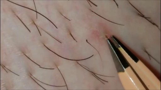
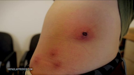
What is an ingrown hair cyst? An ingrown hair cyst refers to an ingrown hair that turns into a cyst — a large bump that extends between the skin’s surface and deep underneath it. The appearance is a cross between a regular ingrown hair and an acne cyst, though this is a different condition. These types of cysts are common among people who shave, wax, or use other methods to remove their hair. Although you may be eager to get rid of these cysts simply because of their appearance, it’s also important to watch for signs of an infection. Keep reading to learn what causes these cysts to form, plus how to treat them and prevent them from returning.


If you have an upcoming procedure at UC Davis Children’s Surgery Center, this video provides information and details of what you and your family can expect from arrival to check-in through to surgery and after care.
This video is also available in these languages:
Arabic: https://youtu.be/ERPikb0prlI
Dari: https://youtu.be/UW5fT433IGQ
Punjabi: https://youtu.be/Xq6PV2qtOMo
Russian: https://youtu.be/v223nDdN1b4
Spanish: https://youtu.be/4Jr4dkzAaWA
——
At UC Davis Children’s Hospital, we put your child at the center of everything that we do. It’s personalized care, uniquely sized for your child. You’ll see it in our child-friendly designs throughout the hospital, our farm-to-fork approach to dining, our playrooms and teen rooms and our team that feels like family. UC Davis Children’s Hospital is Sacramento’s only nationally ranked, comprehensive hospital for children, serving infants, children, adolescents and young adults with primary, subspecialty and critical care.
UC Davis Children’s Hospital: https://children.ucdavis.edu
Children’s Surgery Center: https://health.ucdavis.edu/chi....ldren/services/child
Child Life and Creative Arts Therapy: https://health.ucdavis.edu/chi....ldren/services/child
Fetal Care and Treatment Center: https://health.ucdavis.edu/chi....ldren/services/fetal
See the latest news from UC Davis Health: https://health.ucdavis.edu/newsroom
Kids Considered podcast: https://www.youtube.com/playli....st?list=PLM7qvIv8N9R
Facebook: https://www.facebook.com/UCDavisChildrensHospital
Instagram: https://www.instagram.com/ucdavischildren
Twitter/X: https://twitter.com/UCDavisChildren
——
#surgery #childrenshospital #surgeryrecovery #ucdavis

Watch that Real Human Body Decomposing Process On Video

Dealing with Anxiety and Panic Attacks
