- Physical Examination
- Surgical Examination
- Ophthalmology
- Clinical Skills
- Orthopedics
- Surgery Videos
- Laparoscopy
- Pediatrics
- Funny Videos
- Cardiothoracic Surgery
- Nursing Videos
- Plastic Surgery
- Otorhinolaryngology
- Histology and Histopathology
- Neurosurgery
- Dermatology
- Pediatric Surgery
- Urology
- Dentistry
- Oncology and Cancers
- Anatomy Videos
- Health and Fitness
- Radiology
- Anaesthesia
- Physical Therapy
- Pharmacology
- Interventional Radiology
- Cardiology
- Endocrinology
- Gynecology
- Emergency Medicine
- Psychiatry and Psychology
- Childbirth Videos
- General Medical Videos
- Nephrology
- Physiology
- Diet and Food Health
- Diabetes Mellitus
- Neurology
- Women Health
- Osteoporosis
- Gastroenterology
- Pulmonology
- Hematology
- Rheumatology
- Toxicology
- Nuclear Medicine
- Infectious Diseases
- Vascular Disease
- Reproductive Health
- Burns and Wound Healing
- Other
Top videos
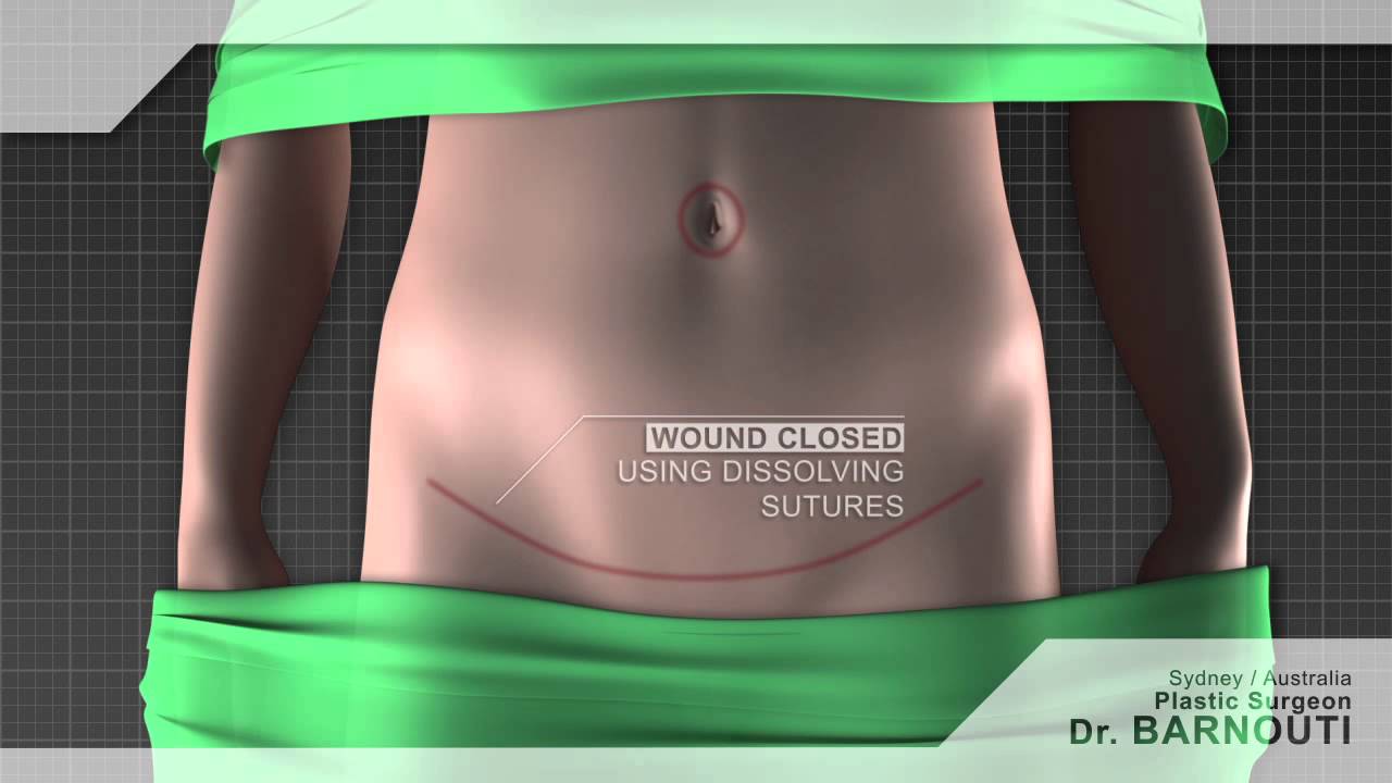
Tummy tuck Sydney Dr Barnouti. Call us on 02-9561 0222 or 1300 002 006
Broadway, Chatswood, Burwood NSW Australia
email:drbarnouti@australiaplasticsurgery.com.au
https://www.plasticsurgery-syd....ney.com.au/abdominop
What is a tummy tuck?
A tummy tuck operation is also known as abdominoplasty. It involves removing excess skin and fat from the stomach area, mainly the lower part of the tummy through surgical procedure. A tummy tuck operation is intended to leave the patient with a flatter tummy and to remove any signs of an 'apron' stomach or an overhang which is sometimes visible above underwear. The skin on this area tends to be stretched and of poor quality. A tummy tuck operation will usually focus on the lower part of the stomach, below the belly button and may require the belly button to be repositioned in some cases. The procedure is often carried out on women or men who have suffered from stretched skin in the stomach area after pregnancy, giving birth, excess fat deposition or weight loss.
What happens during a tummy tuck?
During a tummy tuck procedure the aim of the surgeon is to cut away fat and excess skin. To do this Dr Barnouti will make in incision on the lowest part of the stomach, where a fold will be visible above the pubic bone. He will take out as much excess fat as can be removed and will then cut the skin to fit back over the place where the fat has been removed from. It is important to have realistic expectations of a tummy tuck. Taking too much fat and skin away can result in folds at each end of the resulting scar which are sometimes referred to as "dog ears". Dr Barnouti will make sure you will not have this problem.
Who should have a tummy tuck?
Tummy tucks are recommended for either men or women who have an excess of fat and skin around their abdomen which cannot be removed by weight loss, exercise or liposuction. Tummy tuck operations in women are usually reserved for those who are not likely to have children as it is inadvisable to get pregnant again after having skin removed, this can cause the wound to stretch and scar.
The cost of a tummy tuck in Sydney Australia
The total cost is $7,900 if the patient's health fund cover the hospital's fees. In case the health fund does not cover the hospital's fee, the total cost will be around $12,000 inclusive of the Surgeon, assistant surgeon, Anaesthetist, hospital, operating theatre and follow ups visit.
Payment plans are alos available from Dr Barnouti's office in Chatswood, Burwood or Broadway.
A tummy tuck is a cosmetic procedure that removes excess skin and fatty tissue in order to give a flatter appearance to the stomach. Tummy tucks, also known as abdominoplasties, are ideal for patients who are not excessively overweight but suffer from an overhang of skin around the abdomen.
Performed under general anaesthetic, tummy tucks involve a horizontal incision being made just above the pubic area between the hip bones. Skin and fatty tissue is separated from the muscle and the area is tightened, with the excess skin and fatty tissues then being pulled downwards and removed.
Following your tummy tuck, there will be a scar present across the lower abdomen, but this will gradually fade. You may experience moderate tissue swelling for several months, but this will disappear with time. There may also be a sensation reduction just above the pubic area.
Once your tummy tuck recovery is complete however, you'll benefit from a more attractive figure and the ability to wear a wider selection of clothes.
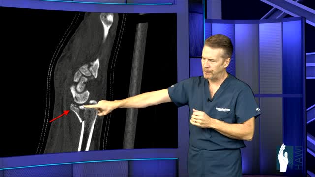
A distal radius fracture almost always occurs about 1 inch from the end of the bone. The break can occur in many different ways, however. One of the most common distal radius fractures is a Colles fracture, in which the broken fragment of the radius tilts upward. This fracture was first described in 1814 by an Irish surgeon and anatomist, Abraham Colles -- hence the name "Colles" fracture.
Meningitis is a common life-threatening medical emergency caused by infectious and non-infectious agents. Rapid and accurate evaluation by history and clinical examination is helpful to guide further specific investigation and treatment. Kernig's sign, Brudzinski's sign, and nuchal rigidity are bedside diagnostic signs used to evaluate suspected cases of meningitis. The presence of meningeal irritation, however, is not pathognomonic for meningitis.
Pediatric 4-Step Basic Technique
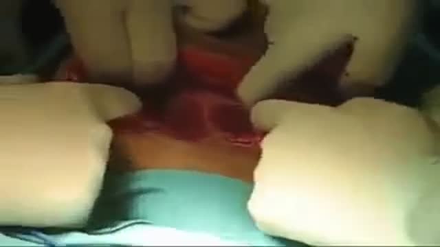
How to remove the Intra Uterine Device (IUD)

© 2023 Elsevier. All rights reserved. What are lymph nodes? Lymph nodes are small secondary lymphoid organs that are found along lymphatic vessels throughout the body.
Find our full video library only on Osmosis Prime: http://osms.it/more.
Join over 3 million current & future clinicians who learn by Osmosis, and over 130 universities around the world who partner with us to make medical and health education more engaging and efficient. We have unparalleled tools and materials to prepare you to succeed in school, on board exams, and as a future clinician. Sign up for a free trial at http://osms.it/more. If you're interested in exploring an institutional partnership, visit osmosis.org/educators to request a personalized demo.
Follow us on social:
Facebook: http://osms.it/facebook
Twitter: http://osms.it/twitter
Instagram for med: http://osms.it/instagram
Instagram for nursing: https://osms.it/ignursing
Linkedin: https://osms.it/linkedin
Our Vision: Everyone who cares for someone will learn by Osmosis.
Our Mission: To empower the world’s clinicians and caregivers with the best learning experience possible. Learn more here: http://osms.it/mission
Medical disclaimer: Knowledge Diffusion Inc (DBA Osmosis) does not provide medical advice. Osmosis and the content available on Osmosis's properties (Osmosis.org, YouTube, and other channels) do not provide a diagnosis or other recommendation for treatment and are not a substitute for the professional judgment of a healthcare professional in diagnosis and treatment of any person or animal. The determination of the need for medical services and the types of healthcare to be provided to a patient are decisions that should be made only by a physician or other licensed health care provider. Always seek the advice of a physician or other qualified healthcare provider with any questions you have regarding a medical condition. © 2023 Elsevier. All rights reserved.
The 12-lead ECG is a vital tool for EMT’s and paramedics in both the prehospital and hospital setting. It is extremely important to know the exact placement of each electrode on the patient. Incorrect placement can lead to a false diagnosis of infarction or negative changes on the ECG.
Level of fundus and exam
In breech position, the baby's bottom is down. There are a few types of breech: Complete breech means the baby is bottom-first, with knees bent. Frank breech means the baby's legs are stretched up, with feet near the head. Footling breech means one leg is lowered over the mother's cervix. You are more likely to have a breech baby if you: Go into early labor Have an abnormally shaped uterus, fibroids, or too much amniotic fluid Have more than one baby in your womb Have placenta previa (when the placenta is on the lower part of the uterine wall, blocking the cervix)

Colorectal surgeon Conor Delaney, MD, explains laparoscopic surgery for colon cancer, including how it works and what patients can typically expect before, during, and after the procedure.
Learn more about colon cancer at http://cancer.org/coloncancer
When you’re trying to conceive a baby it is worth giving anything a go which you think will boost your chances. This includes considering that there may be better positions for getting pregnant. But it pays to bear in mind that the human race has been around for over 200,000 years and most of us were probably conceived without our ancestors investing too much thought into the mechanics. Science has proven that successful conception isn’t so much about sexual position as the frequency of sex between a fertile couple. Basically, if you want to fall pregnant, don’t use contraception and have frequent, active and enjoyable sex. Importantly, don’t stress too much about whether you’re doing it the right way. Women can, and do, conceive in any position. Nature has a way of making sure of that.
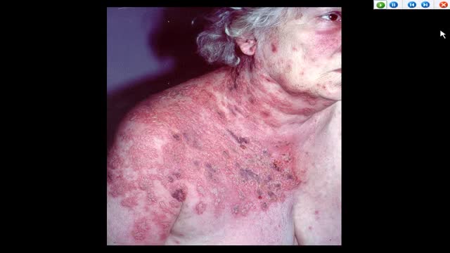
AUTO-HEMOTHERAPY IN HERPES CASES. THE STORY OF A DOCTOR IN FERME-NEUVE. CBC NEWS 1977.

Will you still love me if I have herpes? About 1 in 6 Americans between the ages of 14 and 49 is infected with herpes simplex virus type 2, according to a health survey released by the Centers for Disease Control and Prevention. If you’re living with herpes, HSV, HPV or other STDs, you're recommended to check out the largest STD support site STDdatings.
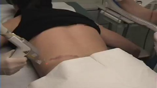
aser treatment for scars reduces the appearance of scars. It uses focused light therapy to either remove the outer layer of the skin’s surface or stimulate the production of new skin cells to cover damaged skin cells. Laser treatment for scars can reduce the appearance of warts, skin wrinkles, age spots, scars, and keloids. It doesn’t completely remove a scar.
plantar fasciitis and calcaneal spur can be treated by EPFR with calcanean drilling - endoscopic plantar fascia release علاج الشوكة العظمية للكعب بالمنظار د. أسامة الشاذلي مدرس جراحة العظام واستشاري جراحات و مناظير القدم والكاحل كلية الطب جامعة عين شمس


► Sign up here and try our FREE content: http://lectur.io/freecontentyt
► If you’re an medical educator or faculty member, visit: http://lectur.io/medytb2u
This video “Respiratory Histology” is part of the Lecturio course “Histology” ► WATCH the complete course on http://lectur.io/respiratoryhistology
► LEARN ABOUT:
- The cellular components of epithelium
- Structure and function of the conchae
- The cellular components of olfactory epithelium
- Components of the true vocal cord
- Function of the epiglottis
- The difference between bronchus, bronchiole, respiratory bronchiole
- Alveolar duct and Alveolar sac
- Components that make up the interalveolar septum
- Type I and Type II of alveolar cells, macrophages and endothelium
- Two separate blood supplies to the lungs and their functions
- Summary of the functions and the system of the respiratory system
► THE PROF: Your lecturer is Professor Geoff Meyer. He is currently teaching at the School of Anatomy, Physiology and Human Biology at the University of Western Australia (UWA). As a leading anatomy and histology expert he is also coordinating the Federative International Program for Anatomical Terminologies (FIPAT) of the International Federation of Associations of Anatomists (IFAA). Besides medical research on the ovarian function, steroidogenesis, corpus luteum, angiogenesis, and microcirculation, Geoff Meyer’s research activities also focus on developing innovative, computer-aided learning and teaching tools. For his inventiveness, Geoff Meyer has received a number of awards, including the Australian University Teaching Award.
► LECTURIO is your single-point resource for medical school:
Study for your classes, USMLE Step 1, USMLE Step 2, MCAT or MBBS with video lectures by world-class professors, recall & USMLE-style questions and textbook articles. Create your free account now: http://lectur.io/respiratoryhistology
► INSTALL our free Lecturio app
iTunes Store: https://app.adjust.com/z21zrf
Play Store: https://app.adjust.com/b01fak
► READ TEXTBOOK ARTICLES related to this video:
Histology of the Airways
http://lectur.io/respiratoryhistoloyarticle
► SUBSCRIBE to our YouTube channel: http://lectur.io/subscribe
► WATCH MORE ON YOUTUBE: http://lectur.io/playlists
► LET’S CONNECT:
• Facebook: https://www.facebook.com/lectu....rio.medical.educatio
• Instagram: https://www.instagram.com/lecturio_medical_videos
• Twitter: https://twitter.com/LecturioMed
• Pinterest: https://www.pinterest.de/lecturiomedical
• LinkedIn: https://www.linkedin.com/company/lecturio-medical/
0:00 Introduction
2:00 Respiratory system Summary of structure and function
3:32 Conducting portion
7:49 The Nasal cavities
11:27 The nasal cavity
13:06 Respiratory mucosa
15:26 Olfactory mucosa
17:13 Olfactory receptor
21:16 The larynx (and the epiglottis)
25:44 The trachea
29:16 The bronchi
31:35 Bronchiole
38:50 Alveolus
40:45 Air blood barrier
43:36 Alveolar macrophages
45:35 Blood supply and lymphatic drainage of the pulmonary lobule
