- Physical Examination
- Surgical Examination
- Ophthalmology
- Clinical Skills
- Orthopedics
- Surgery Videos
- Laparoscopy
- Pediatrics
- Funny Videos
- Cardiothoracic Surgery
- Nursing Videos
- Plastic Surgery
- Otorhinolaryngology
- Histology and Histopathology
- Neurosurgery
- Dermatology
- Pediatric Surgery
- Urology
- Dentistry
- Oncology and Cancers
- Anatomy Videos
- Health and Fitness
- Radiology
- Anaesthesia
- Physical Therapy
- Pharmacology
- Interventional Radiology
- Cardiology
- Endocrinology
- Gynecology
- Emergency Medicine
- Psychiatry and Psychology
- Childbirth Videos
- General Medical Videos
- Nephrology
- Physiology
- Diet and Food Health
- Diabetes Mellitus
- Neurology
- Women Health
- Osteoporosis
- Gastroenterology
- Pulmonology
- Hematology
- Rheumatology
- Toxicology
- Nuclear Medicine
- Infectious Diseases
- Vascular Disease
- Reproductive Health
- Burns and Wound Healing
- Other
Top videos
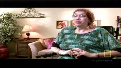

Acromegaly is usually caused by a noncancerous tumor. Middle-aged adults are most commonly affected. Symptoms include enlargement of the face, hands, and feet. Prompt treatment is needed to avoid serious illness. Drugs can reduce the effects of growth hormone. If needed, surgery and radiation may be used to remove tumor cells.
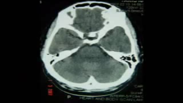
Right PCA Aneurysm Clipping Posterior Cerebral Artery
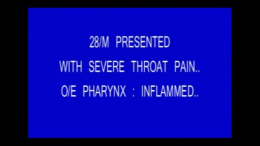
Pharyngitis is caused by swelling in the back of the throat (pharynx) between the tonsils and the voice box (larynx). Most sore throats are caused by colds, the flu, coxsackie virus or mono (mononucleosis). Bacteria that can cause pharyngitis in some cases: Strep throat is caused by group A streptococcus.
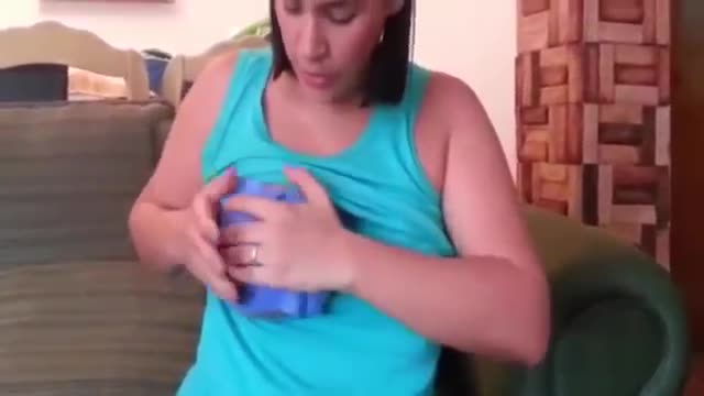
Hand Express Breast Milk,Breastfeeding
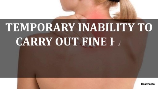
A cervical rib in humans is an extra rib which arises from the seventh cervical vertebra. Sometimes known as "neck ribs", their presence is a congenital abnormality located above the normal first rib. A cervical rib is estimated to occur in 0.2% (1 in 500 people) to 0.5% of the population.
DHI India - patient Ravish speaks about his For Alopecia Diagnosis, call 1800 103 9300 (Toll Free) Book your consultation - https://goo.gl/wBJh1o
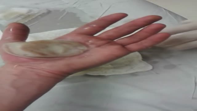
soaking the wound in cool water for five minutes or longer. taking acetaminophen or ibuprofen for pain relief. applying lidocaine (an anesthetic) with aloe vera gel or cream to soothe the skin. using an antibiotic ointment and loose gauze to protect the affected area.
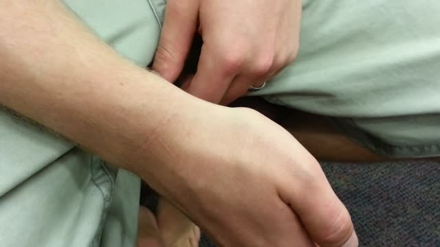
Anatomical snuff box tenderness due to probable scaphoid wrist bone fracture.
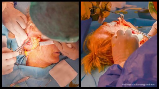
If you have gestational diabetes, your baby may be at increased risk of: Excessive birth weight. Extra glucose in your bloodstream crosses the placenta, which triggers your baby's pancreas to make extra insulin. This can cause your baby to grow too large (macrosomia).
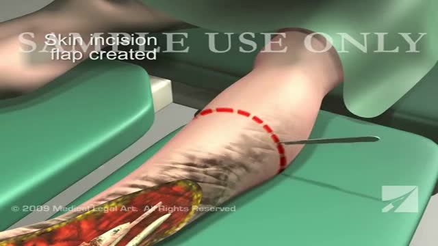
This 3d medical animation features a dramatic operative room overview of a left leg below the knee surgical amputation following severe trauma to the ankle and foot.
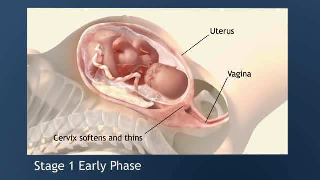
Early labour contractions usually feel like period pain, or you might experience a lower backache at 20 to 30 minute intervals. Sometimes these pains radiate from back to front, or vice versa. There's no need to start timing the contractions straight away – if they are mild contractions, ignore them. The first stage of labor is the longest and involves three phases: Early Labor Phase –The time of the onset of labor until the cervix is dilated to 3 cm. Active Labor Phase – Continues from 3 cm. until the cervix is dilated to 7 cm. Transition Phase – Continues from 7 cm. until the cervix is fully dilated to 10 cm.
http://vene-varicose-rimedi.good-info.co Vene Varicose, Vene Varicose Gambe, Chiva Varici, Laser Per Vene Varicose, Terapia Vene Varicose. Cosa Sono Le Vene Varicose e Quali Sono I Fattori Che Le Determinano? Le vene varicose si verificano quando il sangue non scorre naturalmente attraverso le vene del corpo verso il cuore. Quando ciò accade il sangue si accumula e ristagna nelle vene, di solito nelle vene delle gambe, dando alla pelle un aspetto davvero brutto e sgradevole. Quali Sono Le Vere Cause Delle Vene Varicose? Se si hanno vene varicose probabilmente ci si è già posti questa domanda più di una volta. Le vene varicose possono essere prodotte da vari fattori, quindi analizzeremo i fattori più importanti che possono produrle. 1. Vene Danneggiate. Molte volte le vene non funzionano correttamente, non permettendo al flusso dell sangue di circolare correttamente. Questo a lungo termine causa l'apparizione delle vene varicose. 2. L'Età. Nel corso degli anni molte parti del nostro corpo non funzionano più correttamente e le vene non sono un'eccezione. Gradualmente perdono la loro elasticità e diventano soggette a tutti i tipi di disturbi vascolari. 3. Il Sesso. Le donne sono più propense degli uomini a sviluppare le vene varicose. Le vene varicose compaiono di solito durante la fase di gravidanza, perché in questa fase si produce una maggiore quantità di estrogeni, un ormone che provoca un effetto rilassante nelle vene impedendo la corretta circolazione del sangue in esse. Altri fattori che possono produrre la comparsa delle vene varicose sono la menopausa femminile e le mestruazioni. 4. Stipsi. Le persone che soffrono di stipsi hanno maggiori probabilità di sviluppare vene varicose poiché al tempo dell'evacuazione lo sforzo è maggiore aumenteranno così le forti pressioni sulle vene. Come risultato di questo sforzo, le vene si danneggiano, si stirano e a lungo termine, si produce la comparsa di varici. 5. La Genetica. Se qualcuno nella vostra famiglia ha le vene varicose, è probabile che si svilupperanno in voi a un certo punto della vita. Questo è il motivo per il quale è molto importante sapere la storia della famiglia in modo che si possa essere in grado di identificare il fattore che ha dato origine alle vene varicose e trovare così il corretto trattamento per combatterle. 6. Il Sovrappeso. L’obesità e il sovrappeso possono portare alla comparsa di vene varicose, l'eccesso di peso esercita tensione in molte aree del corpo, includendo le vene. Inoltre, quando una persona è in sovrappeso viene ostacolata la circolazione del sangue, perché si mettono sotto pressione le vene e questo le danneggia. 7. Lesioni Alle Gambe. Le persone che hanno subito traumi a una o entrambe le gambe sono suscettibili allo sviluppo delle vene varicose. Quando si parla di lesioni queste includono: problemi di circolazione nelle gambe, interventi chirurgici, frattura della tibia e perone, ecc. 8. Stile Di Vita Sedentario. La vita sedentaria e le cattive abitudini alimentari possono favorire la comparsa di vene varicose. Una persona che passa seduta lunghi periodi di tempo, magari aggravando la situazione accavallando le gambe, impedisce al sangue di percorrere la sua strada consueta delle gambe al cuore. Il sangue non riesce quindi a lottare contro la forza di gravità e la mancanza di attività nelle gambe causa l'accumulo del sangue nello stesso luogo, portando alla creazione delle vene varicose. 9. Abbigliamento Stretto. Una certa varietà di capi d'abbigliamento e accessori, come ad esempio le scarpe con i tacchi alti, impediscono il flusso corretto del sangue per il corpo e favoriscono l'emergere della varici. Le scarpe chiuse con tacchi alti stringono il piede e impediscono il naturale flusso del sangue verso l'alto, in direzione del cuore. In questo modo il sangue resta all'interno delle vene nelle gambe e a lungo termine possono apparire le vene varicose. Con l'abbigliamento troppo stretto accade la stessa cosa perché impedisce che il sangue scorra correttamente per il corpo. 10. Attività Fisica Ad Alto Impatto. L’Attività fisica ad alto impatto come il sollevamento di pesi è molto popolare perché contribuisce a tonificare in poco tempo i vari gruppi muscolari. Tuttavia il sollevamento di oggetti pesanti, come i pesi, può favorire la comparsa di varici. Tutto questo è ciò che si può imparare in questa guida, Qui scoprirete i migliori metodi naturali per prevenire e contrastare la comparsa di vene varicose! http://vene-varicose-rimedi.good-info.co
Psychological counseling can help parents learn to provide an appropriate environment for the child, especially for homework completion. Psychological counseling may also help children and their families deal more effectively with the social and emotional aspects of TS. Counseling can be an important part of treatment and should not be overlooked.
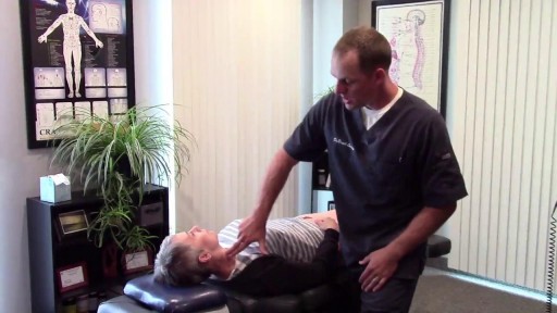
Wisdom teeth extractions can rear their ugly head later in life. This is a video of a patient with neck pain and neck weakness. When we stimulated the nerve fibers in the area of the extracted teeth there was an immediate improvement in her ability to control her neck muscles.
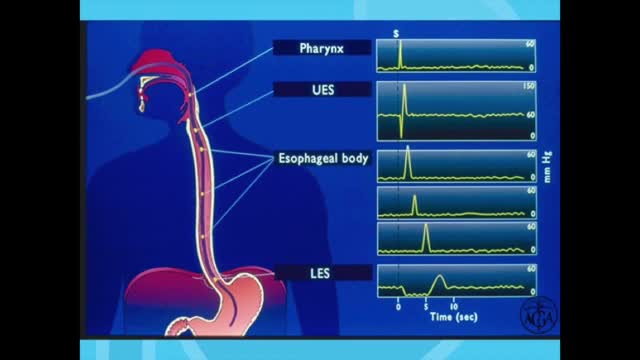
Esophageal manometry is a test used to measure the function of the lower esophageal sphincter (the valve that prevents reflux of gastric acid into the esophagus) and the muscles of the esophagus (see diagram). This test will tell your doctor if your esophagus is able to move food to your stomach normally.
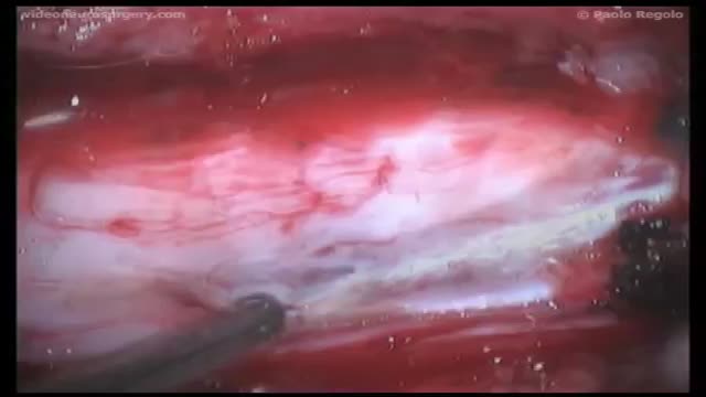
Syringomyelia is a cystic cavitation of the spinal cord associated with Chiari I malformation (70%) or basilar invagination (10%) or tumor. It may be a post-traumatic condition. There are 2 main forms: communicating with the central canal or subarachnoid spaces (Chiari I malformation); non communicating (trauma, tumors).
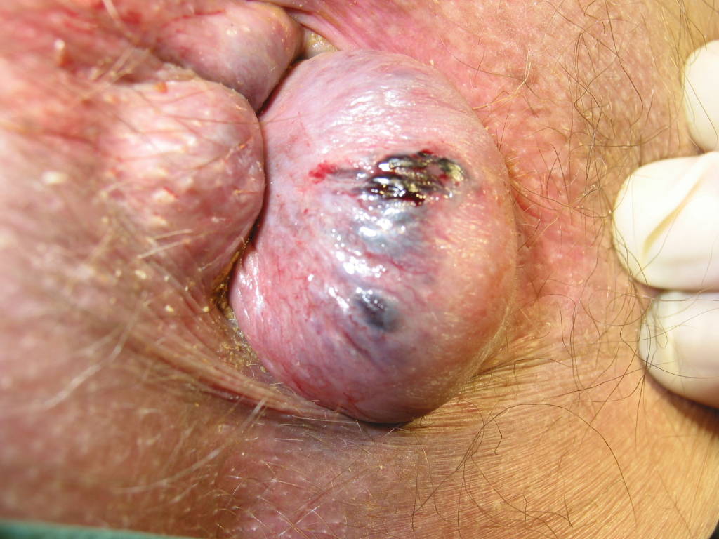
Watch that video to learn everything about Hemorrhoids Repairing Surgery