
- Physical Examination
- Surgical Examination
- Ophthalmology
- Clinical Skills
- Orthopedics
- Surgery Videos
- Laparoscopy
- Pediatrics
- Funny Videos
- Cardiothoracic Surgery
- Nursing Videos
- Plastic Surgery
- Otorhinolaryngology
- Histology and Histopathology
- Neurosurgery
- Dermatology
- Pediatric Surgery
- Urology
- Dentistry
- Oncology and Cancers
- Anatomy Videos
- Health and Fitness
- Radiology
- Anaesthesia
- Physical Therapy
- Pharmacology
- Interventional Radiology
- Cardiology
- Endocrinology
- Gynecology
- Emergency Medicine
- Psychiatry and Psychology
- Childbirth Videos
- General Medical Videos
- Nephrology
- Physiology
- Diet and Food Health
- Diabetes Mellitus
- Neurology
- Women Health
- Osteoporosis
- Gastroenterology
- Pulmonology
- Hematology
- Rheumatology
- Toxicology
- Nuclear Medicine
- Infectious Diseases
- Vascular Disease
- Reproductive Health
- Burns and Wound Healing
- Other
Top videos
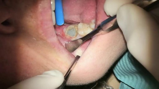

What Is It? Your wisdom teeth (third molars) usually start to erupt (enter your mouth) during the late teen years. Sometimes, there's not enough room for them. They may come into your mouth partially or not at all. Partial eruption of a wisdom tooth can create a flap of gum tissue next to the tooth. The flap can trap bits of food and debris. It can turn into a hotbed for bacteria. It's called pericoronitis if the tissue around the tooth becomes inflamed. Pericoronitis also can occur around a wisdom tooth that is still completely under the gums. Symptoms Symptoms include: Painful, swollen gum tissue in the area of the affected tooth. It can be difficult to bite down comfortably without catching the swollen tissue between your teeth. A bad smell or taste in the mouth Discharge of pus from the gum near the tooth More serious symptoms include: Swollen lymph nodes under your chin (the submandibular nodes) Muscle spasms in the jaw Swelling on the affected side of the face Diagnosis Usually, someone with pericoronitis goes to the dentist, complaining of pain in the area of the back tooth. Pericoronitis is diagnosed during the clinical exam. Your dentist will see inflamed gum tissue in the area of the unerupted or partly erupted wisdom tooth. The gums may be red, swollen or draining fluid or pus. Expected Duration Pericoronitis can be managed with antibiotics and warm salt water rinses. It goes away in about one week. However, it can return. This is likely to happen if the tooth does not completely enter the mouth and food and bacteria keep building up under the gum. Prevention You can help to prevent pericoronitis by brushing any erupting wisdom tooth and flossing around it. This will help make sure that food and bacteria do not build up under the gums. However, sometimes these steps do not work. If pericoronitis returns, you may need to have the flap of gum tissue removed. In some cases, the flap of tissue grows back and the wisdom tooth will need to be extracted. Treatment Pericoronitis can be tricky to treat. That's because the flap of gum tissue won't go away until the wisdom tooth emerges naturally, the tissue is removed or the tooth is removed. Your dentist will clean the area thoroughly by rinsing under the flap with water to remove bits of food and pus. Your dentist also may need to remove damaged tissue. If the area is infected, you'll most likely be given antibiotics. Your dentist will explain how to keep the area clean, which is the best way to prevent the problem from returning. This usually involves brushing and flossing daily and rinsing your mouth with water several times a day. These steps will help to prevent food from getting stuck under the gum flap. In some cases, your dentist may suggest removing the erupting tooth. Or the dentist may want to remove the tooth above it, which bites down on the gum below. If your dentist thinks the tooth may erupt fully into the mouth without problems, he or she may leave it alone. However, if pericoronitis comes back, the tooth may be extracted. Pericoronitis that causes symptoms should be treated as soon as possible. If it is not, the infection can spread to other areas of your mouth. The most severe cases are treated in a hospital. They sometimes require intravenous antibiotics and surgery. When To Call a Professional If you have symptoms of pericoronitis, make an appointment to see your dentist. If your wisdom teeth are coming in, visit your dentist at least twice a year for regular checkups. During those visits, the dentist can check on the progress of your wisdom teeth. Prognosis Pericoronitis does not cause any long-term effects. If the affected tooth is removed or erupts fully into the mouth, the condition cannot return.
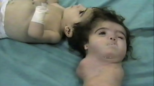
- A baby born with two heads is expected to survive after doctors removed the parasitic twin that was “feeding off” her blood supply. The baby girl, who is yet to be named, was born via C-section at Ram Snehi Hospital in northern India last month
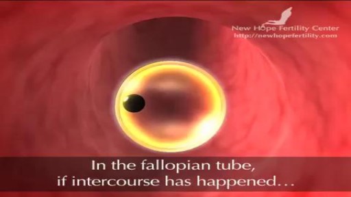
Before ovulation occurs, the average diameter of the dominant follicle is 22 to 24 mm (range 18-36 mm). It is the only marker that can predict ovulation with ease. * In stimulated cycle (hormonal treatment), generally, all or most of the antral follicles grow. The growth rate will be different for each of them.
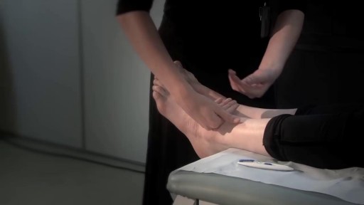
This brief exam will help you to quickly detect major risks and prompt you to refer patients to appropriate specialists.
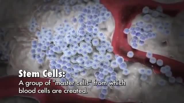
Red blood cells, most white blood cells, and platelets are produced in the bone marrow, the soft fatty tissue inside bone cavities. Two types of white blood cells, T and B cells (lymphocytes), are also produced in the lymph nodes and spleen, and T cells are produced and mature in the thymus gland.

American Andy received a Birmingham hip resurfacing surgery by Dr.Venkatachalam in Chennai India. The BHR is a highly successful treatment for patients for osteo-arthritis seeking relief from hip arthritis.
Hip resurfacing is offered by Madras Joint Replacement center by Dr.Venkatachalam. Visit http://www.hipsurgery.in
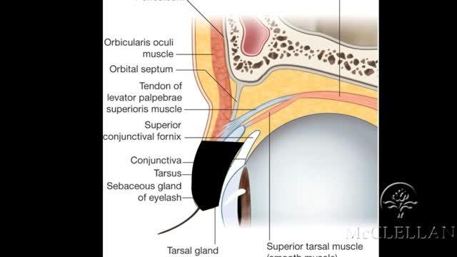
Live Surgery: Upper Eyelid Reconstruction - Tenzel Flap
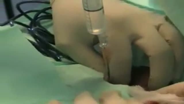
Tracheostomy in the ICU
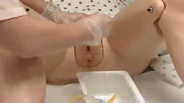
Foley's Catheter in FEMALES : How to?
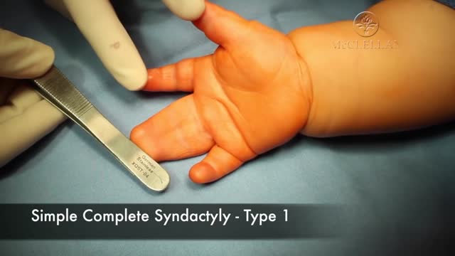
This is a surgical video demonstrating the release and skin grafting of the middle and ring fingers. It demonstrates the marking, dissection, and repair of the fingers.
This is another funny video we made regarding the use of Intermittent catheters. If you would like more information regarding our Service Plus Program( where we ship directly to your home and bill your Health Insurance), call 800-747-0246 or visit www.colonialmed.com
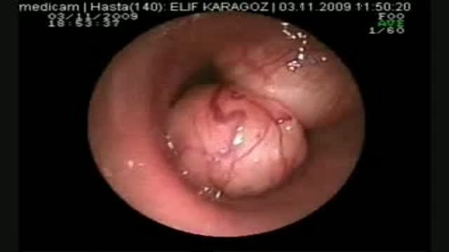
1 yıldır astım tedavisi gören 45 yaşında bayan hasta. Nefes darlığı şikayeti artması üzerine yapılan bronkoskopide trakea lümenini tamayakın tıkayan kitle gözlendi. Coller insizyonu ve parsiyel sternotomi ile yaklaşıldı ve rezeke edildi.
o you have an exercise routine that makes you feel absolutely awesome when you stick to it? Or a healthy eating pattern that makes you crackle with energy and vitality when you actually follow it. Well, today I’m sharing with you what makes me feel even more alive, energetic and vibrant than both of those… My daily sexual self-care practice.
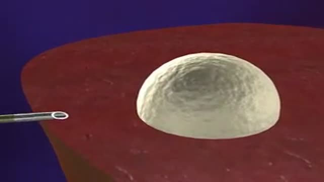
An animation showing the general principle of Radiofrequency Ablation of Hepatocellular carcinoma HCC.
A video discussing the Liver Cancer Treatment with Radiofrequency Ablation
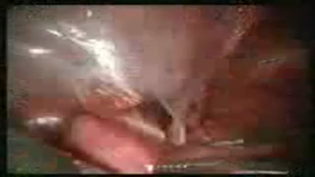
laparoscopic anterior resection
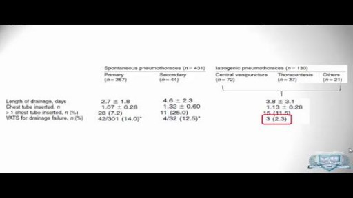
The surgeon may use treatment options such as thoracoscopy, electrocautery, laser treatment, resection of blebs or pleura, or open thoracotomy. Other surgical indications are as follows: Persistent air leak for longer than 7 days. Recurrent, ipsilateral pneumothorax.
The Closure Procedure for Varicose Veins is a clinically proven, minimally invasive procedure that treats varicose veins and their underlying cause, venous reflux, with little or no pain. Closure patients can walk away from the vein procedure and be back to everyday activities – either at home or at work – typically within a day.
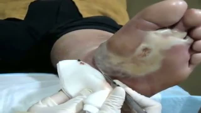
See http://nursing-resource.com for more on debridement.
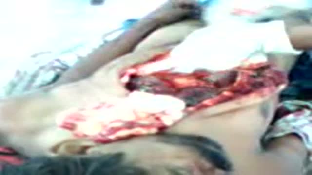
This is the postmortem of a male who died of due to a blunt injury to abdomen n thus the ruptue of the spleen. there was no any scra or wond on epidermis. but the 4th rib was fractured. all the above mentioned conditions are clearly seen in this video.
