- Physical Examination
- Surgical Examination
- Ophthalmology
- Clinical Skills
- Orthopedics
- Surgery Videos
- Laparoscopy
- Pediatrics
- Funny Videos
- Cardiothoracic Surgery
- Nursing Videos
- Plastic Surgery
- Otorhinolaryngology
- Histology and Histopathology
- Neurosurgery
- Dermatology
- Pediatric Surgery
- Urology
- Dentistry
- Oncology and Cancers
- Anatomy Videos
- Health and Fitness
- Radiology
- Anaesthesia
- Physical Therapy
- Pharmacology
- Interventional Radiology
- Cardiology
- Endocrinology
- Gynecology
- Emergency Medicine
- Psychiatry and Psychology
- Childbirth Videos
- General Medical Videos
- Nephrology
- Physiology
- Diet and Food Health
- Diabetes Mellitus
- Neurology
- Women Health
- Osteoporosis
- Gastroenterology
- Pulmonology
- Hematology
- Rheumatology
- Toxicology
- Nuclear Medicine
- Infectious Diseases
- Vascular Disease
- Reproductive Health
- Burns and Wound Healing
- Other
Top videos
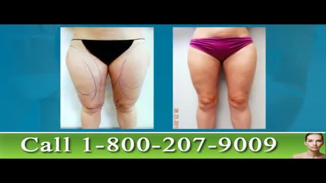
These Tampa Thigh Liposuction before and after photos show the work of renowned liposculpting surgeon Dr. Thomas Su. Dr. Su, of the Artistic Liposculpting Center specializes in body contouring procedures like leg liposuction. Thigh lipo is perhaps one of the most cosmetically rewarding liposuction procedures as it enhances the patient’s overall shape and contour. To learn more about leg and thigh lipo in Tampa, please visit http://www.artlipo.com/leg-liposuction-in-tampa-bay.html

The Principles of Laparoscopic Suturing
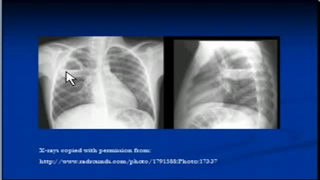
The video will describe how cavitating lesions appear on a chest x-ray. Please see my website for disclaimer.
Peritoneal Dialysis for Kidney Disease
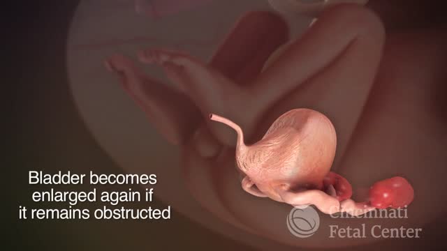
The etiology of BOO is diverse and definitely gender specific. Often anatomic causes induce functional abnormality that remains somewhat unique for each individual, regardless of sex. A full appreciation of the possible etiologies of obstruction is necessary in order to identify overt and more subtle scenarios. In women, iatrogenic causes of obstruction are the most common. Other entities account for far fewer of the cases. The obstruction evaluation in women is somewhat more diverse in terms of modalities used, with no single grouping of techniques that are generally apropos. Individualized evaluation remains a tenet of analysis, and urodynamic criteria used to diagnose BOO in women continue to evolve.
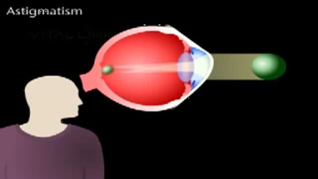
This animated video explains what is meant by astigmatism, which is a very common problem with the eyes.
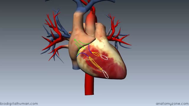
Heart Anatomy - Right Ventricle c
After 11 years of my work on my new migraine surgery, I start to do migraine surgery in all 4 principal places - places # 1 (STA) both sides, and places # 3 - Occipital artery also from both sides. You can see my first patients; he had bifrontal migraine headaches and daily chronic headaches in occipital area and the top of the head. On 30 September I sutured the occipital artery from both sides, and on 2 October I sutured STA in places # 1 from both sides. www.alisultaneh.8m.com
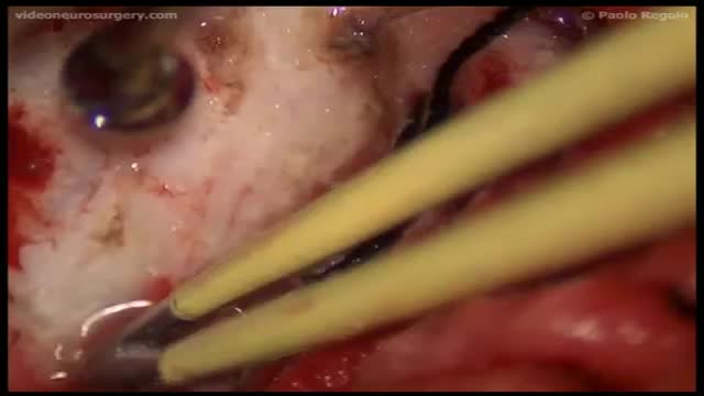
Chordoid meningioma, classified as atypical meningioma according to the World Health Organisation (WHO) classification, is a rare subtype, which represents only 0.5% of all meningiomas and is associated with a high incidence of recurrence. Multiple intracranial meningiomas are rare in non-neurofibromatosis patients. We present a female patient with both of these rare types of meningioma. The patient presented with two concurrent intracranial meningiomas, with one a meningotheliomatous subtype and the other a chordoid meningioma. Given the wide array of histological differential diagnoses in chordoid meningioma, immunohistochemistry has a significant role to play in differentiating them. Recurrence in chordoid meningioma can be generally predicted based on the extent of resection, the percentage of chordoid element, and proliferation indices.
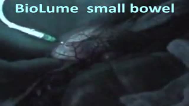
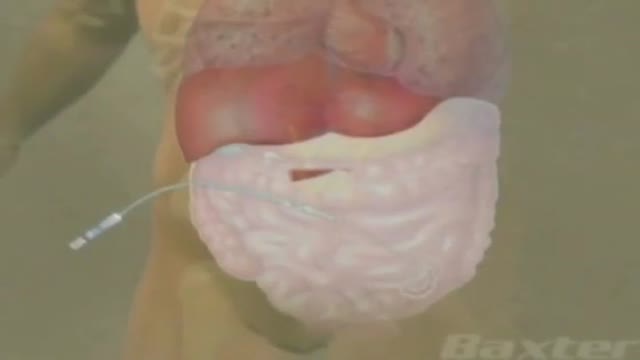
small bowel anastomosis (the luminescent material check for leaks, and good flow)
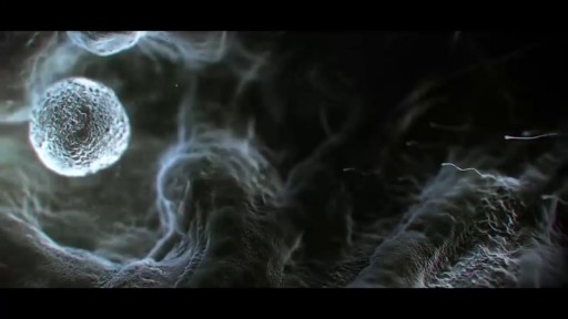
The journey of egg and sperm. There are a lot of casualties (deaths) among the sperm as they swim toward the egg. First, many get lost in the maze of a woman's uterus where they also have to contend with acidic vaginal secretions.
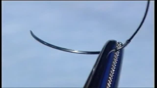
a video showing the correct position for needle holding
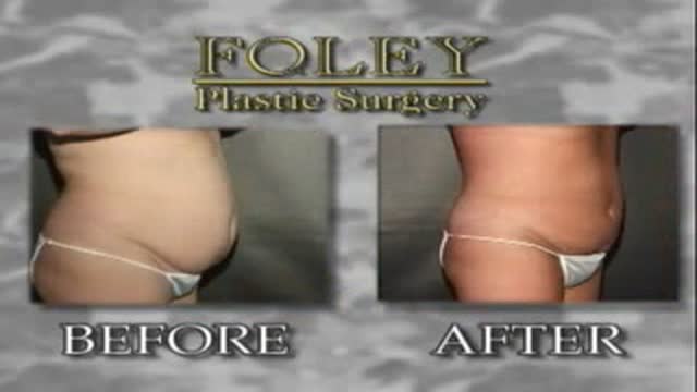
This is an Abdominal Liposuction surgery performed by Dr. Art Foley. Liposuction is a procedure that can help sculpt the body by removing unwanted fat from specific areas including the abdomen, hips, buttocks, thighs, knees, upper arms and neck. Although no type of liposuction is a substitute for dieting and exercise, liposuction can remove stubborn areas of fat that don't respond to traditional weight loss methods.

Five percent of pregnant women have gestational diabetes, so if you're expecting, understanding the basics of this common condition is vital
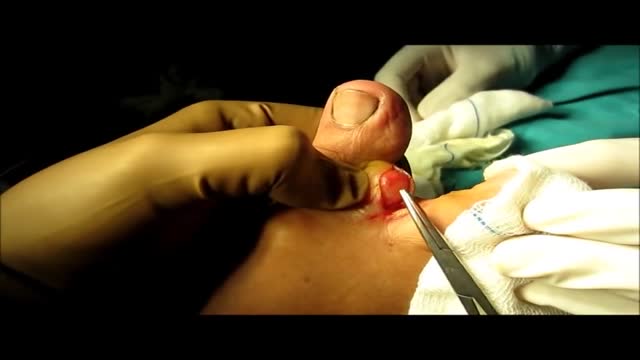
Lipoma From Foot (Inter Digital Web Space) Removal Technique
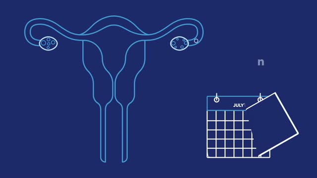
Wondering how pregnancy happens? It’s not as simple as it seems! Pregnancy takes several days, and there are lots of steps. Here are the basics on how pregnancy works.
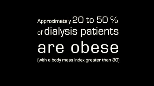
Because of his weight, Jimmie Jones was on the waiting list for a new kidney for 17 years. University of Illinois Hospital surgeons used robotic surgery to give him a life without dialysis.
Laparoscopic Appendectomy Video
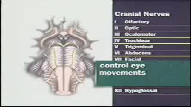
Examination of Cranial nerves III, IV and V: occulomotor,trochlear and trigeminal
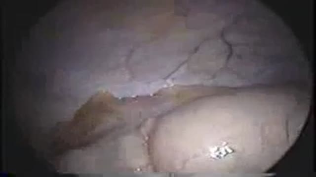
Laparoscopic removal of the spleen
