
- Physical Examination
- Surgical Examination
- Ophthalmology
- Clinical Skills
- Orthopedics
- Surgery Videos
- Laparoscopy
- Pediatrics
- Funny Videos
- Cardiothoracic Surgery
- Nursing Videos
- Plastic Surgery
- Otorhinolaryngology
- Histology and Histopathology
- Neurosurgery
- Dermatology
- Pediatric Surgery
- Urology
- Dentistry
- Oncology and Cancers
- Anatomy Videos
- Health and Fitness
- Radiology
- Anaesthesia
- Physical Therapy
- Pharmacology
- Interventional Radiology
- Cardiology
- Endocrinology
- Gynecology
- Emergency Medicine
- Psychiatry and Psychology
- Childbirth Videos
- General Medical Videos
- Nephrology
- Physiology
- Diet and Food Health
- Diabetes Mellitus
- Neurology
- Women Health
- Osteoporosis
- Gastroenterology
- Pulmonology
- Hematology
- Rheumatology
- Toxicology
- Nuclear Medicine
- Infectious Diseases
- Vascular Disease
- Reproductive Health
- Burns and Wound Healing
- Kidney Zone Podcast
- Other



Brain

Educational only: videos on MedicalVideos.com are for general information and are not medical advice. Always consult your own doctor about your health.
The human brain is the command center for the human nervous system. It receives input from the sensory organs and sends output to the muscles. The human brain has the same basic structure as other mammal brains, but is larger in relation to body size than any other brains.
This video in 30 seconds
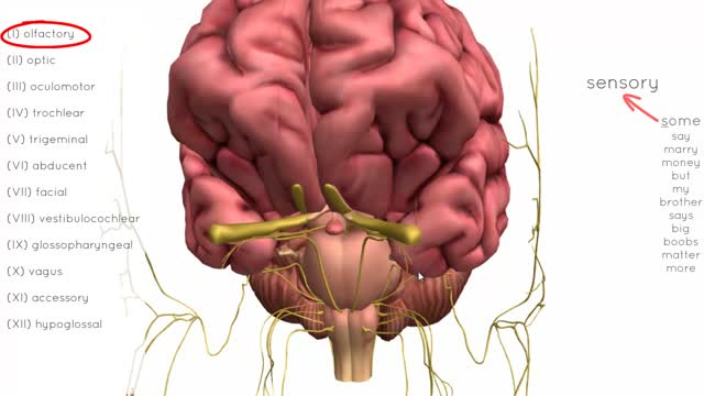
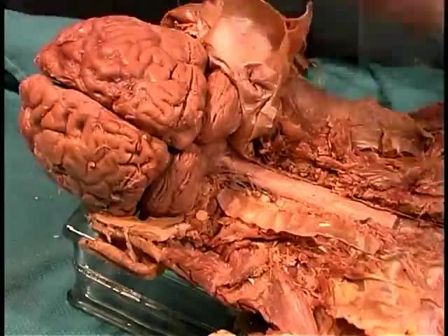
A presenter examines an unfixed human brain removed at autopsy to show how soft it is compared with formalin-fixed teaching specimens. He points out the ventral surface, surface vessels, cerebellum, pons, medulla, optic and olfactory nerves and temporal lobes. He also explains how cerebrospinal fluid cushions the brain and notes no sign of temporal lobe herniation in this specimen.
Key points
- The unfixed brain is described as very soft and easily damaged
- Surface vessels and the ventral surface of the brain are shown
- Cerebrospinal fluid is explained as a cushion for the brain
- The cerebellum, pons, medulla and cranial nerves are pointed out
- The temporal lobes are checked for signs of herniation
Chapters
- 0:10 — Fixed specimens versus the real brain
- 0:37 — The unfixed autopsy specimen
- 0:57 — Ventral surface, vessels and fluid
- 2:35 — Cerebellum, brainstem and nerves
- 3:12 — Checking for temporal lobe herniation
- 4:29 — Base of brain and its arteries
Video Transcript
A presenter examines an unfixed human brain, described as weighing 1,400 grams and removed at autopsy, to show how soft it is compared with formalin-fixed teaching specimens. He points out the ventral surface, surface vessels, cerebellum, pons, medulla, optic and olfactory nerves, and temporal lobes, explains cerebrospinal fluid cushioning the brain, and notes no sign of temporal lobe herniation in this specimen.
Read the full transcript
Students think that the brain is sort of the consistency of a rubber ball. And that's because in the laboratories and teaching specimens we have fixed, formal and fixed brains. However, if you're a trauma surgeon or a neurosurgeon, you realize that the brain is really very, very soft and much more vulnerable than the impression you get looking at the fixed brain. So I would like to show you a 1400 gram brain that has just been removed from an autopsy. And we are fortunate enough to be able to show you what a normal, unfixed,
recently deceased patient's brain would look like. This is the ventral surface of the brain. And what you see are the peel vessels, this nice blush with the clear leptomaninges, and the vessels running in between the arachnoid and the Pia. The cerebral spinal fluid has leaked out through the cisterns, and so that subarachnoid space is no longer visible unless I move the brain. But it's very, very soft. Notice it's totally squishy. It's the consistency. It's much softer than most of the meat you would see in a market.
So if I were to pinch this in either way, I could easily damage this with my thumb. In fact, neurosurgeons, when they are doing surgery, often just evacuate with a vacuum or suck out parts of the brain. This string here has been placed around the basilar artery so it can be suspended in a bucket or formaldehyde in order to denature the protein and harden it. If we didn't, it would sit on the bottom of the bucket and the brain would become deformed as you would see here,
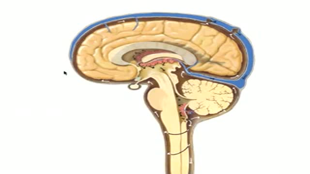
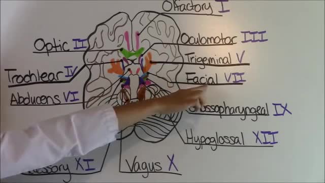
so that it would be compressed in this direction just from the weight of the brain. So that points out one of the purposes of the cerebral spinal fluid is to float the brain inside the calvarium and act as a cushion. The cerebellum is easily seen, the medulla, the pons. Here you can see the arachnoid spanning across the space here at the base of the brain. The optic nerves are very obvious as are the olfactory nerves. Here are our temporal lobes and here is the uncus on either side.
The uncus, remember, is the most mesial part of the temporal lobe. The tentorium that fit in here has been removed, but we can see a slight groove right along here where the tentorium sat. So there is no evidence in this brain of any of the temporal lobe herniating immediately over the edge of the tentorium. It's a beautiful specimen. Unfortunately, this cancer patient died of complications secondary to a cell transplant. Here you can see how soft and pliable the dura is, and we can look down between the hemispheres and see the corpus callosum.
I can also see a bit of the anterior cerebral artery coming over the surface. So this is the inter hemispheric fissure. And if I turn the brain around and we look at the occipital lobe, we can see the top of the cerebellum. And here there is a very small amount of blood that was actually seen on MRI that the patient had before death. If I let the cerebellum hang down a little bit, you can see the superior surface of the cerebellum. Turn it back around. So our very delicate and vulnerable brain with its two vertebral arteries on the medulla,
its basilar artery on the pons, it's branching to the posterior cerebral artery right here where you have the cerebral peduncle. And here we have the inter space here is the inter peduncular cistern. And we can see the two mammillary bodies, the pituitary gland stayed within the body. And here you can see the base of the brain with the optic nerve, the chiasm and the tract. You can see here an impression, a depression just from where I was holding the brain. Just from holding it that small amount of time, the depression from my finger is clearly visible.
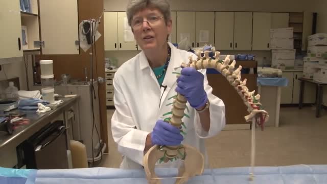
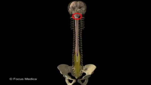
Think how vulnerable the brain is, then think how narrow and small the spinal cord is and what a devastating consequence, just a quick subluxation of the vertebra or a herniation of the disc can cause to the delicate spinal cord, which is about the width of one of these gyri. These gyri are nice, there's no big space between them, but neither does it look particularly swollen for a patient of this age.
Transcript auto-generated from the video’s audio for accessibility and study. Educational content only — not medical advice.
Video Overview
From our Anatomy Videos collection: Anatomy videos: 3D human anatomy, dissections and body systems explained clearly for medical students and curious minds. Browse all Anatomy Videos videos →
Medical Videos
Free medical and surgery videos for patients, students and professionals — trusted since 2007.
Founded by Dr. Mohamed Ibrahim, MD, PhD — physician-scientist.
Follow us on Facebook · Instagram
SORT BY-
Top Comments
-
Latest comments