
- Physical Examination
- Surgical Examination
- Ophthalmology
- Clinical Skills
- Orthopedics
- Surgery Videos
- Laparoscopy
- Pediatrics
- Funny Videos
- Cardiothoracic Surgery
- Nursing Videos
- Plastic Surgery
- Otorhinolaryngology
- Histology and Histopathology
- Neurosurgery
- Dermatology
- Pediatric Surgery
- Urology
- Dentistry
- Oncology and Cancers
- Anatomy Videos
- Health and Fitness
- Radiology
- Anaesthesia
- Physical Therapy
- Pharmacology
- Interventional Radiology
- Cardiology
- Endocrinology
- Gynecology
- Emergency Medicine
- Psychiatry and Psychology
- Childbirth Videos
- General Medical Videos
- Nephrology
- Physiology
- Diet and Food Health
- Diabetes Mellitus
- Neurology
- Women Health
- Osteoporosis
- Gastroenterology
- Pulmonology
- Hematology
- Rheumatology
- Toxicology
- Nuclear Medicine
- Infectious Diseases
- Vascular Disease
- Reproductive Health
- Burns and Wound Healing
- Kidney Zone Podcast
- Other
What Is an Endometrial Biopsy?
What is an endometrial biopsy? How a small sample of the womb lining is taken, step by step, what it can show, and what happens next.
11/03/26 • 5 Views


An endometrial biopsy comes down to one small thing: a tiny sample of the lining of the womb, taken so a laboratory can look at its cells under a microscope. That sample can answer questions that a scan or a blood test often can't — why bleeding has changed, why bleeding has appeared after the menopause, or whether the lining has grown in a way that needs treatment. The test is usually done in a clinic room, without a general anaesthetic, and the sampling itself takes only a short time.
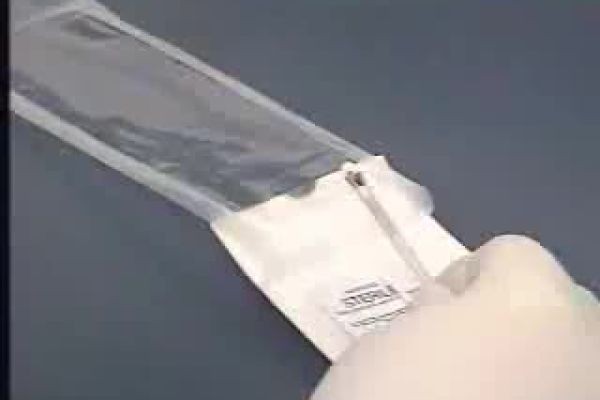
The video at the top of this page, "Endometrial Biopsy of Uterus", runs for about two minutes and twenty seconds and shows the biopsy itself, so you can see the instrument and the setting before reading the steps below. It is a short clinical recording rather than a lecture, and it is best watched as a visual companion to the written steps that follow.
Step 1: A reason to look at the lining directly
Doctors suggest this test when they need information from the tissue itself. The common reasons are bleeding that is heavier, longer or less predictable than usual, bleeding between periods, and — importantly — any bleeding after the menopause. A related video on this site, "Post-Menopausal Bleeding", makes that last point plainly: bleeding after the menopause isn't normal and should be evaluated. An ultrasound scan may come first, and if the lining looks thickened, or bleeding keeps happening without a clear explanation, a biopsy is a direct way to check the cells.
What the laboratory is looking for is a pattern. The lining might be entirely normal for your stage of life. It might show hormonal effects, inflammation, or a polyp-like growth. Sometimes it shows overgrowth of the lining — hyperplasia — and, less often, precancerous changes or cancer. That range is exactly why the test exists: the symptoms alone can't separate those possibilities. Our cancer condition hub gathers the site's cancer-related pages together, and our general guide to biopsy and how results are read explains what happens to tissue samples more broadly.
Step 2: Getting ready, briefly
There is usually little to prepare. You'll be told if the appointment needs to be timed to a particular point in your cycle, and it's worth mentioning every medicine and supplement you take, any chance you could be pregnant, and any previous difficult examination or procedure involving the cervix, so the clinician can plan around it. Some teams suggest an ordinary pain-relief tablet beforehand; follow your own team's advice on that rather than a general rule, because what's suitable depends on you.
Tell the team, too, about symptoms that might point to an infection, such as unusual discharge or pelvic pain. A biopsy is normally postponed if an active infection is suspected, because passing an instrument through the cervix could carry infection upwards. None of this preparation is complicated — it's mostly about giving the clinician the full picture before they begin.
Step 3: The sample is taken
You'll lie on an examination couch, usually with your legs supported, much as for a smear test. The clinician places a speculum so they can see the cervix, may clean the cervix, and then passes a thin, flexible tube through the opening of the cervix into the womb. That tube is the biopsy device: with a small plunger or suction, it draws a narrow strip of the lining into the tube as it's gently moved and rotated. The sampling part is brief, though it may be repeated once or twice to get enough tissue.
Most people describe the uncomfortable part as strong period-type cramping while the tube is inside, easing once it is removed. It isn't usually described as sharp pain, but experiences vary, and you can ask the clinician to pause or stop. Occasionally the cervix is too tight for the tube to pass — this can happen after the menopause, for example — in which case the clinician will stop and discuss other ways of sampling the lining rather than forcing it.
Step 4: The tissue goes to the laboratory
The sample is placed in a small pot of preservative and sent to a pathology laboratory, where it's processed, sliced very thinly, stained, and examined under a microscope by a specialist. That laboratory step, not the clinic appointment, is what produces the answer, which is why results take time: the clinic will tell you how and when to expect them, and it's worth asking before you leave if that hasn't been explained.
While you wait, expect your body to feel fairly normal fairly quickly. Light spotting and mild cramps for a day or so are common. Your team will tell you what to watch for — heavier bleeding, worsening pain, fever, or offensive-smelling discharge are reasons to contact them promptly, as they can be signs of infection or another complication that needs checking.
Step 5: Reading the result, and what follows
A normal or benign result often ends the investigation, or points it back towards hormonal or other non-tissue causes of the bleeding. A result showing inflammation may lead to treatment for infection. Hyperplasia — an overgrowth of the lining — is managed in different ways depending on its type and your circumstances, from monitoring and hormone treatment to surgery, and your specialist will explain which applies and why. If precancerous changes or cancer are found, you'll be referred promptly along the appropriate specialist pathway; the biopsy has done its job by finding the problem at the tissue level, where it can be acted on.
Sometimes the report says the sample was insufficient — there simply wasn't enough tissue to judge. That isn't a result to panic about; it means the question hasn't been answered yet, and the usual next step is another way of looking at the lining. One of those is hysteroscopy, where a thin camera is passed into the womb so the lining can be seen directly and a targeted sample taken; our guide to hysteroscopy explains it, and our article on uterine fibroids covers another common cause of heavy bleeding that investigations may uncover.
Taken together, the five steps are simple: a reason to sample, a short preparation, a brief clinic procedure, a careful laboratory examination, and a result that decides what happens next. For a test that can feel daunting in advance, its value is in how directly it answers the question.
References & further reading
- MedlinePlus — Endometrial biopsy: https://medlineplus.gov/ency/article/003917.htm
- Johns Hopkins Medicine — Endometrial biopsy: https://www.hopkinsmedicine.org/health/treatment-tests-and-therapies/endometrial-biopsy
- NHS — Post-menopausal bleeding: https://www.nhs.uk/symptoms/post-menopausal-bleeding/
Frequently Asked Questions
Does an endometrial biopsy need anaesthetic?
It is usually done in a clinic without a general anaesthetic. Some people take an ordinary pain-relief tablet beforehand if their team suggests it, and the cramping during sampling is usually brief. If the cervix cannot be passed comfortably, the clinician will stop and discuss alternatives rather than continue.
How long does the appointment take?
The sampling itself takes only a few minutes, within a normal clinic appointment. The longer wait comes afterwards, because the tissue has to be processed and examined in the laboratory before a result can be reported.
Can the biopsy miss something?
A small sample can't represent every part of the lining, and sometimes the sample is too scant to judge. If symptoms continue or the sample was insufficient, your team may suggest hysteroscopy, which lets them see the lining directly and take a targeted sample.
When should I seek help after the biopsy?
Light spotting and mild cramps for a day or so are common. Contact your team promptly for heavier bleeding, worsening pelvic pain, a fever, or discharge that smells unpleasant, as those can signal a complication that needs treatment.
MedicalVideos.com has published trusted medical videos since 2007. The site was founded by Dr. Mohamed Ibrahim, MD, PhD — physician-scientist.
Educational only — this article is general information about endometrial biopsy, not medical advice and not a diagnosis. Whether this test, or any investigation, is right for you is a matter for your own care team, who know your results and your history.
Related Articles






Medical Videos
Free medical and surgery videos for patients, students and professionals — trusted since 2007.
Founded by Dr. Mohamed Ibrahim, MD, PhD — physician-scientist.
Follow us on Facebook · Instagram