- Physical Examination
- Surgical Examination
- Ophthalmology
- Clinical Skills
- Orthopedics
- Surgery Videos
- Laparoscopy
- Pediatrics
- Funny Videos
- Cardiothoracic Surgery
- Nursing Videos
- Plastic Surgery
- Otorhinolaryngology
- Histology and Histopathology
- Neurosurgery
- Dermatology
- Pediatric Surgery
- Urology
- Dentistry
- Oncology and Cancers
- Anatomy Videos
- Health and Fitness
- Radiology
- Anaesthesia
- Physical Therapy
- Pharmacology
- Interventional Radiology
- Cardiology
- Endocrinology
- Gynecology
- Emergency Medicine
- Psychiatry and Psychology
- Childbirth Videos
- General Medical Videos
- Nephrology
- Physiology
- Diet and Food Health
- Diabetes Mellitus
- Neurology
- Women Health
- Osteoporosis
- Gastroenterology
- Pulmonology
- Hematology
- Rheumatology
- Toxicology
- Nuclear Medicine
- Infectious Diseases
- Vascular Disease
- Reproductive Health
- Burns and Wound Healing
- Other
Physical Examination
Upper Limb Examination Video
Vital Signs and Chest Examination
Abdomen Exam Video

Musculoskeletal Physical Examination Lecture

Physical Examination Introduction

Neurology Physical Examination Lecture
Cardiology Physical Examination Lecture

Pulmonary Physical Examination Lecture

Abdominal Physical Examination Lecture

Integrative Physical Examination Lecture

Emergency Physical Examination Lecture

ENT Physical Examination Lecture
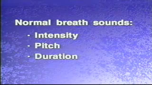
Normal and Adventitious Breath Sounds

Thyroid Exam Physical Exam
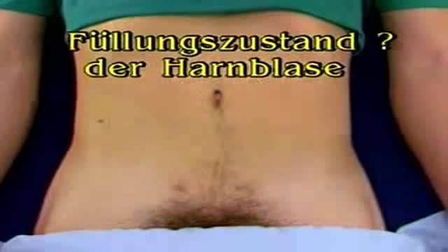
Urinary Bladder Medical Exam
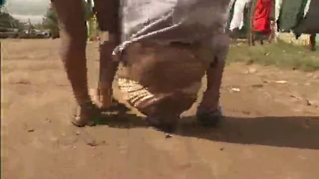
Giant Swollen Leg Elephantiasis Filariasis

When it comes to our health men over the age of 45 are in need of regular doctor visits and testing, as a large percentage of medical decisions are based on the lab test results.

Doctor Amneris Luque talks about HIV Prevention in Africa. STDdatings .com was created to help people to learn HIV / AIDS prevention, share medical treatments, find trusted people to talk, get help and advice.

In this medical video: This 72-year-old patient was unable to resist blinking when we tapped on the glabella. This is the glabellar reflex or Myerson's sign . It is often an early sign of Parkinson's disease, but can also be seen in early dementia as well as other progressive neurologic illness. Note the left (i.e., asymmetrical) hand resting tremor.

Piles Treatment
contact : drjamil79@yahoo.com
Rubber band application around the pile is a pain free procedure.Patient is put to sleep for a few minutes and can go home after a few hours.In this procedure anal fissure was also treated with the transparent anoscope that comes with the PPH gun set.
Piles Treatment piles: HAL Hemorrhoidal Artery Ligation new fast and painless treatment of haemorrhoids dr jamil ahmad hashmi -PainlessRubber band application around the pile is pain free procedure.Patient put to sleep for few minutes can go home after hours.In this procedure anal fissure was also treated with transparent anoscope that comes PPH gun set. Category: health
