- Physical Examination
- Surgical Examination
- Ophthalmology
- Clinical Skills
- Orthopedics
- Surgery Videos
- Laparoscopy
- Pediatrics
- Funny Videos
- Cardiothoracic Surgery
- Nursing Videos
- Plastic Surgery
- Otorhinolaryngology
- Histology and Histopathology
- Neurosurgery
- Dermatology
- Pediatric Surgery
- Urology
- Dentistry
- Oncology and Cancers
- Anatomy Videos
- Health and Fitness
- Radiology
- Anaesthesia
- Physical Therapy
- Pharmacology
- Interventional Radiology
- Cardiology
- Endocrinology
- Gynecology
- Emergency Medicine
- Psychiatry and Psychology
- Childbirth Videos
- General Medical Videos
- Nephrology
- Physiology
- Diet and Food Health
- Diabetes Mellitus
- Neurology
- Women Health
- Osteoporosis
- Gastroenterology
- Pulmonology
- Hematology
- Rheumatology
- Toxicology
- Nuclear Medicine
- Infectious Diseases
- Vascular Disease
- Reproductive Health
- Burns and Wound Healing
- Other
Latest videos
Tracheostomy in the ICU
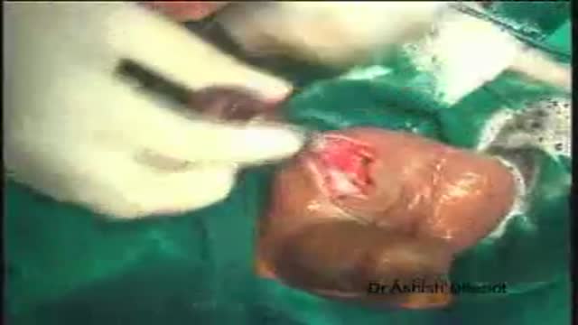
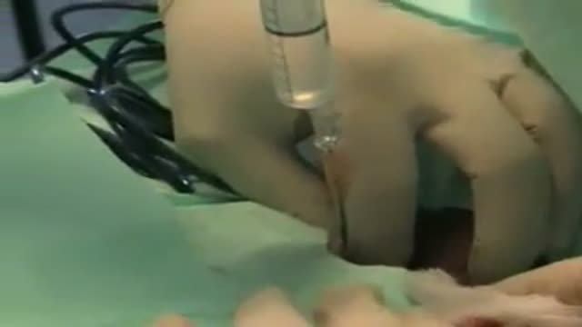
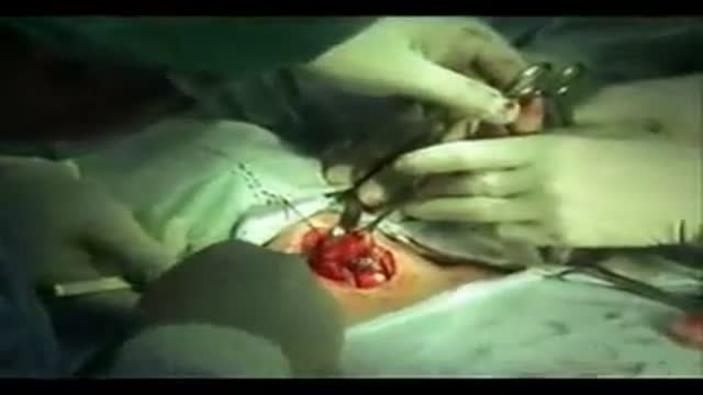
Orchidectomy and Orchidopexy in Testicular Torsion
Difficult Laparoscopic Cholecystectomy for Gall bladder Stones
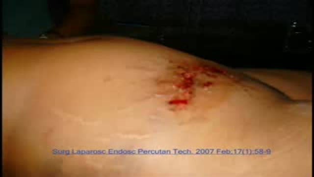
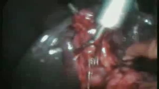
Laparoscopic Repair of Rupture Urinary Bladder
A video showing thyroidectomy surgery

In this video Erin K, a tubal reversal patient, explains the symptoms she experienced while suffering from Post Tubal Ligation Syndrome (PTLS). After having tubal reversal surgery her symptoms were relieved. Although numerous women suffer from adverse symptoms after having a tubal ligation, many physicians do not believe PTLS exists. In an ongoing study of over 300 patients reporting Post Tubal Ligation symptoms more than 90% have found relief after tubal reversal at Chapel Hill Tubal Reversal Center.
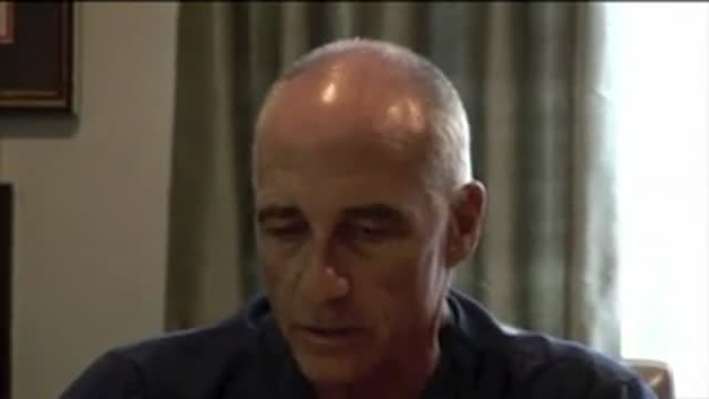
Dr. Berger, Medical Director of Chapel Hill Tubal Reversal Center discusses the pros and cons of tubal reversal vs. IVF with a couple wanting a baby after a tubal ligation.
The video will shed light on left upper lobe collapse
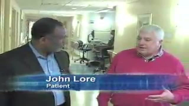
DMC Heart Imaging Specialist Doctor Hamid Sattar uses the 64-slice Coronary CTA to find coronary artery disease before symptoms even appear. ~ Detroit Medical Center

DMC Podiatrist Dr. Charles Kissel perfoms a bunionectomy to help a woman get back on her feet and back to work ~ Detroit Medical Center

Secondary Cataract

The video will shed some light on fungal infections. Please see disclaimer on my website.

American Andy received a Birmingham hip resurfacing surgery by Dr.Venkatachalam in Chennai India. The BHR is a highly successful treatment for patients for osteo-arthritis seeking relief from hip arthritis.
Hip resurfacing is offered by Madras Joint Replacement center by Dr.Venkatachalam. Visit http://www.hipsurgery.in
Lecture presented by Dr. Mostafa Yakoot, to the European Multicongress of parasitology Valencia, Spain
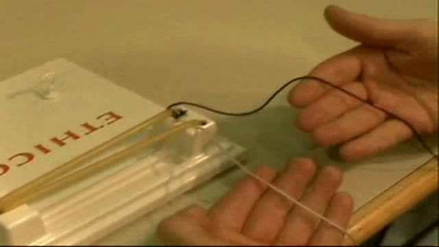
One Handed Knot Tie with Right Hand
very funny
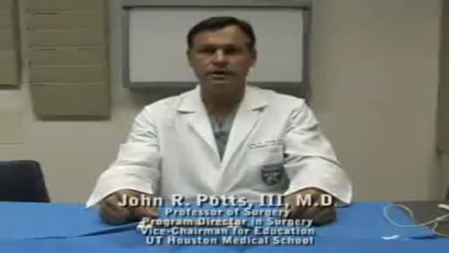
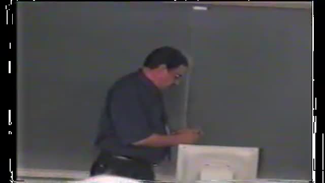
A video by UT Houston Student Surgical Association (SSA) illustrating the 2 handed not.
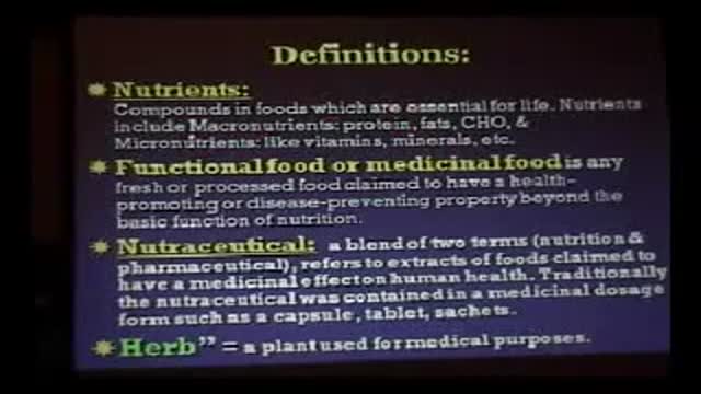
This is part 2 Herbal Medicine. Lecture presented to the International Congress of Pediatric Hepatology Sharm 2009. It is one of a series of lectures discussing the Alternative medicine practices with critical appraisal and measure the evidence.
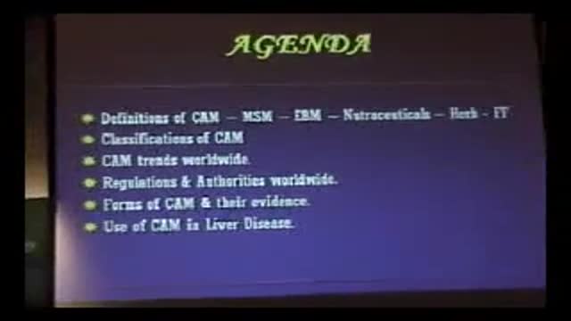
Lecture presented by Dr. "Yakoot M" to the International Congress of Pediatric Hepatology Sharm 2009. It is one of a series of lectures discussing the Alternative medicine practices with critical appraisal and measure the evidence.
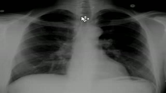
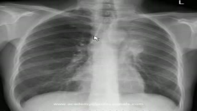
Presentation of histoplasmosis on a chest x-ray. Please see disclaimer on my website. www.academyofprofessionals.com
