סרטונים אחרונים

The dilatation and Curettage procedure that is commonly performed (D and C)
arthroscopic medial menisectomy and plica excision of knee
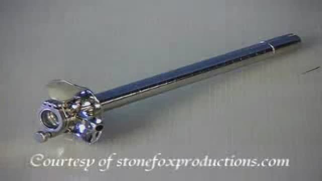
Proctoscope rectal examination
examination of the recturm
Inguinal hernia Diagram of an indirect, scrotal inguinal hernia ( median view from the left). Diagram of an indirect, scrotal inguinal hernia ( median view from the left). By far the most common hernias (up to 75% of all abdominal hernias) are the so-called inguinal hernias. For a thorough understanding of inguinal hernias, much insight is needed in the anatomy of the inguinal canal. Inguinal hernias are further divided into the more common indirect inguinal hernia (2/3, depicted here), in which the inguinal canal is entered via a congenital weakness at its entrance (the internal inguinal ring), and the direct inguinal hernia type (1/3), where the hernia contents push through a weak spot in the back wall of the inguinal canal. Inguinal hernias are more common in men than women while femoral hernias are more common in women.
26 week uterus using Gyrus PKS Cutting Forcep, PKS Lyons Dissecting Forceps & PKS Needle.

A Documentary on Uterine Transplantation. Dr. Edwin Ramirez pioneers this new breakthrough medical procedure, destined to change the world.
The thyroid is a butterfly shaped gland overlying the voice box and the windpipe. Adjacent to the thyroid are the parathyroid glands which control the body's calcium and the recurrent laryngeal nerves that control the voice box muscles. The thyroid is removed while preserving the recurrent laryngeal nerves and the parathyroids.
Total thyroidectomy is the treatment of choice for all types of thyroid cancer(papillary, follicular, medular and anaplastic).
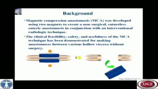
A novel technique of magnetic compression anastomosis for canalization in patients with severe biliary stricture
Surgery video of transgastric cholecystectomy

approach to Peripheral venous access
Thyroid status assessment and thyroid gland examination
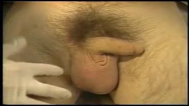
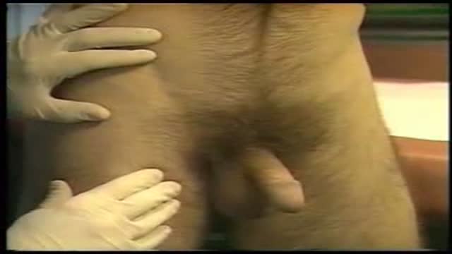
Inserteing a foley catheter in the male's urethra

a video showing Phlebootomy

a video showing Phlebotomy steps

Arterial Blood Gas Sampling

Child birth in squatting positions. The most comfortable position for the mother
Two methods to reduce the shoulder are demonstrated and the need for analgesia or anesthesia discussed

the technique of retrograde intubation to maintain the patient's airway.
