
- Physical Examination
- Surgical Examination
- Ophthalmology
- Clinical Skills
- Orthopedics
- Surgery Videos
- Laparoscopy
- Pediatrics
- Funny Videos
- Cardiothoracic Surgery
- Nursing Videos
- Plastic Surgery
- Otorhinolaryngology
- Histology and Histopathology
- Neurosurgery
- Dermatology
- Pediatric Surgery
- Urology
- Dentistry
- Oncology and Cancers
- Anatomy Videos
- Health and Fitness
- Radiology
- Anaesthesia
- Physical Therapy
- Pharmacology
- Interventional Radiology
- Cardiology
- Endocrinology
- Gynecology
- Emergency Medicine
- Psychiatry and Psychology
- Childbirth Videos
- General Medical Videos
- Nephrology
- Physiology
- Diet and Food Health
- Diabetes Mellitus
- Neurology
- Women Health
- Osteoporosis
- Gastroenterology
- Pulmonology
- Hematology
- Rheumatology
- Toxicology
- Nuclear Medicine
- Infectious Diseases
- Vascular Disease
- Reproductive Health
- Burns and Wound Healing
- Other
Top videos


Welcome to the latest episode of HT Physio Quick Tips!
In this episode, Farnham's leading over-50's physiotherapist, Will Harlow, reveals the most common knee injuries that can be sustained from a fall. You'll learn the 5 most common knee injuries from falls, how to differentiate between them and the key signs to look for before getting help.
To register your interest for the upcoming Optimum Knee Health course and to be among the first to know when it is released, reach out to Will@ht-physio.co.uk
To get a copy of Will's new book, Thriving Beyond Fifty, you can find it on Amazon below:
UK link: https://amzn.to/3mAISFv
US link: https://amzn.to/43TE5Q8
(Amazon Affiliate links)
If you're suffering from nagging knee pain that hurts in the morning and stops you from walking as far as you'd like, you can take our free knee pain guide - which will give you 5 expert tips to put a stop to knee pain at home - by visiting here: https://ht-physio.co.uk/knee-pain-guide-download/
If you're over-50 with a painful problem in the Farnham, Surrey area, you can learn more about how Will Harlow and HT Physio can help you overcome a painful problem here: https://ht-physio.co.uk/
**Any information in this video should not be used as a substitute for individual medical advice. Please seek advice from your local healthcare professional before taking action on the information in this video.**
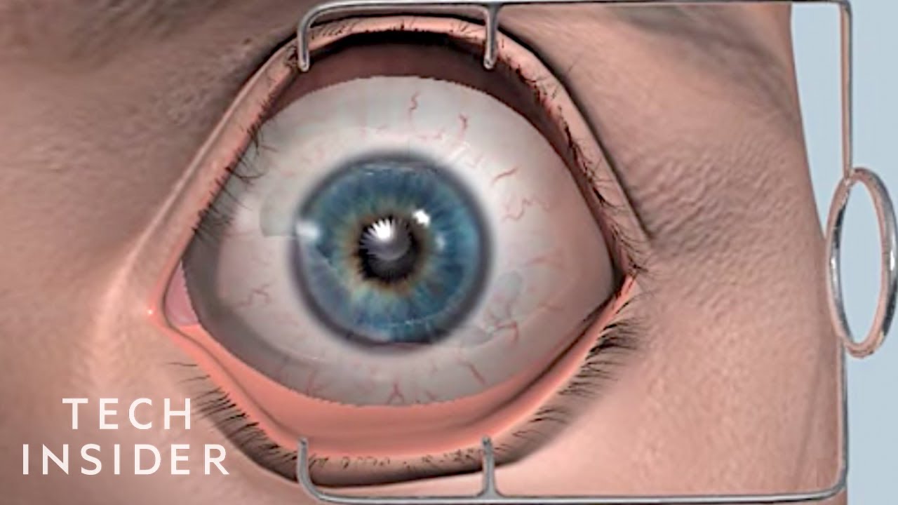
Medical technology continues to advance and help doctors continue to treat patients. From face transplants to LASIK eye surgery watch the video below for 8 medical procedures that are improving lives.
MORE MEDICAL CONTENT:
Meet The 24-Year-Old Whose Prosthetic Limbs Are Changing Lives
https://www.youtube.com/watch?v=E0dbagxeMT0
Lifelike Medical Robot Actually Bleeds
https://www.youtube.com/watch?v=IjnhmcCQLsc
Medical Tourniquet Works Like A Zip Tie
https://www.youtube.com/watch?v=k9q27yVxPL8
------------------------------------------------------
#Medicine #Surgery #TechInsider
Tech Insider tells you all you need to know about tech: gadgets, how-to's, gaming, science, digital culture, and more.
Visit us at: https://www.businessinsider.com
TI on Facebook: https://www.facebook.com/techinsider
TI on Instagram: https://www.instagram.com/tech_insider/
TI on Twitter: https://twitter.com/techinsider
TI on Amazon Prime: http://read.bi/PrimeVideo
INSIDER on Snapchat: https://insder.co/2KJLtVo
------------------------------------------------------
8 Medical Procedures That Are Improving Lives

Subscribe to the Nucleus Biology channel to see new animations on biology and other science topics, plus short quizzes to ace your next exam: https://bit.ly/3lH1CzV
For Employees of Hospitals, Schools, Universities and Libraries: Download 8 FREE medical animations from Nucleus by signing up for a free trial: http://nmal.nucleusmedicalmedi....a.com/free-trial-mem
This animation by Nucleus shows you the function of plant and animal cells for middle school and high school biology, including organelles like the nucleus, nucleolus, DNA (chromosomes), ribosomes, mitochondria, etc. Also included are ATP molecules, cytoskeleton, cytoplasm, microtubules, proteins, chloroplasts, chlorophyll, cell walls, cell membrane, cilia, flagellae, etc.
0:07 What is a cell?
0:35 What are the 2 categories of cells?
1:22 What is an Organelle? DNA, Chromatin, Chromosomes
2:06 Organelles: Ribosomes, Endoplasmic Reticulum
2:59 Organelles: ER function, Vesicles, Golgi Body (Apparatus)
3:50 Organelles: Vacuole, Lysosome, Mitochondrion
4:45 Organelles: Cytoskeleton
5:04 Plant Cell Chloroplast, Cell Wall
5:43 Unique Cell Structures: Cilia
Watch another version of this video, narrated by biology teacher Joanne Jezequel here: https://youtu.be/cbiyKH9uPUw
#cell #nucleus #biology
----
Watch other Nucleus Biology videos:
- Controlled Experiments: https://youtu.be/D3ZB2RTylR4
- Independent vs. Dependent Variables: https://youtu.be/nqj0rJEf3Ew
- Active Transport: https://youtu.be/ufCiGz75DAk
----
Learn more about the company that created this video: http://www.nucleusmedicalmedia.com/
https://www.instagram.com/nucleusmedicalmedia
This animation won a Platinum Best of Show Aurora Award in 2016.

Make sure to subscribe to our channel to get regular updates on other interesting legal animations.
Interested in other medical videos? Click the link below to watch a surgery animation!
https://www.youtube.com/watch?v=Cc6-Y_CIHx4&t=53s&ab_channel=CourtroomAnimation
Or, if you want to watch a video about a diskectomy, a common but major surgery, watch this video:
https://www.youtube.com/watch?v=PVoOVUPhdaM&t=8s&ab_channel=CourtroomAnimation
--------------------------------------------------------------------------------------------------------
Learn more about medical animations from our in-depth guide:
https://courtroomanimation.com..../qa-everything-about
Learn more about medical graphics in general from our free eBook:
https://info.courtroomanimatio....n.com/ebook-complete
Check out our blog page for other topics on legal graphics and 3D medical illustrations:
https://courtroomanimation.com/blog/
--------------------------------------------------------------------------------------------------------
Connect with us online!
Facebook: https://www.facebook.com/courtroomanimation
LinkedIn: https://www.linkedin.com/compa....ny/courtroom-animati
Twitter: https://twitter.com/courtanimation
Instagram: https://www.instagram.com/courtroomanimation/
Or give us a call to see how we can create a trial animation for your plaintiff's civil case, and help you land a more favorable verdict or settlement!
(424) 321-7488
--------------------------------------------------------------------------------------------------------
#discectomy #medicalanimation #surgery #forensicanimation #legaltechnology #legaltech #medicalvideos #lumbar #lumbarpain

Surgical cutting and removal of a deep skin cyst Medical Videos

Thought a snake in your boot was bad? That old 19th-century idiom is nothing compared to one in your ear.
Shocking footage captured the alleged moment that a “surgeon” tried to remove a live snake that infiltrated a woman’s ear. Video of the herpetological surgery has racked up more than 125,000 views as viewers speculate whether or not the squirm-inducing footage is authentic.
“The snake has gone in the ear,” reads the caption to the bizarre Facebook clip, which was posted Sept. 1 by an India-based social media star named Chandan Singh to his 20,126 followers. However, it’s unclear where, when or how this unfortunate event transpired, local outlet the Economic Times reported.
In the nearly four-minute clip, an alleged medical practitioner can be seen using tweezers in a desperate attempt to extract a black and yellow serpent that’s peeking its head out from a female patient’s ear.

https://bit.ly/3HIStRc #shorts
Coloscopy | Colon Polyp Resection | Polypectomy
Colonoscopies are essential for detecting colorectal abnormalities, including colon polyps. Polypectomy, the surgical removal of these growths, can prevent them from becoming cancerous. This article offers a brief overview of colonoscopies, colon polyps, and polypectomy procedures.
A colonoscopy is an endoscopic examination allowing healthcare providers to visualize the colon and rectum using a colonoscope. The colonoscope, a flexible tube with a camera and light source, helps detect abnormalities, including polyps or tumors.
Colon polyps are abnormal growths arising from the colon's inner lining. While most polyps are benign, some can become malignant. Adenomatous polyps have a higher potential to become cancerous, whereas hyperplastic and inflammatory polyps pose a lower risk.
Polypectomy involves removing colon polyps during a colonoscopy. Two primary techniques include snare polypectomy, using a wire loop to cut the polyp, and cold forceps polypectomy, which employs forceps to grasp and remove smaller polyps.
Following a polypectomy, patients may experience mild discomfort or bleeding. Regular surveillance is crucial to minimize colorectal cancer risk. The frequency of surveillance colonoscopies depends on the number, size, and type of polyps found, as well as the patient's overall risk factors.
Colonoscopies and polypectomies play vital roles in detecting and removing colon polyps, reducing the risk of colorectal cancer, and maintaining optimal colon health.
Do you want to learn more about colon polyps and colonoscopy? check our:
Article @ https://bit.ly/41w5Ooq
For more free resources, find us on Pinterest & Facebook pages:
https://www.pinterest.ca/medicalartsofficial/
https://www.facebook.com/Medicalartsofficial
https://www.youtube.com/@medic....alarts?sub_confirmat
https://www.instagram.com/medicalartsofficial/
https://www.tiktok.com/@medicalarts
#endoscopicsurgery #digestivesystem
coloscopy
polyp
colon polyp
polypectomy
colonoscopy
colon polyp animation
gi endoscopy
@MedicalArts , 2023.
You've come to the perfect YouTube Video if you want to learn hospital and medical English while watching shows of "The Good Doctor." Watch medical English talks from the TV show "the good doctor" to acquire new terminology about ailments that people experience but don't know the names of.
Medical English Lesson 2 with the good doctor - https://youtu.be/gU107Q9Jerw
Hospital English Vocabulary Lesson 3 with Dr. House - https://youtu.be/lE4i1pY53Us
Illness English used in Hospitals Lesson 4 with Chicago Med - https://youtu.be/u3VPRdierKA
So keep learning and watching our video lessons to learn and improve your English to a great level.
Other English lessons through The Big Bang Theory
Learn English through Big Bang Theory Lesson 1 - https://youtu.be/iM-o5EKK5pg
Funny English Lesson through Big Bang Theory Lesson 2 - https://youtu.be/O6CRNi6OJ1k
Enjoy Learning English through Big Bang Theory Lesson 3 - https://youtu.be/-iSDlbReAxk
Want to Learn English through Big Bang Theory Lesson 4 - https://youtu.be/AZSkyjk-Ioo
Learn Romantic English with The Big Bang Theory - Lesson 5 - https://youtu.be/k5EqRArT41w
Visit our Instagram page: https://www.instagram.com/englishfluencymission/
Our Facebook Page: https://www.facebook.com/englishfluencymission/
To be fluent in English, we should have a vocabulary of between 3000 and 4000 important English words. Once we attain this objective, we can be guaranteed to sound like a native speaker.
The links to a few books I've collected for you are listed below; if you truly enjoy, these could be excellent starting points for your quest for greater English fluency.
Oxford Learner's Pocket Word Skills: Pocket-sized, topic-based English vocabulary - https://amzn.to/34LKv7a
Word power made easy - https://amzn.to/38Ht8Fy
Cambridge Grammar for IELTS - https://amzn.to/34OUdWa
Unbroken - The incredible true story of Louis Zamperini, now a major motion picture directed by Angelina Jolie. - https://amzn.to/3rqtyZH
THE INTERNATIONAL NUMBER ONE BESTSELLER -
Other Video Lessons:
Learn English with Wonder Woman - https://youtu.be/6F8oqQWErU0
Learn English with Stuart Little - https://youtu.be/EIeOooR8vas
Learn English with Avengers 2012 - https://youtu.be/u97FZWkd4A8
Learn English with Forrest Gump - https://youtu.be/uH_kTF8QAZc
Learn English with Spider-Man 2 (2004) - https://youtu.be/DHy-2g-N7SQ
Your Queries
learn English with movies
learning English from movies
English vocabulary through Hollywood movies
hospital English conversation
hospital English words
hospital English movie
hospital English video
hospital English vocabulary
hospital English speaking
hospital English sentences
medical English speaking practice
medical English conversation
medical English words
medical English listening practice with the good doctor
learn hospital English with the good doctor
#thegooddoctor #learnenglishwithtvseries

Intestinal obstruction.....
This video is only educational purposes and this is not for entertainment....this is surgery time

From our beginnings in 1990 in primary healthcare, Healthway Medical has grown to become a respected medical group in Singapore. With over 100 clinics and medical centres, Healthway Medical has a wide network of medical centres and clinics in Singapore.
We offer comprehensive services including GP & family medicine clinics, health screening, adult specialists, baby & child specialists, dental services and allied healthcare services.

This video provides a guide peforming a respiratory examination in an OSCE station, including real-time auscultation sounds of common pathology such as coarse crackles, fine crackles, wheeze and stridor.
You can access our step-by-step OSCE guide to accompany this video here: https://geekymedics.com/respiratory-examination-2/
Check out our other awesome clinical skills resources including:
• 🔥 Geeky Medics Bundles (discounted products): https://app.geekymedics.com/purchase/bundles/
• ✨ 1000+ OSCE Stations: https://app.geekymedics.com/pu....rchase/osce-stations
• 🏥 Geeky Medics OSCE Revision Book: https://app.geekymedics.com/purchase/book/
• 📝 150+ PDF OSCE Checklists: https://geekymedics.com/pdf-osce-checklists/
• 🗂️ 3000+ OSCE Flashcards: https://app.geekymedics.com/pu....rchase/flashcard-col
• 📱 Geeky Medics OSCE App: https://geekymedics.com/geeky-medics-app/
• 🩺 Medical Finals SBA Question Pack: https://app.geekymedics.com/pu....rchase/medical-stude
• 💊 PSA Question Pack: https://app.geekymedics.com/pu....rchase/prescribing-s
Chapters:
- Introduction 00:00
- General inspection 00:40
- Inspection of the hands 00:50
- Schamroth's window test 01:09
- Heart rate and respiratory rate 01:50
- Jugular venous pressure 02:02
- Face, eyes and mouth 02:13
- Anterior chest inspection 02:36
- Trachea and cricosternal distance 03:01
- Palpation of apex beat 03:16
- Chest expansion 03:28
- Lung percussion 03:50
- Auscultation of lungs 04:21
- Vocal resonance 05:03
- Lymph node palpation 05:32
- Inspection of posterior chest 06:04
- Posterior chest expansion 06:10
- Percussion of posterior chest 06:32
- Auscultation of posterior chest 06:55
- Sacral and pedal oedema 08:04
- Summary of findings 08:39
Subscribe to our newsletter to be the first to know about our latest content: https://geekymedics.com/newsletter/ ✉️
Join the Geeky Medics community: 👩👩👧👧
Twitter: http://www.twitter.com/geekymedics
Instagram: https://instagram.com/geekymedics
Facebook: http://www.facebook.com/geekymedics
Always adhere to your medical school/local hospital guidelines when performing examinations or clinical procedures. DO NOT perform any examination or procedure on patients based purely upon the content of these videos. Geeky Medics accepts no liability for loss of any kind incurred as a result of reliance upon the information provided in this video.
Some people have found this video useful for ASMR purposes.
Special thanks to www.easyauscultation.com and Andy Howes for providing some of the respiratory sounds.

Clinical Examination of the respiratory system

General Examination - Clinical Skills OSCE - Dr Gill
The general examination is one of those early exams, which is essentially used to start medical students off with their clinical skills studies.
In the real world, it's mainly used with regard to gaining an overview of a patient, such as for a medical check up, or a baseline examination, for example, a health report.
They have been a couple of comments about the pulse monitor used in the video. For those who are interested. I’ve reached out to the manufacturer, and they’ve requested that the following code is provided to viewers, in order to get 20% off, if they decide on themselves.
Product model number: Vibeat SP20
Official Website: https://vibeatstore.com/produc....ts/sp20-handheld-pul
Special 20% OFF code: JAMES
--------------
Different medical schools, nursing colleges and other health professional courses will have their own preferred approach to a clinical assessment - you should concentrate on THEIR marks schemes for your assessments.
Some people watching this video may experience an ASMR effect
#DrGill #Asmr #Clinicalskills
#drgill #clinicalskills #asmr

This video demonstrates how to perform an abdominal examination in an OSCE station.
You can access our step-by-step OSCE guide to accompany this video here: https://geekymedics.com/abdominal-examination/
Check out our other awesome clinical skills resources including:
• 🔥 Geeky Medics Bundles (discounted products): https://app.geekymedics.com/purchase/bundles/
• ✨ 1000+ OSCE Stations: https://app.geekymedics.com/pu....rchase/osce-stations
• 🏥 Geeky Medics OSCE Revision Book: https://app.geekymedics.com/purchase/book/
• 📝 150+ PDF OSCE Checklists: https://geekymedics.com/pdf-osce-checklists/
• 🗂️ 3000+ OSCE Flashcards: https://app.geekymedics.com/pu....rchase/flashcard-col
• 📱 Geeky Medics OSCE App: https://geekymedics.com/geeky-medics-app/
• 🩺 Medical Finals SBA Question Pack: https://app.geekymedics.com/pu....rchase/medical-stude
• 💊 PSA Question Pack: https://app.geekymedics.com/pu....rchase/prescribing-s
Chapters:
- Introduction 00:00
- General inspection 00:35
- Hands 00:47
- Asterixis 01:20
- Arms and axilla 01:32
- Face, eyes & mouth 01:45
- Lymph node palpation 02:19
- Chest inspection 02:50
- Inspection of abdomen 03:02
- Palpation of abdomen 03:34
- Percussion of abdomen 05:36
- Shifting dullness 06:30
- Auscultation of abdomen 06:55
- Summary 07:29
Subscribe to our newsletter to be the first to know about our latest content: https://geekymedics.com/newsletter/ ✉️
Join the Geeky Medics community: 👩👩👧👧
Twitter: http://www.twitter.com/geekymedics
Instagram: https://instagram.com/geekymedics
Facebook: http://www.facebook.com/geekymedics
Always adhere to your medical school/local hospital guidelines when performing examinations or clinical procedures. DO NOT perform any examination or procedure on patients based purely upon the content of these videos. Geeky Medics accepts no liability for loss of any kind incurred as a result of reliance upon the information provided in this video.
Some people have found this video useful for ASMR purposes.

Examination of Peripheral Vascular System - Clinical Skills OSCE Revision - Dr Gill
In this video, we demonstrate the peripheral vascular examination - a less common examination, but still vitally important, particularly amongst the older population
Starting with the examination of the hands looking for clinical signs of vascular compromise, we then check the pulses of the major arteries of the upper body - the radial, brachial and carotid arteries, before moving down to assess for an abdominal aortic aneurysm.
At this point, I feel it's a practical step to check the femoral pulses before doing the overview of the legs.
After visually assessing we must examine the major vascular areas of leg.- namely the popliteal pulses, before wrapping up around the ankle with the posterior tibial and dorsalis pedis pulses
For completeness, the cardiovascular examination is demonstrated here
https://www.youtube.com/watch?v=ECs9O5zl6XQ&t=2s
#PeripheralVascular #ClinicalSkills #DrGill

Am I missing something?
Subscribe to my fun weekly newsletter (for free!): http://eepurl.com/iaYycn
To check out a previous newsletter, click here: https://mailchi.mp/a9909f90cac....a/why-are-you-having
For more Doc Schmidt content, check out my website: https://www.docschmidt.org/
Check out my children's book here: https://www.amazon.com/Night-Before-Med-School-Medical/dp/B0B193KWXT/ref=sr_1_1?keywords=doc+schmidt&qid=1653339841&sprefix=doc+sc%2Caps%2C202&sr=8-1
Logo and graphics designed by iamlindaayoade.com and loigraphics.com (LOI Graphics Inc.)
Want me to make you a personalized video for you or your friend? Check me out on Cameo!
https://v.cameo.com/DFKBSe2HSib
Want to connect with me and watch more content?
Find me on TikTok!
https://vm.tiktok.com/ZMRFmqKts/
And Instagram!
https://instagram.com/docschmidtig?r=nametag
All content is intended as medical education or entertainment and is NOT intended to be medical advice. If you have any symptoms concerning you, please schedule an appointment with your doctor.
Join my channel to get access to perks! Click link below:
https://www.youtube.com/channe....l/UCLbidg2ZT49dWrxDk

Elbow Exam - Orthopaedic OSCE - Clinical Skills - Dr Gill
The elbow examination is a core skill - in this video, we demonstrate how to perform an elbow EXAM for an Orthopaedic Clinical Skills OSCE, which should be one of the more accessible examination stations for medical students.
For a passing grade in your Clinical Skills OSCE, an elbow assessment should follow the LOOK, FEEL, MOVE approach
Initially looking for erythema, scars, swelling and position
Palpating the elbow - specifically the olecranon, medial and lateral epicondyles, and radial head for heat, oedema and crepitus
Finally assess range of movement with flexion and extension at the elbow, before determining for tennis and golfers' elbows
Watch further orthopaedic examinations for your OSCE revision:
The Elbow - Deep Dive
https://youtu.be/SX5buhtCVDw
The Spine Examination:
https://youtu.be/pJxMHa6SCgU
The Knee examination
https://youtu.be/oyKH4EYfJDM
The Hip examination
https://youtu.be/JC9GKq5nSdQ
The GALS examination
https://youtu.be/5qJaf7gW-B0 - Gait, Arms, Legs, Spine - GALS screen
------------
Please note that there is no ABSOLUTE way to perform a clinical examination. Different institutions and even clinicians will have differing degrees of variations - the aim is the effectively identify medically relevant signs.
However during OSCE assessments. Different medical schools, nursing colleges and other health professional courses will have their own preferred approach to a clinical assessment - you should concentrate on THEIR marks schemes for your assessments.
The examination demonstrated here is derived from Macleods Clinical Examination - a recognised standard textbook for clinical skills.
Some people viewing this medical examination video may experience an ASMR effect
#clinicalskills #Elbow #DrGill

#GIT#Abdominalexamination#AETCM
