
- Physical Examination
- Surgical Examination
- Ophthalmology
- Clinical Skills
- Orthopedics
- Surgery Videos
- Laparoscopy
- Pediatrics
- Funny Videos
- Cardiothoracic Surgery
- Nursing Videos
- Plastic Surgery
- Otorhinolaryngology
- Histology and Histopathology
- Neurosurgery
- Dermatology
- Pediatric Surgery
- Urology
- Dentistry
- Oncology and Cancers
- Anatomy Videos
- Health and Fitness
- Radiology
- Anaesthesia
- Physical Therapy
- Pharmacology
- Interventional Radiology
- Cardiology
- Endocrinology
- Gynecology
- Emergency Medicine
- Psychiatry and Psychology
- Childbirth Videos
- General Medical Videos
- Nephrology
- Physiology
- Diet and Food Health
- Diabetes Mellitus
- Neurology
- Women Health
- Osteoporosis
- Gastroenterology
- Pulmonology
- Hematology
- Rheumatology
- Toxicology
- Nuclear Medicine
- Infectious Diseases
- Vascular Disease
- Reproductive Health
- Burns and Wound Healing
- Other
Top videos
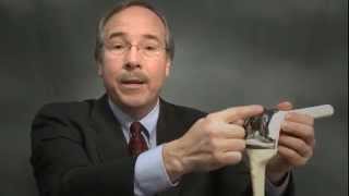

Timothy Lovell, MD, an orthopedic surgeon, talks to Spokane, WA knee replacement surgery patients about the procedure, possible risks and complications of surgery, and about your recovery time.
Dr. Lovell addresses anesthesia, the size and location of the incision, and shows you what the knee replacement ball and socket joint looks like. He'll talk about the recovery process; using a crutches, a walker or a cane to get around; movements to avoid; and how long it takes to feel better and return to your normal, active life.
To learn more about Dr. Lovell, visit http://washington.providence.o....rg/find-a-provider/l
And, to learn more about having orthopedic surgery in Spokane, WA, visit http://washington.providence.o....rg/clinics/providenc

New research from Mount Sinai Health System says these surgeries have limited effectiveness and can be economically unjustifiable when they're done on patients with less severe symptoms.
» Subscribe to NBC News: http://nbcnews.to/SubscribeToNBC
» Watch more NBC video: http://bit.ly/MoreNBCNews
NBC News is a leading source of global news and information. Here you will find clips from NBC Nightly News, Meet The Press, and original digital videos. Subscribe to our channel for news stories, technology, politics, health, entertainment, science, business, and exclusive NBC investigations.
Connect with NBC News Online!
Visit NBCNews.Com: http://nbcnews.to/ReadNBC
Find NBC News on Facebook: http://nbcnews.to/LikeNBC
Follow NBC News on Twitter: http://nbcnews.to/FollowNBC
Follow NBC News on Google+: http://nbcnews.to/PlusNBC
Follow NBC News on Instagram: http://nbcnews.to/InstaNBC
Follow NBC News on Pinterest: http://nbcnews.to/PinNBC
New Study Questions Effectiveness Of Knee Replacement Surgery | NBC Nightly News

Knee replacement involves replacing a knee joint that has been damaged or worn away, usually by arthritis or injury. Find out more here: http://bit.ly/MAdfmE
The content is intended for general information only and does not replace the need for personal advice from a qualified health professional.
#surgical #arthritis #surgery #medical #ortho #health #orthopaedic #medicine #ortopedia #medstudent #physicaltherapy #medschool #medicalstudent #medlife #sports
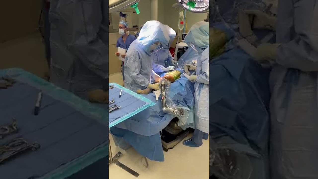
✍️Dr. Matthew Harb talks about knee replacement surgery
https://www.MatthewHarbMD.com/links
👨⚕️Orthopedic Hip and Knee Surgeon
📍Located in Washington DC, and Maryland
📚Education and Insight
🛠Minimally invasive, outpatient, hip and knee replacement surgery
👉Visit me Online: https://www.MatthewHarbMD.com
☎️Schedule a virtual or in office appointment: https://www.MatthewHarbMD.com/links
📲Follow me on Social Media:
💥TIKTOK: https://www.tiktok.com/@matthewharbmd
💥INSTAGRAM: https://www.instagram.com/thebonesurgeon
📲Please SUBSCRIBE for new #shorts #doctor videos!
https://www.youtube.com/channel/UCbte-7tl7kaAm1xfWgz6xvQ?app=desktop&sub_confirmation=1
✍️QUESTION — Have a question or comment about medicine, orthopedic surgery, or musculoskeletal conditions. Post in the comment sections and let me know!
Dr. Matthew Harb specializes in minimally invasive, muscle sparing, hip and knee replacement surgery. Minimally invasive surgery allows patients to recover faster and have less pain post operatively. Implants are tailored and custom fit to each patient to allow for improved performance. Dr. Harb’s expertise in rapid recovery protocols allow for quick recovery after surgery and excellent outcomes in patients with hip and knee arthritis. With minimally invasive, muscle sparing surgery patients can return to their lifestyles and get back to doing the things they love sooner. Dr. Harb performs outpatient joint replacement surgery with many of his patients walking independently and going home the day of surgery.
“My focus is excellence in patient care, expedited recovery after surgery, and getting people back to the normal activities they love. Our team focused approach is committed to superb outcomes, improving lives, and returning patients to living pain free.”

In this video, we have explained the procedure of total #knee #replacement #surgery in patient in 3D animation.
Learn more: https://ecgkid.com
_____________________________________________________________________
Knee replacement, commonly known as complete knee replacement or knee arthroplasty, is a surgical treatment that resurfaces a knee that has been destroyed by arthritis. The extremities of the bones that make up the knee joint, as well as the kneecap, are capped with metal and plastic pieces. Someone with severe arthritis or a major knee injury may benefit from this procedure.
The knee joint can be affected by a variety of arthritis forms. The degradation of joint cartilage and neighboring bone in the knees can be caused by osteoarthritis, a degenerative joint disease that primarily affects middle-aged and older persons. Rheumatoid arthritis produces pain and stiffness by inflaming the synovial membrane and resulting in an excess of synovial fluid. Traumatic arthritis, or arthritis caused by an injury, can harm the joints.
The purpose of knee replacement surgery is to resurface damaged areas of the knee joint and cure knee discomfort that has not responded to prior therapies.

Watch Dr. Robert Thomas, of Panorama Orthopedics & Spine Center, perform a Mako Knee replacement. He narrates each step of the process.

Each person's knee is different. This is why UC San Diego Health offers several surgical options for knee replacements to tailor care to each person's injury and health. Each surgical approach has benefits for the right surgical candidate. Our surgeons can discuss what option is most appropriate for each person.
To learn more about knee replacement options at UC San Diego Health, visit:
https://health.ucsd.edu/specia....lties/orthopedics/jo
Francis Gonzales, MD, is a board-certified orthopedic surgeon who specializes in adult hip and knee joint replacement surgery. Learn more about Dr. Gonzales: https://providers.ucsd.edu/det....ails/11935/orthopedi
UC San Diego Health is repeatedly ranked among the nation's best in orthopedic care by U.S. News & World Report. We are also a Blue Distinction Center recognized for our treatment expertise and better overall patient results for knee replacement, as well as a designated Center of Excellence for orthopedic care by Optum. This means you'll receive expert, safe and cost-effective care.
UC San Diego Health's orthopedic surgeons are the first and only in San Diego to offer customized knee replacements with the ROSA knee system — for a faster recovery and more natural feeling knee. Talk to one of our surgeons about whether a ROSA knee replacement is right for you. https://health.ucsd.edu/specia....lties/orthopedics/jo#a

Mohs Surgeon Dr. Leslie Christenson shows entire Mohs Surgery from start to finish. This is the full procedure and includes the entire surgery. Dr. Christenson talks about the procedure as she removes the skin cancer.
Learn more about Dr. Christenson: https://www.mcfarlandclinic.co....m/doctors/leslie-chr
Learn more about Mohs Surgery: https://www.mcfarlandclinic.co....m/doctors/specialtie
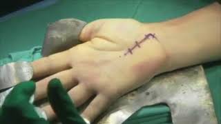
Carpal tunnel release (part 1). Skin incision and retraction. Procedure performed by Deepak Kapila, MD, Broward Health, Fort Lauderdale, FL. Courtesy of BroadcastMed (http://ortho.broadcastmed.com/....4229/videos/carpal-t
There are hundreds more procedural videos as well as news, features, resources and references on Medscape.com. Join today for free.
High Quality Surgical videos and uncut stories ▶ https://surgeoncut.com

Surgeons at St Mary's Hospital, part of Imperial College Healthcare have come up with a new surgical procedure that cures heartburn with a device called RefluxStop.
Mr Ahmed Ahmed, a consultant surgeon, says surgery should now be seen as an alternative to life-long drug treatment - as Sky's Thomas Moore reports.
Read more: https://news.sky.com/story/new....-nhs-heartburn-surge
#heartburncure #surgery #skynews
SUBSCRIBE to our YouTube channel for more videos: http://www.youtube.com/skynews
Follow us on Twitter: https://twitter.com/skynews
Like us on Facebook: https://www.facebook.com/skynews
Follow us on Instagram: https://www.instagram.com/skynews
Follow us on TikTok: https://www.tiktok.com/@skynews
For more content go to http://news.sky.com and download our apps: Apple https://itunes.apple.com/gb/ap....p/sky-news/id3163919 Android https://play.google.com/store/apps/details?id=com.bskyb.skynews.android&hl=en_GB
Sky News Daily podcast is available for free here: https://podfollow.com/skynewsdaily/
Sky News videos are now available in Spanish here/Los video de Sky News están disponibles en español aquí: https://www.youtube.com/channe....l/UCzG5BnqHO8oNlrPDW
To enquire about licensing Sky News content, you can find more information here: https://news.sky.com/info/library-sales

Surgical site infections (SSIs) remain a prevalent threat to patient safety. Proper surgical hand scrub or rub techniques are essential to decreasing the incidence of SSIs. This video provides instructions on the anatomical surgical hand scrub procedure using the brushstroke method. Learn more from the Department of Hospital Epidemiology and Infection Control (HEIC) at The Johns Hopkins Hospital: http://www.hopkinsmedicine.org/heic

Gastric bypass, also called Roux-en-Y gastric bypass surgery, is considered a “metabolic” procedure because it changes how your body absorbs fat, calories and nutrients. This metabolic change occurs because your gastrointestinal tract is altered when your gastric bypass surgeon attaches the smaller section of your stomach directly to your small intestine. As a result, your appetite changes and you feel full faster.

In this video, Dr. Robert Rozbruch, chief of Limb Lengthening and Complex Reconstruction at Hospital for Special Surgery performs an osseointegration after a primary amputation. The patient, a 40 year old woman, had chronic nerve pain and compromised function of her residual limb.
For more information, visit: https://www.limblengthening.com/
https://www.hss.edu/limblengthening
https://www.hss.edu/LSARC
https://www.facebook.com/limblengtheningNYC
https://www.instagram.com/limblengthening
https://www.twitter.com/limblengthen
https://www.youtube.com/channe....l/UC-JL_X6ALjZXiXtcP
key words: Osseointegration, Amputee, Amputation, Limb Replacement, Tibia, Osseointegration

If a fetal lung lesion is causing heart failure, fetal surgery may be performed to remove the CCAM before birth. http://fetalsurgery.chop.edu
N. Scott Adzick, MD, Mark Johnson, MD, and Holly Hedrick, MD, experts from the Center for Fetal Diagnosis and Treatment at Children’s Hospital of Philadelphia, explain when fetal intervention for CCAM is recommended, the various approaches that may be used to treat the most complex fetal lung lesions before birth, and how these procedures are performed.
One concern with fetal lung lesions is that they take up space in the chest. If the lung mass grows and pushes the heart and other organs out of place, it can lead to complications such as fetal hydrops (heart failure in the fetus). If this happens, a fetal surgery procedure may be performed to remove the CCAM before birth.
In other cases, an EXIT procedure may be performed to partially deliver the baby, so the team can remove the mass before the baby is fully delivered.
In this video series, parents, nurses and doctors from Children’s Hospital of Philadelphia’s Center for Fetal Diagnosis and Treatment talk about the different types of fetal lung lesions like congenital cystic adenomatoid malformation (CCAM) and bronchopulmonary sequestration (BPS), the importance of accurate diagnosis and monitoring, and the most advanced treatment options currently available. They also discuss follow-up care and long-term outcomes for babies diagnosed with fetal lung lesions.

Our mission: Empower you with the tools and support you need for weight loss and live a healthier life. Get started on your weight loss journey today: https://bit.ly/2Ms4JaX
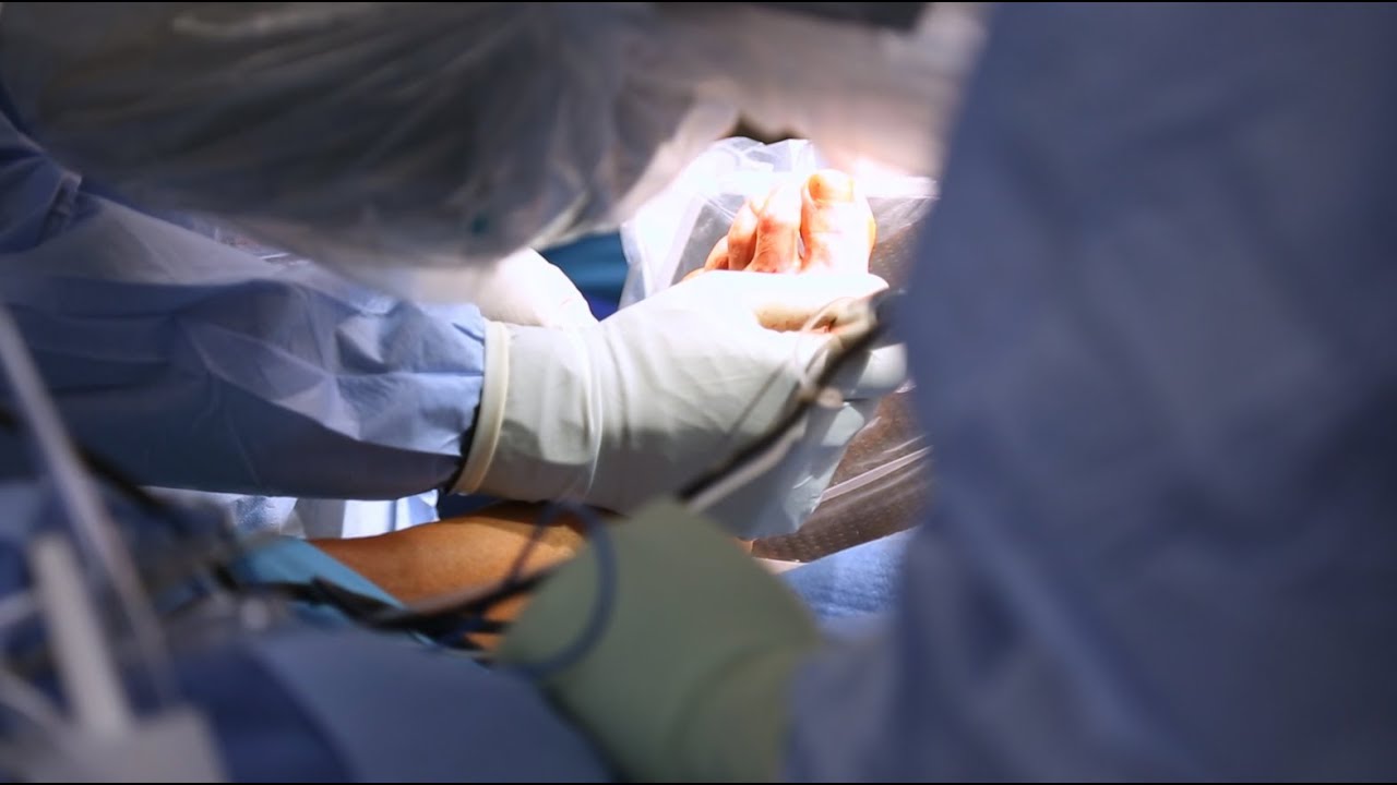
Ettore Vulcano, MD, Foot and Ankle Orthopedic Surgeon at Mount Sinai West, discusses a new minimally invasive bunion surgery that has patients walking immediately after surgery, and getting back to an active lifestyle much quicker than with the traditional surgery.

Christopher J. Rapuano, MD, Director of the Cornea Service at Wills Eye Institute describes his surgical approach of a Combined Penetrating Keratoplasty (PK) and Cataract Surgery

The video demonstrates complete excision of endometrosis in a variety of challenging situations.

OB_A_1013
3D animation depicting the operating room and initial procedure preparing the patient for a laparoscopic hysterectomy. The patient is prepped and draped in the usual fashion and surrounded by the surgeon and surgical assistants. The skin is elevated, an infraumbilical incision is made, a trocar port is inserted through the incision and the abdomen is insufflated. Finally, a laparoscope is inserted into the port to allow for direct visualization of the uterus and the surgery can begin.
To view more animations and exhibits, visit our medical library: https://www.trialexhibitsinc.c....om/library/multimedi
Contact us on your next case for consulting, trial graphics, animations, medical illustrations or presentation services. 800-591-1123 [a]www.trialex.com[/a]
This video is for reference only. The video may not be otherwise used, reproduced nor modified. For more information to purchase a copy or permission to use this animation on your next case, project, website or TV, contact us at [a]www.trialex.com[/a] or 800-591-1123.
Copyright @ Trial Exhibits, Inc.
