- Physical Examination
- Surgical Examination
- Ophthalmology
- Clinical Skills
- Orthopedics
- Surgery Videos
- Laparoscopy
- Pediatrics
- Funny Videos
- Cardiothoracic Surgery
- Nursing Videos
- Plastic Surgery
- Otorhinolaryngology
- Histology and Histopathology
- Neurosurgery
- Dermatology
- Pediatric Surgery
- Urology
- Dentistry
- Oncology and Cancers
- Anatomy Videos
- Health and Fitness
- Radiology
- Anaesthesia
- Physical Therapy
- Pharmacology
- Interventional Radiology
- Cardiology
- Endocrinology
- Gynecology
- Emergency Medicine
- Psychiatry and Psychology
- Childbirth Videos
- General Medical Videos
- Nephrology
- Physiology
- Diet and Food Health
- Diabetes Mellitus
- Neurology
- Women Health
- Osteoporosis
- Gastroenterology
- Pulmonology
- Hematology
- Rheumatology
- Toxicology
- Nuclear Medicine
- Infectious Diseases
- Vascular Disease
- Reproductive Health
- Burns and Wound Healing
- Other
Top videos

Diastasis recti often occurs during pregnancy and can persist after pregnancy. It affects core strength and the appearance of the abdominal muscles.
Dr. Erick Sanchez repairs the abdominal muscles with every tummy tuck. This short video shows the muscle repair portion of the surgery with a bonus after photo at the end!
To request a consultation with Dr. Sanchez, visit sanchezplasticsurgery.com and click Request a Consultation. Fill out the form and someone will get in touch with you to answer all your questions.
Expected cost can be found at the bottom of each procedure page on our website.

Full Tummy Tuck 3D Video - http://drlandsman.com
Look great... feel great
•Smart Liposuction + Liposculpture
•Abdominplasty (Tummy Tuck)
+ Full Mini Modified
•Brazilian Lift with Fat Transfer
•Vaginal Aesthetics & Rejuvenation
•Laser Hair Removal
•Full Body Lift
•Thigh lift
•Brachioplasty (Arm Lift) + Short Scar
Expertise in Body Contouring
Board Certified Plastic Surgeon
Expertise in body contouring combines skin excision techniques and advanced fat contouring technology
Weight control personalized training and smoking cessation results in a healthier lifestyle improved shape and longer lasting results
With over 2 decades of experience Dr Lloyd Landsman provides state of the art cosmetic and plastic surgery
Dr Landsman integrates the finest and safest products with the newest procedures
A customized treatment plan is created for each patient utilizing classic surgical and minimally invasive techniques for optimal results
Call for your complimentary consultation to learn how Dr Landsman can help you look your very best
Visit http://drlandsman.com Call 631 864 4111
Main Office 994 W Jericho Tpke Smithtown NY 11787
Affiliates East Islip • Westbury • Jackson Heights • Manhattan
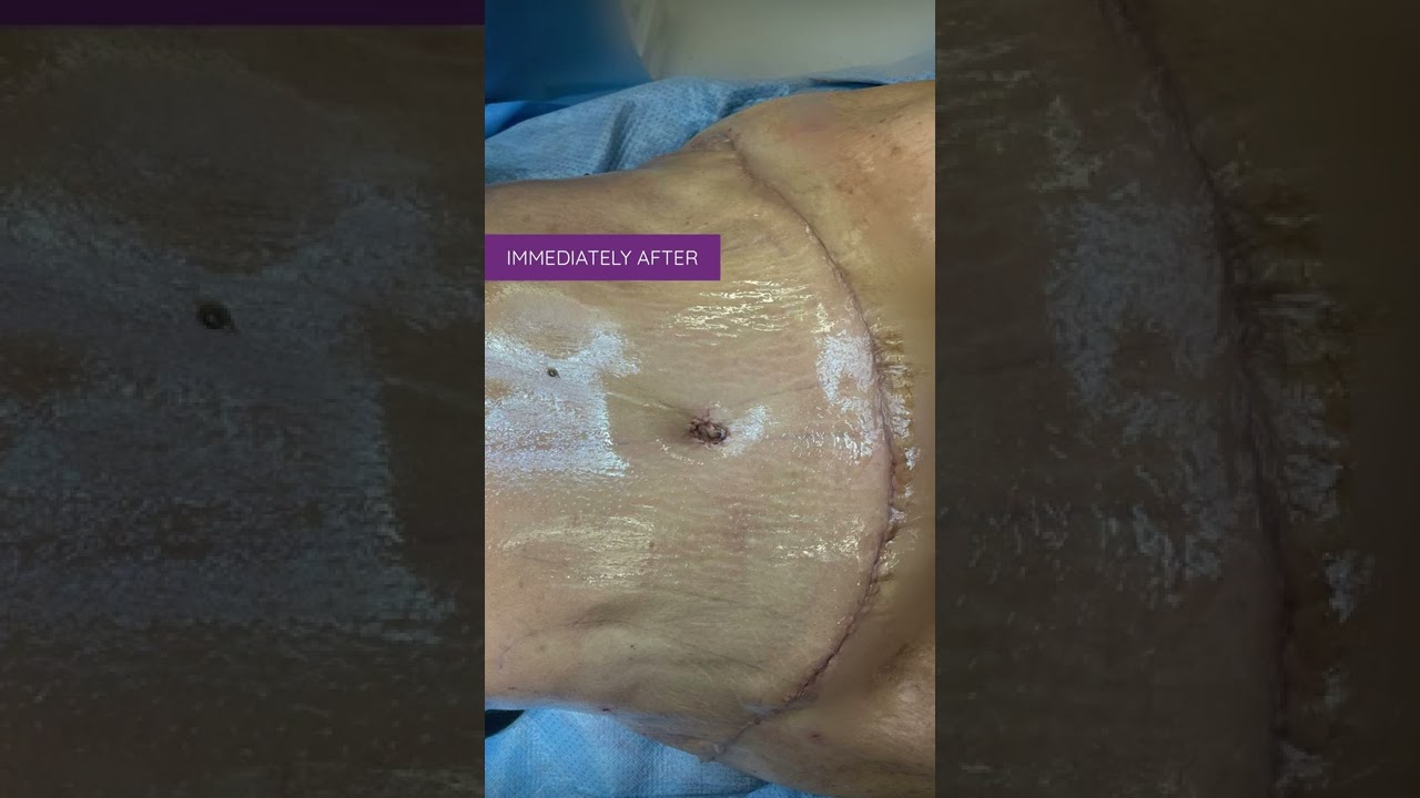
Liposuction in tummy tuck requires special planning and technique. I need to ensure that the blood circulation is well maintained for good healing. Yet proper liposuction is important to have a nice flat and contoured tummy.
#hdliposuction #tummytuck #lipoabdominoplasty #surgicalplanning #skinremovalsurgery #imeediatelyafter #plasticsurgeondubai #cocoonaclinic #drsanjayparashar #dubai
For more information visit www.drsanjayparashar.com
For more content, follow me on my social media
Instagram : https://www.instagram.com/drsanjayparashar/
Facebook : https://www.facebook.com/drsanjayparashar

#tummytucksurgery #tummytuckcost #tummytuckresult #drprashantyadav #dezireclinicindia #cosmeticsurgery #plasticsurgery #weightloss #tummytuck #abdominoplasty
Tummy tuck surgery | Weight Loss
Tummy Tuck Marking before surgery
Tummy Tuck Surgery , Tummy Tuck Surgery Cost, Weight Loss with Tummy tuck surgery, tummy tuck surgery result , tummy tuck cost, Abdominoplasty
📞 Delhi - +91 8956880644 | Pune - +91 9222122122 | Bangalore- 8971224700 | Gurugram - 9272007896 | Ahmedabad - 9711162746
Subscribe to our Channel https://www.youtube.com/dezireclinic
💸Cost: call for free consultation | Zero % interest finance options
Subscribe on YouTube : https://youtube.com/dezireplas....ticsurgerycenter?sub
Subscribe on YouTube https://youtube.com/dezireclin....ic?sub_confirmation=
📸 https://www.instagram.com/drprashantdezireclinic/
🌐 http://dezireclinic.in/
Facebook: https://www.facebook.com/drprashantmch/
Twitter: https://twitter.com/drprashantmch
Email: dezireclinicindia@gmail.com
WHY choose Dezire plastic surgery center?
We are leading cosmetic surgery centre in India leading by Dr Prashant Yadav . See hundreds of various cosmetic surgery and real feedback videos. Watch live surgery to gain confidence before deciding surgery
Dr. Prashant Yadav
M.S., M.Ch. (Plastic & Cosmetic Surgery)
#plasticsurgery #cosmeticsurgery #dezireclinicindia #drprashantyadav #dezireplasticsurgerycenter

This animation shows you how a tummy tuck is performed at Boerhaave Medical Centre. Curious? Watch the video!
Boerhaave Medical Centre sets itself the goal of providing the highest quality care. Quality not just in terms of treatment, but also in terms of our staff and the before and after care given. By providing thorough information and clear advice in advance, carefully supporting our patients through the procedure and caring for them afterwards, we believe this quality is assured.
Although we are one of the largest clinics in the Netherlands and have built up many years of experience, we continuously strive to improve. After all, the Boerhaave Medical Centre intends to remain a pioneer in the healthcare sector, by working in accordance with the latest medical findings and techniques both now and in the future.
We offer the highest standard of plastic surgery in our cosmetic care department. For 365 days a year, you can also come to us for non-surgical treatments, such as injectables, permanent hair removal and gastric balloons.
We have been awarded the ZKN quality mark and are certified to ISO 9001-2008 for giving advice and carrying out plastic surgery, including after care.
Visit our website for more information: https://www.boerhaave.com/all-....treatments/upperbody
Follow us:
Facebook: https://www.facebook.com/boerhaavemc
Google+: https://plus.google.com/+BoerhaaveNl-Kliniek
Pinterest: https://nl.pinterest.com/BoerhaaveMC/
Instagram: https://www.instagram.com/BoerhaaveMC/

Access my FREE Online Membership today → https://www.thenotedanatomist.com
___
Unlock my Premium Tutoring Memberships → https://www.thenotedanatomist.com/premium-memberships
Lifetime Access to Online Anatomy Course
Foundational Q&A Cards Per Video
Notes and Key Takeaways
Downloadable Documents
Flashcards for Each Course
Weekly Group Tutoring Sessions
Direct Tutoring Sessions
___
Discover A Simplified Approach to Master the Complexity of Anatomy with me, Dr. David Morton ... The Noted Anatomist!
This video tutorial discusses an Introduction to Histology (study of tissues):
0:00. Intro
0:35. Hierarchical organization of living matter
1:56. H&E stains
3:00. Epithelium overview (characteristics and classifying scheme)
- 9:12. Simple squamous epithelium
- 11:05. Simple cuboidal epithelium
- 12:20. Simple columnar epithelium
- 13:36. Stratified squamous epithelium
- 15:51. Urinary epithelium (transitional epithelium)
- 16:45. Pseudo-stratified ciliated columnar epithelium (respiratory epithelium)
18:55. Connective tissue overview (characteristics and classifying scheme)
- 21.14. Connective tissue proper (loose CT, dense irregular CT, dense regular CT, adipose tissue)
- 24:50. Cartilage (hyaline cartilage, elastic cartilage, fibrocartilage)
- 26:04. Bone (osteoblasts, osteocytes, osteoclasts, calcium ...)
- 27:34. Blood (RBC, WBC, platelet, plasma)
28:54. Muscle tissue (skeletal muscle, cardiac muscle, smooth muscle)
32:54. Nervous tissue (neurons and glial cells)
36:58. In-a-Nutshell
37:07. Acknowledgements
For a more detailed study of histology go to The Histology Wizard: https://www.youtube.com/channe....l/UCAeLLruy9RkUWaW_r

A complete organized library of all my videos, digital slides, pics, & sample pathology reports is available here: https://kikoxp.com/posts/5084 (dermpath) & https://kikoxp.com/posts/5083 (bone/soft tissue sarcoma pathology)
Topics discussed:
Epidermis:
Layers of epidermis: 0:10
Melanocytes vs Keratinocytes: 5:16
Langerhans cells: 10:10 & 33:30 & 57:30
Dermis:
Papillary and reticular dermis: 11:50
Three types of white empty spaces on a slide: vessels, glands/ducts/cysts, or artifact: 15:25
Blood vessels & nerves: 18:24 & 48:50 & 58:59
Arrector pili & other dermal smooth muscle: 20:00
Adnexal:
Sebaceous gland: 21:10
Hair follicle 23:14
Eccrine sweat glands and ducts 24:45 & 50:00
Gland/duct vs blood vessel 27:20 & 48:50
Apocrine glands: this video https://kikoxp.com/posts/7837 (at 12:30)
Acrosyringium: this video https://kikoxp.com/posts/7837 (at 10:00)
Three types of pink bundles: smooth muscle, nerve, dense connective tissue: 27:50
Acral skin (palm sole) with contact dermatitis 29:37
Parakeratosis 30:00
Perivascular lymphocytes 30:40
Eosinophils vs neutrophils 31:20
Spongiosis with desmosome keratinocyte spines 32:10
Spongiotic vesicles with Langerhans cells 33:30
Normal acral skin (palm & sole) with stratum lucidum 34:20
Normal glomus body/apparatus (canal of Sucquet-Hoyer) 35:40
Nerve 36:46 & 51:50
Adipose tissue (white fat cells) in subcutis with Lochkern 37:55
Normal scalp skin with large anagen hair follicles: 39:30
Hair follicle anatomy (bulb/matrix, inner root sheath, outer root sheath, hair shaft, isthmus, infundibulum): 40:55 (labeled images):
https://kikoxp.com/posts/3661 & https://kikoxp.com/posts/7899
Pacinian corpuscle 50:40
Meissner corpuscle 1:02:28
Dense regular connective tissue (Fascia/Tendon/Ligament) vs Smooth Muscle 53:00
Basic Normal Skin Immunohistochemistry:
-cytokeratin in epidermis: 55:33
-S100 in melanocytes and Langerhans cells and adipocytes: 57:30
-Desmin in smooth muscle (arrector pili and blood vessels): 58:59
-CD31 in endothelial cells of blood vessels: 59:33
-SOX-10 in melanocytes: 1:00:40
Digit/Finger/Toe histology (amputation for subungual acral melanoma) 1:04:10 & 1:08:30
-bone 1:05:40
-glomus body 1:05:15
-tendon/ligament 1:06:10
-artery 1:06:58
-fingernail/toenail 1:08:54
-acrosyringium 1:10:45
Solar elastosis (what wrinkles look like microscopically!) 1:11:50
Other videos you might like:
Tendon vs Nerve Histology Made Simple with the Ramen Noodle Sign (of Fulton) video: https://kikoxp.com/posts/4466
Melanocytes vs Keratinocytes made easy video: https://kikoxp.com/posts/3802
Blood Vessel vs Gland vs Artifact Made Easy video: https://kikoxp.com/posts/4808
The basic normal structures of the skin discussed and described by a dermatopathologist. This material is intended for use by medical students, junior pathology or dermatology residents, or for anyone else studying normal human histology. Special thanks to two of my medical students at UAMS for helping make this video possible. Miki Lindsey convinced me that I really needed to sit down and record this video. Akash Patel took time to edit the video and make it ready for YouTube. My sincere thanks to both of them for helping me overcome procrastination.
Huge thanks to Abigail Cline, a medical student at Medical College of Georgia, for volunteering to type a transcript of this ENTIRE video (over 14,000 words!) so that I could provide closed caption subtitles for those with hearing impairments and for those who may need assistance in understanding spoken English (particularly given how quickly I speak!). You can access a text version of her transcript of my video here: https://kikoxp.com/posts/5390
Correction - I made a mistake in the video. I said that sebaceous gland secretions are turned into smelly substances by bacteria and that this makes body odor. That is incorrect. That is actually true of APOCRINE gland secretions not sebaceous secretions.
Also, in the past I used "keratinocyte" and "squamous cell" interchangeably (this is because in dermatopathology, we see and talk about squamous cell carcinomas all the time, and those tumors are composed of keratinocytes). But technically, in normal skin histology, "squamous cell" refers only to the flattened keratinocytes in the superficial epidermis. Thankfully, a histology PhD colleague pointed this out to me and corrected my lazy nomenclature!
Please check out my Soft Tissue Pathology & Dermatopathology survival guide textbooks: http://bit.ly/2Te2haB
This video is geared towards medical students, pathology or dermatology residents, or practicing pathologists or dermatologists. Of course, this video is for educational purposes only and is not formal medical advice or consultation.
Presented by Jerad M. Gardner, MD. Please subscribe to my channel to be notified of new pathology teaching videos.
Follow me on:
Snapchat: JMGardnerMD
Twitter: @JMGardnerMD
Instagram: @JMGardnerMD
Facebook: https://www.facebook.com/JMGardnerMD/


The spleen is the largest lymphoid organ. It receives blood from the splenic artery and is the only lymphoid organ that primarily filters blood instead of lymph.
Find our full video library only on Osmosis Prime: http://osms.it/more.
Join over 3 million current & future clinicians who learn by Osmosis, and over 130 universities around the world who partner with us to make medical and health education more engaging and efficient. We have unparalleled tools and materials to prepare you to succeed in school, on board exams, and as a future clinician. Sign up for a free trial at http://osms.it/more. If you're interested in exploring an institutional partnership, visit osmosis.org/educators to request a personalized demo.
Follow us on social:
Facebook: http://osms.it/facebook
Twitter: http://osms.it/twitter
Instagram for med: http://osms.it/instagram
Instagram for nursing: https://osms.it/ignursing
Linkedin: https://osms.it/linkedin
Our Vision: Everyone who cares for someone will learn by Osmosis.
Our Mission: To empower the world’s clinicians and caregivers with the best learning experience possible. Learn more here: http://osms.it/mission
Medical disclaimer: Knowledge Diffusion Inc (DBA Osmosis) does not provide medical advice. Osmosis and the content available on Osmosis's properties (Osmosis.org, YouTube, and other channels) do not provide a diagnosis or other recommendation for treatment and are not a substitute for the professional judgment of a healthcare professional in diagnosis and treatment of any person or animal. The determination of the need for medical services and the types of healthcare to be provided to a patient are decisions that should be made only by a physician or other licensed health care provider. Always seek the advice of a physician or other qualified healthcare provider with any questions you have regarding a medical condition. © 2023 Elsevier. All rights reserved.

How to approach histology for Human Anatomy students. Using a key will help get you through it! Add some penguin fairy dust will help too!
Please note: I mis-spoke and said "striated" instead of "stratified epithelium" a couple of times... apologies!
There are lots of histology keys out there, but the one I showed in the video is here: http://www.penguinprof.com/upl....oads/8/4/3/1/8431323
Want more?
Subscribe: http://www.youtube.com/user/ThePenguinProf
FB Page: https://www.facebook.com/ThePenguinProf
Twitter: https://twitter.com/penguinprof
Web: http://www.penguinprof.com/
---------------------------------------------------------------------------------------------------
Details:
Tissue in the human body:
Epithelial: Is made of cells arranged in a continuous sheet with one or more layers, has apical & basal surfaces.
A basement membrane is the attachment between the basal surface of the cell & the underlying connective tissue.
Two types of epithelial tissues: (1) Covering & lining epithelia and (2) Glandular Epithelium.
The number of cell layers & the shape of the cells in the top layer can classify epithelium.
Simple Epithelium - one cell layer
Stratified epithelium - two or more cell layers
Pseudostratified Columnar Epithelium - When cells of an epithelial tissue are all anchored to the basement Membrane but not all cells reach the apical surface.
Glandular Epithelium -- (1) Endocrine: Release hormones directly into the blood stream and (2) Exocrine - Secrete into ducts.
Connective: contains many different cell types including: fibroblasts, macrophages, mast cells, and adipocytes. Connective Tissue Matrix is made of two materials: ground substance - proteins and polysaccharides, fiber -- reticular, collagen and elastic.
Classification of Connective Tissue:
Loose Connective - fibers & many cell types in gelatinous matrix, found in skin, & surrounding blood vessels, nerves, and organs.
Dense Connective - Bundles of parallel collagen fibers& fibroblasts, found in tendons& ligaments.
Cartilage - Cartilage is made of collagen & elastin fibers embedded in a matrix glycoprotein & cells called chondrocytes, which was found in small spaces.
Cartilage has three subtypes:
Hyaline cartilage -- Weakest, most abundant type, Found at end of long bones, & structures like the ear and nose,
Elastic cartilage- maintains shape, branching elastic fibers distinguish it from hyaline and
Fibrous Cartilage - Strongest type, has dense collagen & little matrix, found in pelvis, skull & vertebral discs.
Muscle: is divided into 3 categories, skeletal, cardiac and smooth.
Skeletal Muscle -- voluntary, striated, striations perpendicular to the muscle fibers and it is mainly found attached to bones.
Cardiac Muscle -- involuntary, striated, branched and has intercalated discs
Smooth Muscle -- involuntary, nonstriated, spindle shaped and is found in blood vessels & the GI tract.
Nervous: Consists of only two cell types in the central nervous system (CNS) & peripheral nervous system (PNS):
Neurons - Cells that convert stimuli into electrical impulses to the brain, and Neuroglia -- supportive cells.
Neurons -- are made up of cell body, axon and dendrites. There are 3 types of neurons:
Motor Neuron -- carry impulses from CNS to muscles and glands,
Interneuron - interpret input from sensory neurons and end responses to motor neurons
Sensory Neuron -- receive information from environment and transmit to CNS.
Neuroglia -- is made up of astrocytes, oligodendrocytes, ependymal cells and microglia in the CNS, and schwann cells and satellite cells in the PNS.
Today on Crash Course Anatomy & Physiology, Hank breaks down the parts and functions of one of your body's unsung heroes: your epithelial tissue.
Pssst... we made flashcards to help you review the content in this episode! Find them on the free Crash Course App!
Download it here for Apple Devices: https://apple.co/3d4eyZo
Download it here for Android Devices: https://bit.ly/2SrDulJ
Chapters:
Introduction 00:00
Proper Epithelium & Glandular Epithelium 1:38
We're All Just Tubes! 2:12
Cell Shapes: Squamous, Cuboidal, or Columnar 3:34
How Form Relates to Function 4:15
Layering: Simple or Stratified 5:26
Epithelial Cells: Apical & Basal Sides 7:06
Glandular Epithelial Tissue Forms Endocrine & Exocrine Glands 8:20
Review 9:16
Credits 9:54
***
Crash Course is on Patreon! You can support us directly by signing up at http://www.patreon.com/crashcourse
Want to find Crash Course elsewhere on the internet?
Facebook - http://www.facebook.com/YouTubeCrashCourse
Twitter - http://www.twitter.com/TheCrashCourse
Instagram - https://www.instagram.com/thecrashcourse/
CC Kids: http://www.youtube.com/crashcoursekids

Give away to who ever that will correctly mention the sample specimen used to make this video? (Be specific)
DROP ON THE COMMENT BOX
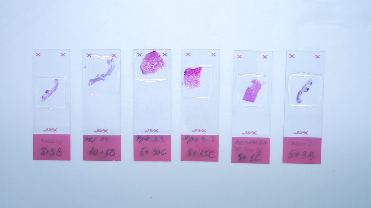
The most popular and one of the principal stains in histology is hematoxylin and eosin stain. It gives us an overview of the tissue and its structure. Hematoxylin binds with basophilic structures – for example DNA and RNA. So we can observe nuclei stained in blue or purple color. Eosin binds to acidophilic substances such as positively charged amino acid side chains. So as the result cytoplasm is pink or orange. All samples in laboratory are stained with H&E. There are several different types of hematoxylins and eosins used in histology which will give us different results.
In this video you will see, how we stain slides with different types of hematoxylins and eosins. Finally, we will compare the results.
• Subscribe to our channel: https://www.youtube.com/c/BioVitrumEN
• Watch other videos about histological process: https://www.youtube.com/playli....st?list=PLw4LQHit0MU
• Our website: http://en.biovitrum.ru/

In this video, I am talking about the best histology resources available on the internet. All the links to the resources I talked are here -
1. Amit's lectures - https://www.youtube.com/channe....l/UCwdAyZnA6FEE0Iqsw
2. VIBS histology - https://www.youtube.com/c/VIBSHistology/featured
3. Dr. Eman Sadek Histology Queen - https://www.youtube.com/channe....l/UCHXGb5GphBKKN-xD3
4. BIOC 21 Histology lectures - https://www.youtube.com/playli....st?list=PLKnI3Jl97pW
5. https://medicalschoolpathology.com/
7. Udemy - https://clnk.in/qfEB
Buy this awsm book for Histology - https://amzn.to/3wSX1Oh


In order to be able to look at tissues under a microscope, we need to first stain them with the right technique. Learn the main staining techniques used in histology today on our full video: https://khub.me/aux9w
Oh, are you struggling with learning anatomy? We created the ★ Ultimate Anatomy Study Guide ★ to help you kick some gluteus maximus in any topic. Completely free. Download yours today: https://khub.me/e0th1
As you probably know, histology is the study of the microscopic anatomy of cells and tissues. So we use staining methods to visualize and distinguish the different parts of cells and tissues since cells and their structures are usually transparent or colorless. The types of dyes used to color cells and their components can either be specific to particular structures, chemical groups or even molecules, and it can also be non-specific in which case most of the cell is stained in the same way.
When staining tissue samples, dyes that are used are either acidic or basic or a combination of the two. And why is that, you might be asking. Well, cellular structures such as nucleic acids or proteins have charged groups which are known as phosphate groups or carboxyl groups, just to name a couple. The dyes used in histology are colored organic compounds which also have a charge. Acidic dyes carry a negative charge and so they bind to positively-charged cell structures.
In the full version of this tutorial, we will cover some of the most common types of dyes used in histological staining of cells and their structures:
- basic dyes vs acidic dyes vs neutral dyes;
- hematoxylin and eosin;
- PAS - staining;
- Golgi method;
- Toluidine blue;
- Masson's trichrome;
- Osmium tetroxide;
To master this topic, click on the link and carry on watching the full video (available to Premium members): https://khub.me/aux9w !
Want to test your knowledge on the different types of cells and tissues? Take this quiz: https://khub.me/3g19f
Read more on how to interpret different histological sections on this complete article which goes through the different stains used in histology https://khub.me/saimh
For more engaging video tutorials, interactive quizzes, articles and an atlas of Human anatomy and histology, go to https://khub.me/pkvz2

The dentin is a hard tissue that forms the bulk of the tooth. It is similar to bone but is slightly harder, although softer than enamel. The dentin has numerous dentinal tubules that run across its length. Each dentinal tubule houses the cytoplasmic process of an odontoblast (odontoblastic process).
📄Notes for the video: https://www.hackdentistry.com/....bundles/revision-nin
💻Website: https://www.hackdentistry.com/
📰Blog: https://hackdentistry.substack.com/
Study resources on our website-
📖Oral pathology Revision Ninja (Notes, Videos & MCQs): https://www.hackdentistry.com/bundles/oral-pathology-revision-ninja
📖Oral Histology Revision Ninja (Notes, Videos & MCQs): https://www.hackdentistry.com/....bundles/revision-nin
📖Periodontics Revision Ninja (Notes & MCQs): https://www.hackdentistry.com/bundles/perio-rn
📖Question Bank: https://www.hackdentistry.com/bundles/question-bank
📖Access all content: https://www.hackdentistry.com/bundles/all-access-premium
References and further reading:
💡Berkovitz BKB, Hollan GR, Moxham BJ. Oral Anatomy, Histology and Embryology. 4th ed. Mosby Elsevier; 2009.
💡Nanci A. Tencate’s Oral Histology. Development, Structure and Function. 8th ed. Elsevier; 2013.
💡Kumar GS. Orban’s Oral Histology and Embryology.13th ed. Elsevier; 2011.
💡Avery JK. Oral development and Histology. 3rd ed. Thieme Medical Publishers; 2002.
Log in to https://www.hackdentistry.com and get access to:
I) Numerous Notes/Cheatsheets and Videos II) Thousands of quiz questions from our vast Question Bank!
HackDentistry is an edtech company that aims make learning dentistry fun,engaging and light hearted.
1) It focuses on helping students understand and retain core concepts in dentistry through highly visual sketch/whiteboard style video animations.
2) The platform helps improve exam performance by providing Revision Bundles and allowing students to test themselves using thousands of Practice Questions from a vast Question Bank. (multiple choice format).
3) It also provides for a community platform where students can come together, and engage with fellow dental students and dentists across the globe!
Facebook:
https://www.facebook.com/hackdentistry
Instagram:
https://www.instagram.com/hack.dentistry/
Twitter:
https://twitter.com/hckdentistry

Join the Amoeba Sisters a they explore different muscle tissues and then focus on the sliding filament theory in skeletal muscle! This video also briefly talks about muscle naming, some vocabulary (such as agonists and antagonists) before focusing on the sliding filament model. Video also mentions general roles of tropomyosin and troponin.
---------------------------------------------------------
Table of Contents:
00:00 Intro
0:39 Muscle Tissue Types
1:58 Muscle Characteristics
2:33 Skeletal Muscle Naming and Arrangement
3:26 Actin Myosin and Sarcomere
4:32 Sliding Filament Model
6:55 Tropomyosin an Troponin
---------------------------------------------------------
Factual References:
Betts, J. Gordon, et al. “10.3 Muscle Fiber Contraction and Relaxation - Anatomy and Physiology 2e | OpenStax.” Openstax.org, 20 Apr. 2022, openstax.org/books/anatomy-and-physiology-2e/pages/10-3-muscle-fiber-contraction-and-relaxation.
Urry, Lisa A, et al. Campbell Biology. 11th ed., New York, Ny, Pearson Education, Inc, 2017.
---------------------------------------------------------
Further Reading Recommendations:
What about I and A bands? What actually initiates the power stroke? How does calcium get released and from where? Remember, there is a lot more detail! We recommend this page from Openstax to learn more:
https://openstax.org/books/bio....logy-2e/pages/38-4-m
-----------------------------------------------
The Amoeba Sisters videos demystify science with humor and relevance. The videos center on Pinky's certification and experience in teaching biology at the high school level. Amoeba Sisters videos only cover concepts that Pinky is certified to teach, and they focus on her specialty: secondary life science. Learn more about our videos here: https://www.amoebasisters.com/our-videos
Support Us? https://www.amoebasisters.com/support-us
Our Resources and Handouts: https://www.amoebasisters.com/handouts
Biology Video Playlist: https://www.youtube.com/playli....st?list=PLwL0Myd7Dk1
GIFs: https://www.amoebasisters.com/gifs.html
Comics: https://www.amoebasisters.com/....parameciumparlorcomi
Unlectured Series: https://www.amoebasisters.com/unlectured
Connect with us!
Website: https://www.AmoebaSisters.com
Twitter: https://www.twitter.com/AmoebaSisters
Facebook: https://www.facebook.com/AmoebaSisters
Tumblr: https://www.amoebasisters.tumblr.com
Pinterest: https://www.pinterest.com/AmoebaSisters
Webtoon: https://www.webtoons.com/en/challenge/amoeba-sisters-sisterhood/list?title_no=289479&page=1
Instagram: https://www.instagram.com/amoebasistersofficial/
TikTok: https://www.tiktok.com/@amoebasistersofficial
Visit our Redbubble store at https://www.amoebasisters.com/store
TIPS FOR VIEWING EDU YOUTUBE VIDEOS:
Want to learn tips for viewing edu YouTube videos including changing the speed, language, viewing the transcript, etc? https://www.amoebasisters.com/....pinkys-ed-tech-favor
MUSIC:
Our intro music designed and performed by Jeremiah Cheshire.
End music in this video is listed free to use/no attribution required from the YouTube audio library.
COMMUNITY:
We take pride in our AWESOME community, and we welcome feedback and discussion. However, please remember that this is an education channel. See YouTube's community guidelines and how YouTube handles comments that are reported by the community. We also reserve the right to remove comments.
TRANSLATIONS:
Spanish Subtitles Translated by Jeremy García
Hindi Subtitles: Translated by Alisha Aggarwal
We gladly accept subtitle translations from our community. Learn more here: https://www.amoebasisters.com/....pinkys-ed-tech-favor We want to thank our amazing community for the generosity of their time in continuing to create translated subtitles.
We also have videos dubbed in Spanish and Portuguese using an artificial voice via https://aloud.area120.google.com to increase accessibility. See our Amoeba Sisters en Español channel https://www.youtube.com/channe....l/UC1Njo3LBy53cOPngz and Amoeba Sisters em Português https://www.youtube.com/channe....l/UCYTQPX2X_mXe0ZMPi

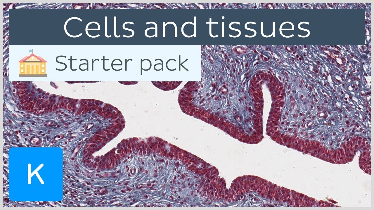
This tutorial is an introduction to the histology of the different tissues in the human body and the cells they are made of. Test yourself on our cells and tissue histology quiz at https://khub.me/jnhny
Oh, are you struggling with learning anatomy? We created the ★ Ultimate Anatomy Study Guide ★ to help you kick some gluteus maximus in any topic. Completely free. Download yours today: https://khub.me/1fcwd
A tissue is a group of cells that has a similar structure and acts together to perform one or more specific functions. In this tutorial, we will introduce you to the 4 main types of tissues in the human body: epithelial tissue, connective tissue, muscle tissue and nervous tissue. Epithelial tissue creates protective boundaries and is involved in the diffusion of ions and molecules, whereas connective tissue underlies and supports other tissue types.
Muscle tissue contracts to initiate movement in the body and nervous tissue transmits and integrates information through the central and peripheral nervous systems.
In this video tutorial we will take a closer look at the histology of the main cells and tissues under the microscope.
- 0:33 introduction to histology
- 1:22 epithelial tissue histology and types
- 5:45 function of the basement membrane
- 6:20 connective tissue histology and structure
- 10:53 muscle tissue and types of muscle cells
- 13:11 basics of the nervous system
Want to test your knowledge on the cells and tissues of the human body? Take this quiz: https://khub.me/jnhny
Why don't you jump into the introduction of the cell and its components with our free article next? Find it here: https://khub.me/apv1d
For more engaging video tutorials, interactive quizzes, articles and an atlas of Human anatomy and histology, go to https://khub.me/wcyx7
