En iyi videolar

Join Dr. Parsia Vagefi, Chief of Surgical Transplantation and Dr. Steven Hanish, Surgical Director of Liver Transplantation, as they grant unprecedented access to the OR while performing a #Liver #Transplant #Surgery.
To find out more about UT Southwestern's transplant programs visit:
https://www.utswmed.org/transplant
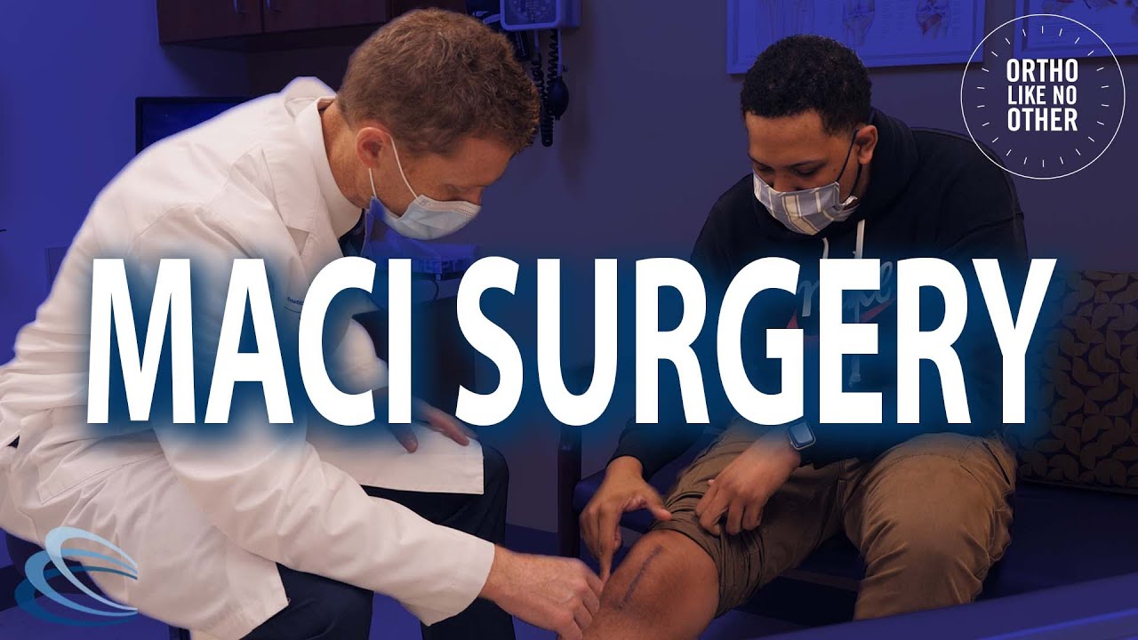
Lattrell Wells was a perfect candidate for the MACI procedure. Dr. Michael O'Malley is a sports medicine surgeon at Carilion Clinic, "It’s a two stage procedure. So what we do is we actually harvest a small portion of the patient's cartilage and bone cells and we send it to a lab where the lab then that grows additional cartilage cells. It comes back to us in a little sheet and six weeks after that initial surgery, we re-implant the cartilage in a second surgery where we implant that sheet depending on the size of lesion right where his defect. This the only option where there’s virtually no risk of any kind of graft rejection or anything of that nature.

In this video, Dr. Robert Rozbruch, chief of Limb Lengthening and Complex Reconstruction at Hospital for Special Surgery performs an osseointegration after a primary amputation. The patient, a 40 year old woman, had chronic nerve pain and compromised function of her residual limb.
For more information, visit: https://www.limblengthening.com/
https://www.hss.edu/limblengthening
https://www.hss.edu/LSARC
https://www.facebook.com/limblengtheningNYC
https://www.instagram.com/limblengthening
https://www.twitter.com/limblengthen
https://www.youtube.com/channe....l/UC-JL_X6ALjZXiXtcP
key words: Osseointegration, Amputee, Amputation, Limb Replacement, Tibia, Osseointegration
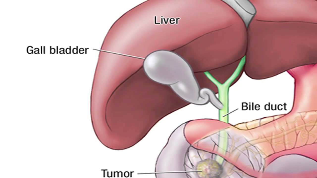
Dr. Horacio Asbun, Mayo Clinic in Florida, explains the Whipple procedure using this animated graphic of a pancreas. Cancer of the pancreas affects 45,000 people every year in the U.S., and it is the fourth leading cause of cancer-related deaths. The five-year overall survival rate if a tumor is detected early and surgically removed is 22 percent, versus 6 percent without early detection and surgery. To learn more, visit http://mayocl.in/2zk7FDi.
This video in Spanish/español: https://www.youtube.com/watch?v=N_zWboNMKWk
Cleft palate is among the most common birth defects affecting children in North America. The incomplete formation of the roof of the mouth can occur individually, or in addition to cleft lip. Cleft palate repair is a type of plastic surgery to correct this abnormal development both to restore function and a more normal appearance. This video explains what to expect for families scheduled for cleft palate surgery at the Craniofacial Anomalies Program at University of Michigan C.S. Mott Children's Hospital.
Learn more about our program at http://www.mottchildren.org/craniofacial

The video is about the evolution of the anatomic UCLA laparoscopic technique over 1325 cases and demonstrates the key steps of our operation to improve patient safety and outcomes.
Learn more at http://urology.ucla.edu

Although it demands an advanced set of skills that remain substantially hard to do, many of the salient steps of “open” surgery, including suturing, are credibly “replicated” in its laparoscopic counterpart with the intention of achieving similar optimal results. This video demonstrates how to tie Laparoscopic Roeder's Knot. Laparoscopic Roeder's Knot is one of the oldest knots used in laparoscopic surgery. It is used most commonly during laparoscopic appendectomy surgery. Recent literature, though abundant with numerous reports pertaining to a variety of endoscopic knotting techniques and technologies, appears to lack scientific data but Roeder's knot is a time tasted extracorporeal slip knot that is secure for 6-8 mm diameter tubular structure.
For more information please contact:
World Laparoscopy Hospital
Cyber City, Gurugram, NCR DELHI
INDIA 122002
Phone & WhatsApp: +919811416838, + 91 9999677788

For more videos, please visit:
http://surgicalfilmatlas.mssm.edu/

Having surgery can be frightening for anyone, but it's especially scary for kids who don't always understand what's going on, or what the grown-ups are saying. We're here to help!
Join Avrie, who had surgery at the Sacred Heart Children's Hospital pediatric surgery center in Spokane, WA. Maybe after watching and hearing her story, you and your kiddo will feel better about having surgery in the hospital.
Follow Avrie's trip - from check-in, vital signs and pre-op checks; meeting the doctor who will do his surgery, along with the anesthesiologist, surgery nurse and the Child Life Specialist; the trip to the Operating Room; waking up in the recovery room with his mom by his side; and getting ready to go home.
To learn more about the pediatric surgery center at Sacred Heart Children's Hospital, visit https://washington.providence.....org/locations-direct
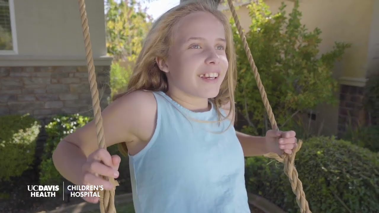
If you have an upcoming procedure at UC Davis Children’s Surgery Center, this video provides information and details of what you and your family can expect from arrival to check-in through to surgery and after care.
This video is also available in these languages:
Arabic: https://youtu.be/ERPikb0prlI
Dari: https://youtu.be/UW5fT433IGQ
Punjabi: https://youtu.be/Xq6PV2qtOMo
Russian: https://youtu.be/v223nDdN1b4
Spanish: https://youtu.be/4Jr4dkzAaWA
——
At UC Davis Children’s Hospital, we put your child at the center of everything that we do. It’s personalized care, uniquely sized for your child. You’ll see it in our child-friendly designs throughout the hospital, our farm-to-fork approach to dining, our playrooms and teen rooms and our team that feels like family. UC Davis Children’s Hospital is Sacramento’s only nationally ranked, comprehensive hospital for children, serving infants, children, adolescents and young adults with primary, subspecialty and critical care.
UC Davis Children’s Hospital: https://children.ucdavis.edu
Children’s Surgery Center: https://health.ucdavis.edu/chi....ldren/services/child
Child Life and Creative Arts Therapy: https://health.ucdavis.edu/chi....ldren/services/child
Fetal Care and Treatment Center: https://health.ucdavis.edu/chi....ldren/services/fetal
See the latest news from UC Davis Health: https://health.ucdavis.edu/newsroom
Kids Considered podcast: https://www.youtube.com/playli....st?list=PLM7qvIv8N9R
Facebook: https://www.facebook.com/UCDavisChildrensHospital
Instagram: https://www.instagram.com/ucdavischildren
Twitter/X: https://twitter.com/UCDavisChildren
——
#surgery #childrenshospital #surgeryrecovery #ucdavis

Have you heard any medical lingo you've thought is strange? Funny healthcare speaker Dr. Brad Nieder discusses funny medical terminology he's learned in his medical career. He brings his medical comedy to a healthcare conference, describing how he didn't know what "stat" meant.
He goes on about how he thought up many funny terms he could say in return to the doctor who introduced him to the word. His healthcare comedy makes the crowd burst with laughter.
Dr. Brad knows how to adapt his hilarious real-life stories into customized presentations for any in-person or virtual event. Watch more of his videos as a medical comedian and all-around funny guy by browsing his videos.

Funny Video from hospital waiting room

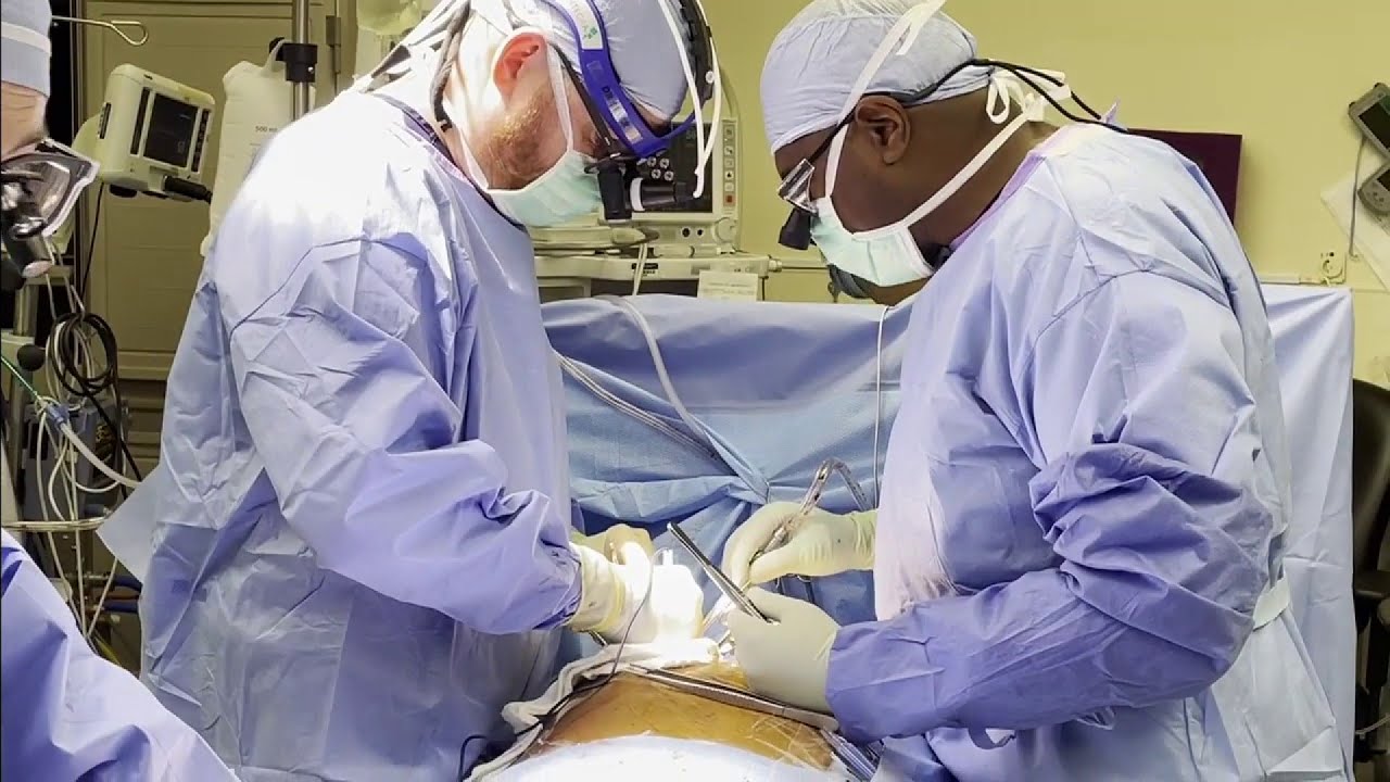
Dr. Ailawadi, M.D., the Chair of Cardiac Surgery at Michigan Medicine, specializes in minimally invasive valve surgery as well as complex cardiac operations. This video shows step by step footage of a Coronary Artery Bypass Graft (CABG) in a complex patient. In this case, CABG was performed through a sternotomy (through the breast bone) using the internal thoracic artery and saphenous leg veins to bypass obstructed coronary arteries. In this video, Dr. Ailawadi will perform a triple vessel bypass (CABG) which has been shown to minimize the risk of future heart attack and help patients live longer in the setting of complex coronary artery disease.
To learn more about cardiac surgery at Michigan Medicine, visit: https://medicine.umich.edu/dept/cardiac-surgery
To learn more about Frankel Cardiovascular Center, visit: https://www.umcvc.org/
To watch the full playlist, visit: https://www.youtube.com/playli....st?list=PLNxqP-XbH8B
-------------------------------------------------------
Subscribe to Michigan Medicine’s YouTube channel for upcoming videos and future live streams featuring our experts answering your questions.
-------------------------------------------------------
Follow Michigan Medicine on Social:
Twitter: https://twitter.com/umichmedicine
Instagram: https://www.instagram.com/umichmedicine/
Facebook: https://www.facebook.com/MichiganMedicine/
Follow the U-M Frankel Cardiovascular Center on Social:
Twitter: https://twitter.com/umichcvc
Facebook: https://www.facebook.com/Unive....rsityofMichiganCardi
#MichiganMedicine #MedEd #CardiacSurgery #UniversityOfMichiganHealth #FrankelCardiovascularCenter #Cardiology #CardiacSurgeon
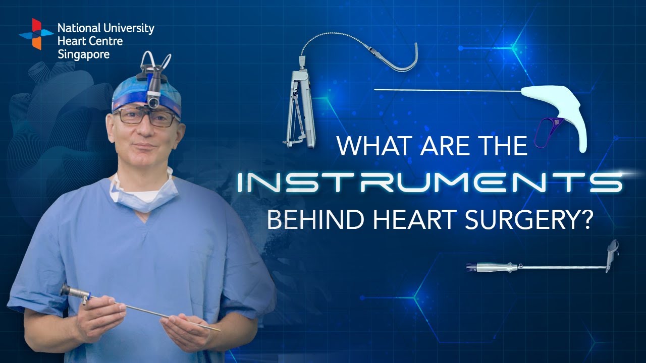
Instruments at work, innovation at play. 🔍
Watch on to discover the behind-the-scenes instruments utilised by our NUHCS cardiac surgery expert, A/Prof Theodoros Kofidis, Head of NUHCS' Department of Cardiac, Thoracic & Vascular Surgery (CTVS), for keyhole heart operations. 🔑
To find out more about Minimally Invasive Heart Surgery @ NUHCS, visit: https://[a]www.nuhcs.com.sg%2FOur-Services%2FSpecialties%2FPages%2FMinimally-Invasive-Cardiac-Surgery-Programme.aspx[/a]
Connect with us:
Instagram: @nuhcsofficial
Facebook: www.facebook.com/nuhcs
Website: www.nuhcs.com.sg
LinkedIn: www.linkedin.com/company/nuhcs
To make an appointment with the NUHCS Heart Clinic, email us at appointment@nuhs.edu.sg
#NUHCS #cardiacsurgery #heartsurgery #keyholesurgery #minimallyinvasive
Dr. Erik Beyer, Florida Medical Center's chief of cardiac surgery, discusses performed a procedure called a micro-thoracotomy.

***SUBSCRIBE WITHIN THE NEXT 28 DAYS FOR A CHANCE TO WIN $1,000!***
Did you know only 20% of our video content is on YouTube? Try out our membership for FREE today! → https://bit.ly/3yRDykI
Try our NCLEX Prep FREE → https://bit.ly/3sRRjvY
Head to https://bit.ly/3yRDykI to get access to the other 80%, along with 800+ study guides, customizable quiz banks with 3,000+ test-prep questions, and answer rationales!
This video illustrates an IM injection for deltoid muscle.
Note that vaccines and other medications can be administered through the deltoid muscle. I will give you some tips through this video.
It is important to check your client’s details such as their medication, time, dose, and the route to be used. Different research works are subject to change the protocols for insertion thus, it is necessary to be up to date with the current changes.
Assemble all the supplies and conduct hand sanitation. Usually, I wear gloves before giving any injection in as much as the CDC may state it is optional unless the patient has an open lesion and contact of body fluids is likely to happen.
Use the acromion process landmark to locate the deltoid muscle. Move your fingers about two widths below the landmark. The patient’s adipose tissue determines the choice of needle length. Note that the needle gauge is determined by the type of medication you plan to give to the patient.
The Z-track technique is recommended rather than pinching the patient’s skin. Pull the patient’s skin to the side using one hand. Use a 90 degree angle to insert the needle to the patient’s skin. At the rate of 10 seconds per mL gently depress the plunger.
Remove the needle carefully and engage the safety precautions then dispose of the needle appropriately in the sharps container. Gauzing helps to cover the injection site.
Nursing School Membership - Try it FREE → https://bit.ly/3yRDykI
New NCLEX Prep - Try it FREE → https://bit.ly/3sRRjvY
Popular Playlists:
NCLEX Fluid & Electrolytes: https://bit.ly/39BSHXs
Heart Failure (CHF): https://bit.ly/2u5zfDm
Myocardial Infarction (MI): https://bit.ly/3bN9AAk
Addison’s vs. Cushing: https://bit.ly/2STvute
Diabetes Mellitus & DKA vs HHNS: https://bit.ly/37D8nbs
Cardiomyopathy: https://bit.ly/38CwcSg
IV Fluids: Hypertonic, Hypotonic & Isotonic: https://bit.ly/2P45BWx
SIADH vs Diabetes Insipidus: https://bit.ly/2wq6Bhb
Follow us on social media for more EXCLUSIVE content 👋
More Videos: https://bit.ly/37CRttH
Instagram: https://www.instagram.com/simplenursing.com_
TikTok: https://www.tiktok.com/simplenursing
Thank you for the support & for tuning in!
Remember… don’t be scared, BE PREPARED!

Vital signs help us assess patients in the nursing profession, and there are six common vital signs that we assess as nurses:
1. Heart Rate (Pulse)
2. Respiration Rate
3. Temperature
4. Blood Pressure
5. Pain Rating
6. Oxygen Saturation
This video will demonstrate how to check vital signs (live) on a patient, along with normal rates for each assessment. I also give you a few tips for taking vital signs as a nurse, CNA, or other healthcare profession.
🟣ABG eBook: https://registerednursern.creator-spring.com/
🟣ABG physical book: https://amzn.to/3EsF0Mc (affiliate link)
More nursing skills: https://www.youtube.com/watch?v=G5-Rp-6FMCQ&list=PLQrdx7rRsKfUhd_qQYEbp0Eab3uUKhgKb
Website: https://www.registerednursern.com/
More Videos: https://www.youtube.com/watch?v=R2XMro13dD0&list=UUPyMN8DzkFl2__xnTEiGZ1w
Nursing Gear: https://teespring.com/stores/registerednursern
Instagram: https://www.instagram.com/registerednursern_com/
Facebook: https://www.facebook.com/RegisteredNurseRNs
Twitter: https://twitter.com/NursesRN
Popular Playlists:
NCLEX Reviews: https://www.youtube.com/playli....st?list=PLQrdx7rRsKf
Fluid & Electrolytes: https://www.youtube.com/playli....st?list=PLQrdx7rRsKf
Nursing Skills: https://www.youtube.com/playli....st?list=PLQrdx7rRsKf

Vial medication administration nursing skill. Learn techniques to withdraw medication from a vial using a syringe with a needle.
Medications can come in different forms, such as ampules, vials, tablets, capsules, and so forth. When withdrawing medication from a vial, there are a few things you'll want to know as a nursing student or nurse.
First, there are different needles that can be attached to the syringe. You can use a traditional needle with a beveled tip; you can use a blunt-tip needle to reduce the risk of needle sticks; or you can use a filter needle, which is sometimes required or recommended when drawing medication from a vial, particularly in cases of reconstituted medication.
When withdrawing from a vial, you'll want to do these things (assuming they fit with the protocols and manufacturer's instructions):
NOTE: Some medications or vaccines may require a different technique, so always consult with the manufacturer's instructions.
-gather your supplies
-perform hand hygiene
-clean the vial's top with alcohol prep
-attach the appropriate needle
-stick the needle using a technique to prevent coring of the rubber on the vial (start with 45 degree angle, and as you puncture the vial, rotate the needle to a 90 degree angle in one smooth motion).
-push air into the vial equal to the amount of medication you plan to draw
-invert the vial to withdraw medication
-remove air bubbles
-and much more
See more Nursing Skills: https://www.youtube.com/playli....st?list=PLQrdx7rRsKf
Notes: https://www.registerednursern.....com/how-to-withdraw-
Website: https://www.registerednursern.com/
More Videos: https://www.youtube.com/watch?v=R2XMro13dD0&list=UUPyMN8DzkFl2__xnTEiGZ1w
Nursing Gear: https://teespring.com/stores/registerednursern
Instagram: https://www.instagram.com/registerednursern_com/
Facebook: https://www.facebook.com/RegisteredNurseRNs
Twitter: https://twitter.com/NursesRN
Popular Playlists:
NCLEX Reviews: https://www.youtube.com/playli....st?list=PLQrdx7rRsKf
Fluid & Electrolytes: https://www.youtube.com/playli....st?list=PLQrdx7rRsKf

Pass your tests and improve your grades with the below FREE resources:
1) A FREE 140 Must Know Meds book
Click here to get your FREE copy of the 140 Must Know Meds Book: https://bit.ly/41rxSt0
2) A FREE test-taking tips webinar
Join us for our free test-taking tips webinar to boost your exam scores: https://bit.ly/nursingtesttaking
You can now test your knowledge with a free lesson quiz on NURSING.com!
Click here for your free quiz: https://bit.ly/3uyTWEu
Learn what's working for other Nursing Students! Check out our Top 10 Most Popular Lessons Here: https://bit.ly/3nda5u3
Dressing Changes- Wet to Dry (Nursing Skills)
FREE Nursing School Cheat Sheets at: http://www.NURSING.com
Get the full Dressing Change lesson here: https://nursing.com/lesson/ski....lls-05-04-wound-care
Click here for the related lesson on Wound Assessment: https://nursing.com/lesson/ski....lls-05-02-wound-care
Welcome to the NURSING Family, we call it the most supportive nursing cohort on the planet.
At NURSING.com, we want to help you remove the stress and overwhelm of nursing school so that you can focus on becoming an amazing nurse.
Check out our freebies and learn more at: (http://www.nursing.com)
Dressing Changes- Wet to Dry (Nursing Skills):
In this video we’re going to look at how to do a wet to dry dressing change. Wound care and dressing changes should be performed at least daily or more often depending on orders. Dressing changes should be sterile to avoid introducing any new bacteria to the wound and to promote wound healing.
Bookmarks:
0:05 Introduction
0:10 Wound Assessment link above
0:24 Dressing Change Prep
1:24 Wet vs Dry Gauze
1:37 Soaking Gauze
2:00 Gauze Ring Out
2:25 Packing the wound
3:00 Covering the wound bed
3:37 Dry gauze barrier
4:00 ABD pad application
4:46 Documentation
4:54 Outro
Visit us at https://nursing.com/medical-disclaimer/ for disclaimer information.
NCLEX®, NCLEX-RN® are registered trademarks of the National Council of State Boards of Nursing, INC. and hold no affiliation with NURSING.com.
