- Physical Examination
- Surgical Examination
- Ophthalmology
- Clinical Skills
- Orthopedics
- Surgery Videos
- Laparoscopy
- Pediatrics
- Funny Videos
- Cardiothoracic Surgery
- Nursing Videos
- Plastic Surgery
- Otorhinolaryngology
- Histology and Histopathology
- Neurosurgery
- Dermatology
- Pediatric Surgery
- Urology
- Dentistry
- Oncology and Cancers
- Anatomy Videos
- Health and Fitness
- Radiology
- Anaesthesia
- Physical Therapy
- Pharmacology
- Interventional Radiology
- Cardiology
- Endocrinology
- Gynecology
- Emergency Medicine
- Psychiatry and Psychology
- Childbirth Videos
- General Medical Videos
- Nephrology
- Physiology
- Diet and Food Health
- Diabetes Mellitus
- Neurology
- Women Health
- Osteoporosis
- Gastroenterology
- Pulmonology
- Hematology
- Rheumatology
- Toxicology
- Nuclear Medicine
- Infectious Diseases
- Vascular Disease
- Reproductive Health
- Burns and Wound Healing
- Other
Top videos

Thank you so much for watching❤
If you enjoyed this video ▶Please leave a LIKE👍 ▶SHARE this video ▶【SUBSCRIBE】my channel for more new videos And click the BELL 🔔so you don't miss any of my videos HERE
https://www.youtube.com/c/nurs....eminder?sub_confirma
You can support my work by purchasing your NurseMinder Merch https://teespring.com/stores/nurseminder-nation (or click on merch pics under the video)
Or simply do your Amazon shopping after clicking on one of the links below
-------------------------------------------------------------------------
Thank you so much! I appreciate you!♥♥
------------------------------------------------------------------------
Nurses often prime IV lines with the hopes that there are no air bubbles. In this video, I will share a couple of tips to help reduce the risk or frequency of air bubbles during line priming. I will also talk about how to troubleshoot the air bubbles when they appear during an infusion
Providing patient care and influencing safe patient outcomes requires that registered nurses and licensed practice nurses maintain air free IV lines. Learn the strategies and tips to decrease the risk of air bubbles appearing in your primary or secondary medication line as well as troubleshooting tips to remove those alarming bubbles. Your patients will thank you!
Whether you are providing normal saline, a medication, or a combination, ensure that all fluids are compatible.
Supplies used in this video include the Alaris Primary Infusion line, alcohol swabs and a sterile 10 cc syringe ... and a nail in the wall :)
------------------------------------------------------------------------
❤️ ~ You may also be interested in watching ~ ❤️
PICC line assessment https://youtu.be/tnKClpU-J1g
How To Access a PICC line https://youtu.be/SCF6bmk8KWc
Putting on Sterile Gloves https://youtu.be/xNwkKLqDJn4
Organizational Plans for Nursing https://youtu.be/_NATxwPwHzc
Medication Conversions https://youtu.be/TCPBXg2TYCs
------------------------------------------------------------------------
💻COMMENT in the description box below and share your ideas
👍 LIKE the video
🗣 SHARE with your friends
📥 SUBSCRIBE ... hit the BELL 🔔
Subscribe to NurseMinder https://www.youtube.com/c/nurs....eminder?sub_confirma
------------------------------------------------------------------------
Amazon Affiliate Links
------------------------------------------------------------------------
Want to support me in another way? Enter Amazon through my links and continue to do your shopping. Simple and Easy Way to support the work I do.
The following list is the equipment I use (or if my version is no longer sold, a close replica).
📱 Phone 11 Cell Phone https://amzn.to/2WpOJfz
💻 MacBook Pro https://amzn.to/2YyxQC1
👉 Final Cut Video Editing software https://amzn.to/3fqlAd9
🎙️ Rode NT USB microphone (Audio Recording) for post-production voiceover https://amzn.to/2W2RJj1
👉 Neewer Professional Recording Stand – mount microphone and adjust positioning to keep it close but out of the camera’s view: https://amzn.to/3fjB4zs
👉 Manfrotto Tripod (hold cell phone) https://amzn.to/2YKGYUz
💡 Neewer Ring Light to reduce shadows and improve lighting. https://amzn.to/3dk5OP5
Disclaimer: I recommend only products that I know and trust to be of high quality. Links are provided for quick access. Some of the links contained in this checklist are affiliate links and I may receive a commission if make a purchase from the affiliate. This helps me to keep creating and offering free content.

Vital signs help us assess patients in the nursing profession, and there are six common vital signs that we assess as nurses:
1. Heart Rate (Pulse)
2. Respiration Rate
3. Temperature
4. Blood Pressure
5. Pain Rating
6. Oxygen Saturation
This video will demonstrate how to check vital signs (live) on a patient, along with normal rates for each assessment. I also give you a few tips for taking vital signs as a nurse, CNA, or other healthcare profession.
🟣ABG eBook: https://registerednursern.creator-spring.com/
🟣ABG physical book: https://amzn.to/3EsF0Mc (affiliate link)
More nursing skills: https://www.youtube.com/watch?v=G5-Rp-6FMCQ&list=PLQrdx7rRsKfUhd_qQYEbp0Eab3uUKhgKb
Website: https://www.registerednursern.com/
More Videos: https://www.youtube.com/watch?v=R2XMro13dD0&list=UUPyMN8DzkFl2__xnTEiGZ1w
Nursing Gear: https://teespring.com/stores/registerednursern
Instagram: https://www.instagram.com/registerednursern_com/
Facebook: https://www.facebook.com/RegisteredNurseRNs
Twitter: https://twitter.com/NursesRN
Popular Playlists:
NCLEX Reviews: https://www.youtube.com/playli....st?list=PLQrdx7rRsKf
Fluid & Electrolytes: https://www.youtube.com/playli....st?list=PLQrdx7rRsKf
Nursing Skills: https://www.youtube.com/playli....st?list=PLQrdx7rRsKf

Vial medication administration nursing skill. Learn techniques to withdraw medication from a vial using a syringe with a needle.
Medications can come in different forms, such as ampules, vials, tablets, capsules, and so forth. When withdrawing medication from a vial, there are a few things you'll want to know as a nursing student or nurse.
First, there are different needles that can be attached to the syringe. You can use a traditional needle with a beveled tip; you can use a blunt-tip needle to reduce the risk of needle sticks; or you can use a filter needle, which is sometimes required or recommended when drawing medication from a vial, particularly in cases of reconstituted medication.
When withdrawing from a vial, you'll want to do these things (assuming they fit with the protocols and manufacturer's instructions):
NOTE: Some medications or vaccines may require a different technique, so always consult with the manufacturer's instructions.
-gather your supplies
-perform hand hygiene
-clean the vial's top with alcohol prep
-attach the appropriate needle
-stick the needle using a technique to prevent coring of the rubber on the vial (start with 45 degree angle, and as you puncture the vial, rotate the needle to a 90 degree angle in one smooth motion).
-push air into the vial equal to the amount of medication you plan to draw
-invert the vial to withdraw medication
-remove air bubbles
-and much more
See more Nursing Skills: https://www.youtube.com/playli....st?list=PLQrdx7rRsKf
Notes: https://www.registerednursern.....com/how-to-withdraw-
Website: https://www.registerednursern.com/
More Videos: https://www.youtube.com/watch?v=R2XMro13dD0&list=UUPyMN8DzkFl2__xnTEiGZ1w
Nursing Gear: https://teespring.com/stores/registerednursern
Instagram: https://www.instagram.com/registerednursern_com/
Facebook: https://www.facebook.com/RegisteredNurseRNs
Twitter: https://twitter.com/NursesRN
Popular Playlists:
NCLEX Reviews: https://www.youtube.com/playli....st?list=PLQrdx7rRsKf
Fluid & Electrolytes: https://www.youtube.com/playli....st?list=PLQrdx7rRsKf
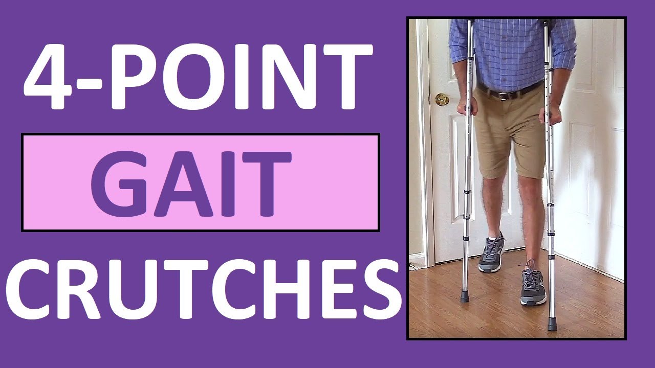
Four-point gait crutches walking pattern demonstration review for
NCLEX assistive devices and nurses.
One of the gaits that you'll have to learn for crutches is the 4-point gait. An example of a four point gait crutch pattern would be the patient moving the right crutch first (on the injured side), followed by the left foot, then the left crutch, and then the right foot. Then, you'll repeat this pattern.
In addition to this video, we have an entire compilation that features the various crutch gait patterns, as well as walkers and canes:
https://www.youtube.com/watch?v=k2-w3LZlCVk
#crutches
#nclex
#nursing
#nurse
Website: https://www.registerednursern.com/
More Videos: https://www.youtube.com/watch?v=R2XMro13dD0&list=UUPyMN8DzkFl2__xnTEiGZ1w
Nursing Gear: https://teespring.com/stores/registerednursern
Instagram: https://www.instagram.com/registerednursern_com/
Facebook: https://www.facebook.com/RegisteredNurseRNs
Twitter: https://twitter.com/NursesRN
Popular Playlists:
NCLEX Reviews: https://www.youtube.com/playli....st?list=PLQrdx7rRsKf
Fluid & Electrolytes: https://www.youtube.com/playli....st?list=PLQrdx7rRsKf
Nursing Skills: https://www.youtube.com/playli....st?list=PLQrdx7rRsKf

► Get a free NCLEX NGN sample test today: http://lectur.io/nclexrnsampletestyt
► Create your free account today: http://lectur.io/nurseregisteryt
► If you’re an nursing educator or faculty member, visit: http://lectur.io/nursytb2u
In this video “How To Do An IM (Intramuscular) Injection” you will learn about:
►the steps in the administration of intramuscular medications
►the angle to position the syringe while administering an intramuscular injection
►the landmark to administer an intramuscular injection in the deltoid muscle
►5 tips for the safe administration of an intramuscular medication
►the steps of the Z-track method for intramuscular injections
►the role of aspirating blood during an intramuscular injection and evaluate whether this practice is currently in use
► This video is part of the Lecturio course “Fundamentals of Nursing: Clinical Skills”
► WATCH the complete course on http://lectur.io/njection
► THE PROF: Samantha Rhea MSN, RN has been a nurse since 2008 and a nursing faculty teacher since 2012. She has been recognized for clinical excellence as an interventional cardiology nurse and also led a Joint Commission Accredited Stroke Center. Ms. Rhea is an award-winning expert in clinical teaching and continues to maintain a current clinical practice and teaches at a University nursing program.
► LECTURIO is your smart tutor for nursing school: Learn the toughest NCLEX® topics with high-yield video lectures, integrated quiz questions, and more. Register now to study anytime and anywhere you want to: https://nursing.lecturio.com/#/
► CHECK OUT ALL NURSING COURSES:
Leadership Nursing: http://lectur.io/leadershipnursing
Dosage Calculation Nursing: http://lectur.io/dosagecalcnursing
Physiology Nursing: http://lectur.io/physiologynursing
Medical Surgical Nursing: http://lectur.io/medsurgnursing
Pharmacology Nursing: http://lectur.io/pharmacologynursing
NCLEX® Pharmacology Nursing: http://lectur.io/pharmnclexnursing
Pediatric Nursing: http://lectur.io/pediatricnursing
Study Skills Nursing: http://lectur.io/studyskillsnursing
Fundamentals of Nursing - Theory: http://lectur.io/fundamentalstheory
Fundamentals of Nursing - Clinical Skills: http://lectur.io/fundamentalsclinicalskills
Nursing Prerequisites: http://lectur.io/nursingprerequisites
Mental Health Nursing: http://lectur.io/mentalhealthnursing
Maternal-Newborn Nursing: http://lectur.io/maternalnewbornnursing
► INSTALL the free Lecturio app
iTunes Store: https://app.adjust.com/z21zrf
Play Store: https://app.adjust.com/b01fak
► SUBSCRIBE to our YouTube channel: http://lectur.io/subscribenursing
► WATCH MORE ON YOUTUBE: http://lectur.io/nursingplaylists
► LET’S CONNECT:
Facebook: www.facebook.com/lecturio.nursing
Instagram: www.instagram.com/lecturio_nursing
Join Discord Community: https://discord.gg/Ue95WDxCrp
TikTok: www.tiktok.com/@lecturio_nursing
LinkedIn: https://www.linkedin.com/company/lecturio-medical/
#nursingschool #nursingeducation #nursingclinicalskills #leadershipnursing #nclex #nursingfundamentals #nursingclinical #nursingskills

Glass ampules are often used to store medication, and as a nurse, you'll need to know how to use them.
In this video, I demonstrate how to clean an ampule using alcohol prep, how to open (or break) an ampule, as well as how to dispose of the ampule.
In addition, I show how to use an ample filter straw while drawing up (withdrawing) medication, how to use the syringe, and how to remove the air bubbles in the syringe.
This is another video in our series on clinical nursing skills.
Notes: https://www.registerednursern.....com/how-to-withdraw-
Website: https://www.registerednursern.com/
More Videos: https://www.youtube.com/watch?v=R2XMro13dD0&list=UUPyMN8DzkFl2__xnTEiGZ1w
Nursing Gear: https://teespring.com/stores/registerednursern
Instagram: https://www.instagram.com/registerednursern_com/
Facebook: https://www.facebook.com/RegisteredNurseRNs
Twitter: https://twitter.com/NursesRN
Popular Playlists:
NCLEX Reviews: https://www.youtube.com/playli....st?list=PLQrdx7rRsKf
Fluid & Electrolytes: https://www.youtube.com/playli....st?list=PLQrdx7rRsKf
Nursing Skills: https://www.youtube.com/playli....st?list=PLQrdx7rRsKf
http://www.utexas.edu
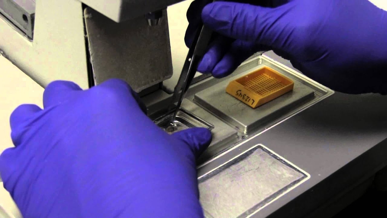
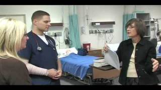
Nursing students practice their skills on mannequins and each other in the Nursing Skills Lab.

Please remember that this video is to be used for educational purposes. You must follow your facility or colleges' policies and procedure checklists to ensure you are completing the skill satisfactorily. Thanks for watching!
Music from #Uppbeat (free for Creators!):
https://uppbeat.io/t/swoop/blue-sea
License code: W9DFUQ4II7YVHA59

Tummy Tuck ( Classic Method ) : Surgery | 3D Animation
How long does tummy tuck last?
Tummy tuck results are considered permanent, insofar that the fat cells and skin removed during an abdominoplasty cannot grow back. Likewise, the internal sutures placed to repair abdominal muscles are designed to remain in place indefinitely.
What is tummy tuck surgery?
A tummy tuck — also known as abdominoplasty — is a cosmetic surgical procedure to improve the shape and appearance of the abdomen. During a tummy tuck, excess skin and fat are removed from the abdomen. Connective tissue in the abdomen (fascia) usually is tightened with sutures as well.
How much does tummy tuck cost?
How much does it cost? It can cost from about £5,000 to £10,000 to have an abdominoplasty in the UK, plus the cost of any consultations or follow-up care.
How painful is a tummy tuck?
A tummy tuck requires significant downtime
At the beginning, you will be fatigued, swollen and sore. It is normal to have moderate pain during these first several days, although this will steadily improve. It is vital to allow yourself time to focus on rest and healing.
What is the disadvantage of tummy tuck?
The cons of a tummy tuck include: A full abdominoplasty is a major operation with a considerable recovery. Expect to postpone strenuous activities for at least 6 weeks. Results take time.
Is tummy tuck more painful than C section?
That's something many women want to know. While patients have different experiences, most plastic surgeons would agree that a cesarean section is more painful than most tummy tucks.
- Tummy tuck
- Abdominoplasty
- Abdominal tuck
- Tummy tuck procedure
- Tummy tuck process
- Tummy tuck surgery
- Tummy tuck operation
- Tummy tuck video
- Tummy tuck recovery
- Tummy tuck before and after
- Abdominoplasty surgery
- Abdominal contouring surgery
- Postpartum tummy tuck
- Post pregnancy tummy tuck
- Mini tummy tuck
- Tummy tuck cost
- Tummy tuck risks
- Tummy tuck complications
- How long does a tummy tuck take
- Tummy tuck scarring
- Tummy tuck skin removal
- Tummy tuck muscle tightening
#tummytuck
#abdominoplasty
#plastic_surgery
#cosmetic_surgery
#body_contouring
#tummy_tuck_surgery
#surgery
#cosmetic_procedure
#beauty
#health
#fitness
#medical_animation
#3d_animation
#medical_video
#explainer_video
#education

Full Tummy Tuck 3D Video - http://drlandsman.com
Look great... feel great
•Smart Liposuction + Liposculpture
•Abdominplasty (Tummy Tuck)
+ Full Mini Modified
•Brazilian Lift with Fat Transfer
•Vaginal Aesthetics & Rejuvenation
•Laser Hair Removal
•Full Body Lift
•Thigh lift
•Brachioplasty (Arm Lift) + Short Scar
Expertise in Body Contouring
Board Certified Plastic Surgeon
Expertise in body contouring combines skin excision techniques and advanced fat contouring technology
Weight control personalized training and smoking cessation results in a healthier lifestyle improved shape and longer lasting results
With over 2 decades of experience Dr Lloyd Landsman provides state of the art cosmetic and plastic surgery
Dr Landsman integrates the finest and safest products with the newest procedures
A customized treatment plan is created for each patient utilizing classic surgical and minimally invasive techniques for optimal results
Call for your complimentary consultation to learn how Dr Landsman can help you look your very best
Visit http://drlandsman.com Call 631 864 4111
Main Office 994 W Jericho Tpke Smithtown NY 11787
Affiliates East Islip • Westbury • Jackson Heights • Manhattan

TUMMY TUCK 🤩 Immediate OR Results
This patient wanted to get her abs back, but unfortunately NO diet or workout can tighten muscles that have been stretched apart from carrying a baby 👀 But we can fix that at Lemmon Avenue Plastic Surgery & Laser Center!
To learn more about the #tummytuck click here: https://drdeuber.com/procedures/tummy-tuck/
For #mommymakeover, click here: https://drdeuber.com/procedures/mommy-makeover/
👙
#MarkDeuberMD

An Abdominoplasty (commonly referred to as a “Tummy Tuck”) removes excess fat and skin around your abdomen to shape and contour your midsection. During surgery, I also restore weakened or separated muscles to help create an abdominal profile that is both; smoother and more firm.
Watch this video as we go from the operating table to her 2-month post-op results!
If you’re interested in learning more about tummy tuck surgery or any other services we offer, please DM us or give us a call today!
☎️(424) 266-4181
🌐DrJohnDiaz.com
#DrJohnDiaz #DrDiaz #BeverlyHills #BeverlyHillsPlasticSurgery #BeverlyHillsPlasticSurgeon #DiazPlasticSurgery #PlasticSurgery #PlasticSurgeon #TummyTuck #Abdominoplasty #BeverlyHillsTummyTuck #TummyTuckBeverlyHills #AbdominoplastyBeverlyHills #BeverlyHillsAbdominoplasty #TummyTuckSurgery

The MINI tummy-tuck is a lesser variant of the classic tummy tuck. The MINI tummy-tuck always involved skin excision (often a scar revision and skin excision of the flabby skin over a C-section scar or hysterectomy or laparotomy scar) but may also involve liposuction, umbilical floating, etc. Commonly it will not include any muscle repair otherwise it it now a classic tummy tuck (aka abdominoplasty). Cost varies depending on the components involved. Here, Toronto Aesthetic Plastic Surgeon Dr Marc DuPéré describes a MINI tummy-tuck done on a patient who had a Brazilian Butt Lift before (and skin harvesting from abdomen) and a recent 20 lbs weight loss, a patient who wants more liposuction to abdomen and flanks and whose skin has now lost elasticity, hence the requirement for this small skin excision. Dr DuPéré also explains what UMBILICAL floating means. Dr DuPéré performs more than 5 different techniques of tummy-tucks in Toronto and the technique chosen reflects the patient’s expectations and anatomy. Call us if interested in learning about YOUR options for a flatter tummy! 📱 416-929-9800
Patient consent obtained. Thank you to my patient.
Visage Clinic Toronto
https://www.visageclinic.com/
(416) 929-9800
101-133 Hazelton Avenue, Toronto, ON M5R 0A6
https://www.facebook.com/VisageClinic/
https://www.instagram.com/VisageClinicDrDuPere/
After MacKenzie Walker lost 100 pounds, her "after" picture remained elusive. So she asked plastic surgeon Dr. Anthony Youn to perform an abdominoplasty.

In this video, we're going to share 11 things you should NOT do after a tummy tuck. These tips will help you recover from your surgery and keep you from having some common post-tummy-tuck complications. If you're considering a tummy tuck, then be sure to follow these post-operative guidelines!
Dr. William will share all the information you need to make the best decisions for your surgery and recovery. So sit back, relax, and enjoy this video on what NOT to do after an abdominoplasty!
#tummytuck #abdominoplastia #drwilliam
Want a Consultation?
Send us your information: https://drwilliammiami.typefor....m.com/YT-consultatio
Learn more about Dr. William Miami at: https://www.drwilliammiami.com
🔔 Subscribe to our Youtube channel, and stay tuned to all the latest information on cosmetic surgery.
Follow us on Social Media:
Instagram: https://www.instagram.com/drwilliammiami/
Facebook: https://www.facebook.com/Drwilliammiami/
Tiktok: https://www.tiktok.com/@drwilliammiami
OnlyFans: https://onlyfans.com/drwilliammiami
Ogee Recovery: https://ogeerecovery.com
305 Plastic Surgery
564 SW 42nd Ave 3rd floor
Coral Gables, FL 33134
Call us at (305) 209-1030

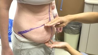
Diastasis recti often occurs during pregnancy and can persist after pregnancy. It affects core strength and the appearance of the abdominal muscles.
Dr. Erick Sanchez repairs the abdominal muscles with every tummy tuck. This short video shows the muscle repair portion of the surgery with a bonus after photo at the end!
To request a consultation with Dr. Sanchez, visit sanchezplasticsurgery.com and click Request a Consultation. Fill out the form and someone will get in touch with you to answer all your questions.
Expected cost can be found at the bottom of each procedure page on our website.

#abdomenliposuction #laserskintightening #drprashantyadav #cosmeticsurgery #plasticsurgery #dezireclinicindia #weightloss #shorts #360degreeabdomenliposuction #lowerbackliposuction
Weight Loss After 360° Abdomen liposuction result, Abdomen Liposuction, lower back liposuction, 360 degree abdomen liposuction
☎️ For more info:
WhatsApp Your Details to know the Cost
Delhi - 8956880644, 9717470550, Pune - 9222122122, Bangalore- 8971224700, Gurugram - 9272007896, Ahmedabad - 9711162746
Why choose Dezire Clinic For Your Cosmetic and plastic surgery treatment ?
Dezire Clinic is a top searched clinic surgical and nonsurgical cosmetic procedure in India when comes to “Cosmetic, Skin ,Laser and Hair transplantation”.
Like and Share the video if you find it useful. Do not forget to Subscribe to our channel to get more updates.
Subscribe on YouTube https://youtube.com/dezireclin....ic?sub_confirmation=
https://youtube.com/dezireplas....ticsurgerycenter?sub
🎦 https://www.youtube.com/dezireclinic
🎦 https://www.youtube.com/DezirePlasticSurgeryCenter
👍🏻 https://www.facebook.com/drprashantmch/
👍🏻 https://www.facebook.com/dezireclinic
📸 https://www.instagram.com/drprashantdezireclinic/
📸 https://www.instagram.com/dezireclinics/
🐥 https://twitter.com/drprashantmch
👍🏻 https://www.linkedin.com/in/drprashantyadav/
🌐 Website: https://www.dezireclinic.in/
📧 dezireclinicindia@gmail.com
📧 info@dezireclinic.in
Dr. Prashant Yadav (M.S., M.Ch. Plastic Surgery ) & Founder of Dezire Clinic
Disclaimer: The content of this channel is for informational and educational purposes only. This content should not be considered a substitute for advice provided by a certified plastic or cosmetic surgeon. Patients must be properly diagnosed by a healthcare professional on an individual basis in order to achieve the desired results. There is no guarantee of getting the results and outcomes shown in videos, as the results can vary at the end. We will not be held liable for any harm caused by someone misusing our name.
#plasticsurgery #cosmeticsurgery #dezireclinic #drprashantyadav

This animation shows you how a tummy tuck is performed at Boerhaave Medical Centre. Curious? Watch the video!
Boerhaave Medical Centre sets itself the goal of providing the highest quality care. Quality not just in terms of treatment, but also in terms of our staff and the before and after care given. By providing thorough information and clear advice in advance, carefully supporting our patients through the procedure and caring for them afterwards, we believe this quality is assured.
Although we are one of the largest clinics in the Netherlands and have built up many years of experience, we continuously strive to improve. After all, the Boerhaave Medical Centre intends to remain a pioneer in the healthcare sector, by working in accordance with the latest medical findings and techniques both now and in the future.
We offer the highest standard of plastic surgery in our cosmetic care department. For 365 days a year, you can also come to us for non-surgical treatments, such as injectables, permanent hair removal and gastric balloons.
We have been awarded the ZKN quality mark and are certified to ISO 9001-2008 for giving advice and carrying out plastic surgery, including after care.
Visit our website for more information: https://www.boerhaave.com/all-....treatments/upperbody
Follow us:
Facebook: https://www.facebook.com/boerhaavemc
Google+: https://plus.google.com/+BoerhaaveNl-Kliniek
Pinterest: https://nl.pinterest.com/BoerhaaveMC/
Instagram: https://www.instagram.com/BoerhaaveMC/

A complete organized library of all my videos, digital slides, pics, & sample pathology reports is available here: https://kikoxp.com/posts/5084 (dermpath) & https://kikoxp.com/posts/5083 (bone/soft tissue sarcoma pathology)
Topics discussed:
Epidermis:
Layers of epidermis: 0:10
Melanocytes vs Keratinocytes: 5:16
Langerhans cells: 10:10 & 33:30 & 57:30
Dermis:
Papillary and reticular dermis: 11:50
Three types of white empty spaces on a slide: vessels, glands/ducts/cysts, or artifact: 15:25
Blood vessels & nerves: 18:24 & 48:50 & 58:59
Arrector pili & other dermal smooth muscle: 20:00
Adnexal:
Sebaceous gland: 21:10
Hair follicle 23:14
Eccrine sweat glands and ducts 24:45 & 50:00
Gland/duct vs blood vessel 27:20 & 48:50
Apocrine glands: this video https://kikoxp.com/posts/7837 (at 12:30)
Acrosyringium: this video https://kikoxp.com/posts/7837 (at 10:00)
Three types of pink bundles: smooth muscle, nerve, dense connective tissue: 27:50
Acral skin (palm sole) with contact dermatitis 29:37
Parakeratosis 30:00
Perivascular lymphocytes 30:40
Eosinophils vs neutrophils 31:20
Spongiosis with desmosome keratinocyte spines 32:10
Spongiotic vesicles with Langerhans cells 33:30
Normal acral skin (palm & sole) with stratum lucidum 34:20
Normal glomus body/apparatus (canal of Sucquet-Hoyer) 35:40
Nerve 36:46 & 51:50
Adipose tissue (white fat cells) in subcutis with Lochkern 37:55
Normal scalp skin with large anagen hair follicles: 39:30
Hair follicle anatomy (bulb/matrix, inner root sheath, outer root sheath, hair shaft, isthmus, infundibulum): 40:55 (labeled images):
https://kikoxp.com/posts/3661 & https://kikoxp.com/posts/7899
Pacinian corpuscle 50:40
Meissner corpuscle 1:02:28
Dense regular connective tissue (Fascia/Tendon/Ligament) vs Smooth Muscle 53:00
Basic Normal Skin Immunohistochemistry:
-cytokeratin in epidermis: 55:33
-S100 in melanocytes and Langerhans cells and adipocytes: 57:30
-Desmin in smooth muscle (arrector pili and blood vessels): 58:59
-CD31 in endothelial cells of blood vessels: 59:33
-SOX-10 in melanocytes: 1:00:40
Digit/Finger/Toe histology (amputation for subungual acral melanoma) 1:04:10 & 1:08:30
-bone 1:05:40
-glomus body 1:05:15
-tendon/ligament 1:06:10
-artery 1:06:58
-fingernail/toenail 1:08:54
-acrosyringium 1:10:45
Solar elastosis (what wrinkles look like microscopically!) 1:11:50
Other videos you might like:
Tendon vs Nerve Histology Made Simple with the Ramen Noodle Sign (of Fulton) video: https://kikoxp.com/posts/4466
Melanocytes vs Keratinocytes made easy video: https://kikoxp.com/posts/3802
Blood Vessel vs Gland vs Artifact Made Easy video: https://kikoxp.com/posts/4808
The basic normal structures of the skin discussed and described by a dermatopathologist. This material is intended for use by medical students, junior pathology or dermatology residents, or for anyone else studying normal human histology. Special thanks to two of my medical students at UAMS for helping make this video possible. Miki Lindsey convinced me that I really needed to sit down and record this video. Akash Patel took time to edit the video and make it ready for YouTube. My sincere thanks to both of them for helping me overcome procrastination.
Huge thanks to Abigail Cline, a medical student at Medical College of Georgia, for volunteering to type a transcript of this ENTIRE video (over 14,000 words!) so that I could provide closed caption subtitles for those with hearing impairments and for those who may need assistance in understanding spoken English (particularly given how quickly I speak!). You can access a text version of her transcript of my video here: https://kikoxp.com/posts/5390
Correction - I made a mistake in the video. I said that sebaceous gland secretions are turned into smelly substances by bacteria and that this makes body odor. That is incorrect. That is actually true of APOCRINE gland secretions not sebaceous secretions.
Also, in the past I used "keratinocyte" and "squamous cell" interchangeably (this is because in dermatopathology, we see and talk about squamous cell carcinomas all the time, and those tumors are composed of keratinocytes). But technically, in normal skin histology, "squamous cell" refers only to the flattened keratinocytes in the superficial epidermis. Thankfully, a histology PhD colleague pointed this out to me and corrected my lazy nomenclature!
Please check out my Soft Tissue Pathology & Dermatopathology survival guide textbooks: http://bit.ly/2Te2haB
This video is geared towards medical students, pathology or dermatology residents, or practicing pathologists or dermatologists. Of course, this video is for educational purposes only and is not formal medical advice or consultation.
Presented by Jerad M. Gardner, MD. Please subscribe to my channel to be notified of new pathology teaching videos.
Follow me on:
Snapchat: JMGardnerMD
Twitter: @JMGardnerMD
Instagram: @JMGardnerMD
Facebook: https://www.facebook.com/JMGardnerMD/