- Physical Examination
- Surgical Examination
- Ophthalmology
- Clinical Skills
- Orthopedics
- Surgery Videos
- Laparoscopy
- Pediatrics
- Funny Videos
- Cardiothoracic Surgery
- Nursing Videos
- Plastic Surgery
- Otorhinolaryngology
- Histology and Histopathology
- Neurosurgery
- Dermatology
- Pediatric Surgery
- Urology
- Dentistry
- Oncology and Cancers
- Anatomy Videos
- Health and Fitness
- Radiology
- Anaesthesia
- Physical Therapy
- Pharmacology
- Interventional Radiology
- Cardiology
- Endocrinology
- Gynecology
- Emergency Medicine
- Psychiatry and Psychology
- Childbirth Videos
- General Medical Videos
- Nephrology
- Physiology
- Diet and Food Health
- Diabetes Mellitus
- Neurology
- Women Health
- Osteoporosis
- Gastroenterology
- Pulmonology
- Hematology
- Rheumatology
- Toxicology
- Nuclear Medicine
- Infectious Diseases
- Vascular Disease
- Reproductive Health
- Burns and Wound Healing
- Other
Top videos

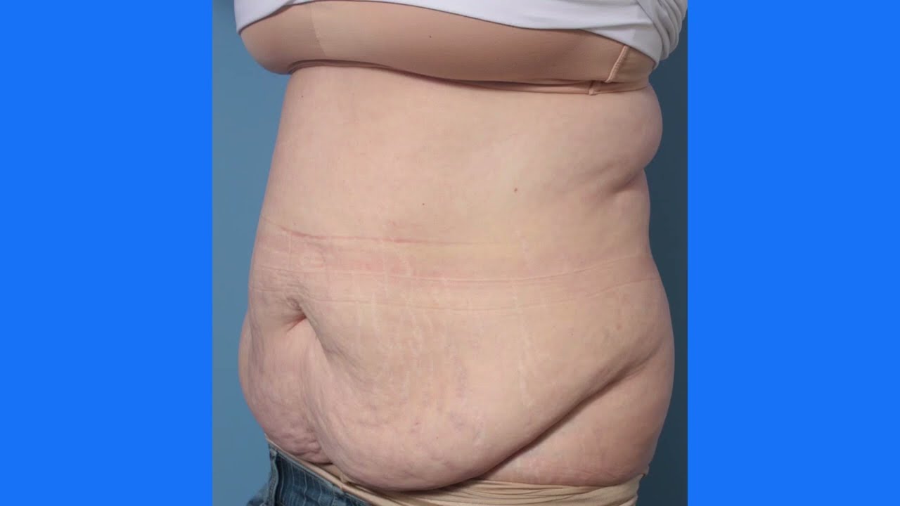
Tummy Tuck Surgery | Immediate Before and After Results | Abdominoplasty
Here are the stunning before and after results of a tummy tuck surgery performed at Divine Cosmetic Surgery.
#tummytuck #tummytuckdelhi #fatreduction #fatreductionlipo #liposuctiondelhi #liposuctionIndia #bodyreshaping
Know more
https://www.divinecosmeticsurg....ery.com/tummy-tuck.p
Tummy Tuck Before & After,
Tummy Tuck Surgery in Delhi,
Abdominoplasty surgeon,
tummy tuck results before and after,
Dr. Amit Gupta,
tummy tuck immediate result,
Tummy fat reduction,
tummy loose skin removal,
𝗗𝗿. 𝗔𝗺𝗶𝘁 𝗚𝘂𝗽𝘁𝗮 (𝗙𝗼𝘂𝗻𝗱𝗲𝗿 & 𝗗𝗶𝗿𝗲𝗰𝘁𝗼𝗿) of Divine Cosmetic Surgery
Skin removal Step 2 of Tummy Tuck - https://www.youtube.com/watch?v=cc9xsk9T_AU
------------------------------------------------------------------------------
A to Z of Tummy Tuck - https://youtu.be/5i6zD0xBkwA
------------------------------------------------------------------------------
Differences between Liposuction & Tummy tuck - https://www.youtube.com/watch?v=jzgeiz4mvc8
------------------------------------------------------------------------------
Tummy tuck surgery with Vaser (A to Z Steps) - https://www.youtube.com/watch?v=6abeUkb1ZcA&t=15s
------------------------------------------------------------------------------
For more details about Tummy tuck Visit - https://www.divinecosmeticsurgery.com/
------------------------------------------------------------------------------
Dr. Amit Gupta
MBBS, M.S., DNB (Plastic & Cosmetic Surgery)
Divine Cosmetic Surgery | Call us at +91 9811994417
info@divinecosmeticsurgery.com | 01141828787
Delhi | Mumbai | Gurgaon
𝗦𝗼𝗰𝗶𝗮𝗹 𝗠𝗲𝗱𝗶𝗮 𝗮𝗻𝗱 𝗬𝗼𝘂𝘁𝘂𝗯𝗲 𝘃𝗶𝗱𝗲𝗼 𝗰𝗵𝗮𝗻𝗻𝗲𝗹 : -
🎦 http://www.youtube.com/c/DrAmi....tGuptaBestPlasticCos
👍🏻 https://www.facebook.com/dramitguptaplasticsurgeon
📷 https://www.instagram.com/divineaesthetics_delhi/
🐥 https://twitter.com/dramitguptajee
🖇️ https://www.linkedin.com/compa....ny/divinecosmeticsur
📌 https://pinterest.com/divinesurgery
#tummytuck #TummyTuckResult #TummyTuckResultBeforeandAfter #dramitgupta #divinecosmeticsurgery #fatreduction #tummytuckdelhi #shorts
Disclaimer: The information on our videos & social media is provided for informational purposes only and is not meant for the advice provided by your surgeon.
We are not responsible for any harm if anyone misguides you from our name. Our all-social media official handles are linked up on our website. All images & content used on our videos & social media are for illustrative concerns only, original results and processes may vary.

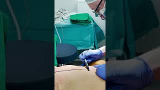
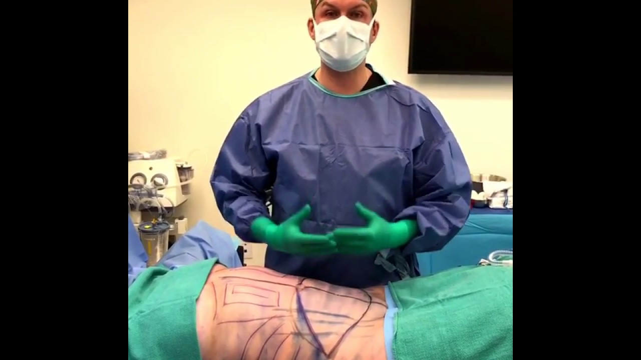
TUMMY TUCK 🤩 Immediate OR Results
This patient wanted to get her abs back, but unfortunately NO diet or workout can tighten muscles that have been stretched apart from carrying a baby 👀 But we can fix that at Lemmon Avenue Plastic Surgery & Laser Center!
To learn more about the #tummytuck click here: https://drdeuber.com/procedures/tummy-tuck/
For #mommymakeover, click here: https://drdeuber.com/procedures/mommy-makeover/
👙
#MarkDeuberMD

An Abdominoplasty (commonly referred to as a “Tummy Tuck”) removes excess fat and skin around your abdomen to shape and contour your midsection. During surgery, I also restore weakened or separated muscles to help create an abdominal profile that is both; smoother and more firm.
Watch this video as we go from the operating table to her 2-month post-op results!
If you’re interested in learning more about tummy tuck surgery or any other services we offer, please DM us or give us a call today!
☎️(424) 266-4181
🌐DrJohnDiaz.com
#DrJohnDiaz #DrDiaz #BeverlyHills #BeverlyHillsPlasticSurgery #BeverlyHillsPlasticSurgeon #DiazPlasticSurgery #PlasticSurgery #PlasticSurgeon #TummyTuck #Abdominoplasty #BeverlyHillsTummyTuck #TummyTuckBeverlyHills #AbdominoplastyBeverlyHills #BeverlyHillsAbdominoplasty #TummyTuckSurgery

It’s not tummy tuck procedure.. it’s liposuction only.. don’t get confused with both procedure..
#beforeandafter #kmc #nose #aesthetic #antiaging #beauty #drhabibhairtransplant #peshawar #nose #islamabad #swat #kohat #nowshehra #karakin #mardan
This is the process of a tummy tuck! This procedure gets rid of the extra skin that has been stretched out due to pregnancy, weight loss, etc. You'll see her before, during, and after surgery!
To download Dr. Youn's FREE ebook, "Ten Things Every Plastic Surgery Patient Must Know," visit http://www.dryoun.com
Please visit Dr. Youn's online store at http://www.dryounonline.com for the latest in skin care products, nutritional supplements, and holistic health aids!

How Liposuction Works in 15 seconds.
See how we illustrated this amazing technology by Alma Lasers.
Curious 🤔 about medical device 3D animation? ➜ http://www.arcreative-media.com

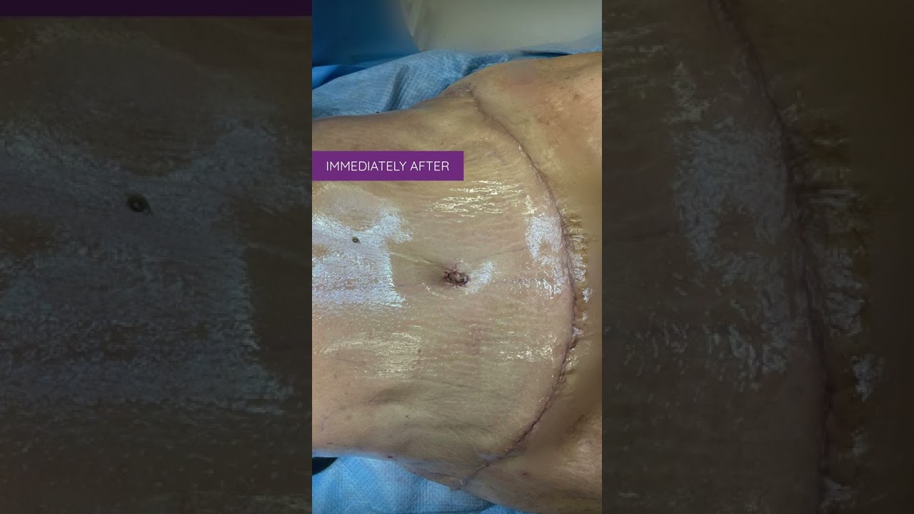
Ever wonder what a drain is for after a Tummy Tuck? Here’s a short explanation by Dr. William.
#tummytuck #abdominoplasty #shorts
WARNING: Explicit and Educational Surgical Content.
Visage Clinic's Dr. Marc DuPéré - located in Toronto, Ontario, Canada discusses Liposuction (upper bra, back rolls, lower back rolls, love handles & abdomen) and "Tummy Tuck" (Abdominoplasty): Skin excision, muscle repair and umbilicoplasty.
For more info and to book a consultation visit www.VisageClinic.com/cosmetic-....surgery/mommy-makeov or call (416) 929-9800.

Dr. Alex Campbell and Dr. Carolina Restrepo of Premium Care Plastic Surgery in Cartagena, Colombia perform a Mommy Makeover on an international patient. Watch the procedure as Dr. Campbell and Dr. Restrepo work together to offer this patient more surgery in less time, which leads to a quicker recovery and better results.

Diastasis recti often occurs during pregnancy and can persist after pregnancy. It affects core strength and the appearance of the abdominal muscles.
Dr. Erick Sanchez repairs the abdominal muscles with every tummy tuck. This short video shows the muscle repair portion of the surgery with a bonus after photo at the end!
To request a consultation with Dr. Sanchez, visit sanchezplasticsurgery.com and click Request a Consultation. Fill out the form and someone will get in touch with you to answer all your questions.
Expected cost can be found at the bottom of each procedure page on our website.
Liposuction in tummy tuck requires special planning and technique. I need to ensure that the blood circulation is well maintained for good healing. Yet proper liposuction is important to have a nice flat and contoured tummy.
#hdliposuction #tummytuck #lipoabdominoplasty #surgicalplanning #skinremovalsurgery #imeediatelyafter #plasticsurgeondubai #cocoonaclinic #drsanjayparashar #dubai
For more information visit www.drsanjayparashar.com
For more content, follow me on my social media
Instagram : https://www.instagram.com/drsanjayparashar/
Facebook : https://www.facebook.com/drsanjayparashar

#abdomenliposuction #laserskintightening #drprashantyadav #cosmeticsurgery #plasticsurgery #dezireclinicindia #weightloss #shorts #360degreeabdomenliposuction #lowerbackliposuction
Weight Loss After 360° Abdomen liposuction result, Abdomen Liposuction, lower back liposuction, 360 degree abdomen liposuction
☎️ For more info:
WhatsApp Your Details to know the Cost
Delhi - 8956880644, 9717470550, Pune - 9222122122, Bangalore- 8971224700, Gurugram - 9272007896, Ahmedabad - 9711162746
Why choose Dezire Clinic For Your Cosmetic and plastic surgery treatment ?
Dezire Clinic is a top searched clinic surgical and nonsurgical cosmetic procedure in India when comes to “Cosmetic, Skin ,Laser and Hair transplantation”.
Like and Share the video if you find it useful. Do not forget to Subscribe to our channel to get more updates.
Subscribe on YouTube https://youtube.com/dezireclin....ic?sub_confirmation=
https://youtube.com/dezireplas....ticsurgerycenter?sub
🎦 https://www.youtube.com/dezireclinic
🎦 https://www.youtube.com/DezirePlasticSurgeryCenter
👍🏻 https://www.facebook.com/drprashantmch/
👍🏻 https://www.facebook.com/dezireclinic
📸 https://www.instagram.com/drprashantdezireclinic/
📸 https://www.instagram.com/dezireclinics/
🐥 https://twitter.com/drprashantmch
👍🏻 https://www.linkedin.com/in/drprashantyadav/
🌐 Website: https://www.dezireclinic.in/
📧 dezireclinicindia@gmail.com
📧 info@dezireclinic.in
Dr. Prashant Yadav (M.S., M.Ch. Plastic Surgery ) & Founder of Dezire Clinic
Disclaimer: The content of this channel is for informational and educational purposes only. This content should not be considered a substitute for advice provided by a certified plastic or cosmetic surgeon. Patients must be properly diagnosed by a healthcare professional on an individual basis in order to achieve the desired results. There is no guarantee of getting the results and outcomes shown in videos, as the results can vary at the end. We will not be held liable for any harm caused by someone misusing our name.
#plasticsurgery #cosmeticsurgery #dezireclinic #drprashantyadav

This animation shows you how a tummy tuck is performed at Boerhaave Medical Centre. Curious? Watch the video!
Boerhaave Medical Centre sets itself the goal of providing the highest quality care. Quality not just in terms of treatment, but also in terms of our staff and the before and after care given. By providing thorough information and clear advice in advance, carefully supporting our patients through the procedure and caring for them afterwards, we believe this quality is assured.
Although we are one of the largest clinics in the Netherlands and have built up many years of experience, we continuously strive to improve. After all, the Boerhaave Medical Centre intends to remain a pioneer in the healthcare sector, by working in accordance with the latest medical findings and techniques both now and in the future.
We offer the highest standard of plastic surgery in our cosmetic care department. For 365 days a year, you can also come to us for non-surgical treatments, such as injectables, permanent hair removal and gastric balloons.
We have been awarded the ZKN quality mark and are certified to ISO 9001-2008 for giving advice and carrying out plastic surgery, including after care.
Visit our website for more information: https://www.boerhaave.com/all-....treatments/upperbody
Follow us:
Facebook: https://www.facebook.com/boerhaavemc
Google+: https://plus.google.com/+BoerhaaveNl-Kliniek
Pinterest: https://nl.pinterest.com/BoerhaaveMC/
Instagram: https://www.instagram.com/BoerhaaveMC/

Access my FREE Online Membership today → https://www.thenotedanatomist.com
___
Unlock my Premium Tutoring Memberships → https://www.thenotedanatomist.com/premium-memberships
Lifetime Access to Online Anatomy Course
Foundational Q&A Cards Per Video
Notes and Key Takeaways
Downloadable Documents
Flashcards for Each Course
Weekly Group Tutoring Sessions
Direct Tutoring Sessions
___
Discover A Simplified Approach to Master the Complexity of Anatomy with me, Dr. David Morton ... The Noted Anatomist!
This video tutorial discusses an Introduction to Histology (study of tissues):
0:00. Intro
0:35. Hierarchical organization of living matter
1:56. H&E stains
3:00. Epithelium overview (characteristics and classifying scheme)
- 9:12. Simple squamous epithelium
- 11:05. Simple cuboidal epithelium
- 12:20. Simple columnar epithelium
- 13:36. Stratified squamous epithelium
- 15:51. Urinary epithelium (transitional epithelium)
- 16:45. Pseudo-stratified ciliated columnar epithelium (respiratory epithelium)
18:55. Connective tissue overview (characteristics and classifying scheme)
- 21.14. Connective tissue proper (loose CT, dense irregular CT, dense regular CT, adipose tissue)
- 24:50. Cartilage (hyaline cartilage, elastic cartilage, fibrocartilage)
- 26:04. Bone (osteoblasts, osteocytes, osteoclasts, calcium ...)
- 27:34. Blood (RBC, WBC, platelet, plasma)
28:54. Muscle tissue (skeletal muscle, cardiac muscle, smooth muscle)
32:54. Nervous tissue (neurons and glial cells)
36:58. In-a-Nutshell
37:07. Acknowledgements
For a more detailed study of histology go to The Histology Wizard: https://www.youtube.com/channe....l/UCAeLLruy9RkUWaW_r

A complete organized library of all my videos, digital slides, pics, & sample pathology reports is available here: https://kikoxp.com/posts/5084 (dermpath) & https://kikoxp.com/posts/5083 (bone/soft tissue sarcoma pathology)
Topics discussed:
Epidermis:
Layers of epidermis: 0:10
Melanocytes vs Keratinocytes: 5:16
Langerhans cells: 10:10 & 33:30 & 57:30
Dermis:
Papillary and reticular dermis: 11:50
Three types of white empty spaces on a slide: vessels, glands/ducts/cysts, or artifact: 15:25
Blood vessels & nerves: 18:24 & 48:50 & 58:59
Arrector pili & other dermal smooth muscle: 20:00
Adnexal:
Sebaceous gland: 21:10
Hair follicle 23:14
Eccrine sweat glands and ducts 24:45 & 50:00
Gland/duct vs blood vessel 27:20 & 48:50
Apocrine glands: this video https://kikoxp.com/posts/7837 (at 12:30)
Acrosyringium: this video https://kikoxp.com/posts/7837 (at 10:00)
Three types of pink bundles: smooth muscle, nerve, dense connective tissue: 27:50
Acral skin (palm sole) with contact dermatitis 29:37
Parakeratosis 30:00
Perivascular lymphocytes 30:40
Eosinophils vs neutrophils 31:20
Spongiosis with desmosome keratinocyte spines 32:10
Spongiotic vesicles with Langerhans cells 33:30
Normal acral skin (palm & sole) with stratum lucidum 34:20
Normal glomus body/apparatus (canal of Sucquet-Hoyer) 35:40
Nerve 36:46 & 51:50
Adipose tissue (white fat cells) in subcutis with Lochkern 37:55
Normal scalp skin with large anagen hair follicles: 39:30
Hair follicle anatomy (bulb/matrix, inner root sheath, outer root sheath, hair shaft, isthmus, infundibulum): 40:55 (labeled images):
https://kikoxp.com/posts/3661 & https://kikoxp.com/posts/7899
Pacinian corpuscle 50:40
Meissner corpuscle 1:02:28
Dense regular connective tissue (Fascia/Tendon/Ligament) vs Smooth Muscle 53:00
Basic Normal Skin Immunohistochemistry:
-cytokeratin in epidermis: 55:33
-S100 in melanocytes and Langerhans cells and adipocytes: 57:30
-Desmin in smooth muscle (arrector pili and blood vessels): 58:59
-CD31 in endothelial cells of blood vessels: 59:33
-SOX-10 in melanocytes: 1:00:40
Digit/Finger/Toe histology (amputation for subungual acral melanoma) 1:04:10 & 1:08:30
-bone 1:05:40
-glomus body 1:05:15
-tendon/ligament 1:06:10
-artery 1:06:58
-fingernail/toenail 1:08:54
-acrosyringium 1:10:45
Solar elastosis (what wrinkles look like microscopically!) 1:11:50
Other videos you might like:
Tendon vs Nerve Histology Made Simple with the Ramen Noodle Sign (of Fulton) video: https://kikoxp.com/posts/4466
Melanocytes vs Keratinocytes made easy video: https://kikoxp.com/posts/3802
Blood Vessel vs Gland vs Artifact Made Easy video: https://kikoxp.com/posts/4808
The basic normal structures of the skin discussed and described by a dermatopathologist. This material is intended for use by medical students, junior pathology or dermatology residents, or for anyone else studying normal human histology. Special thanks to two of my medical students at UAMS for helping make this video possible. Miki Lindsey convinced me that I really needed to sit down and record this video. Akash Patel took time to edit the video and make it ready for YouTube. My sincere thanks to both of them for helping me overcome procrastination.
Huge thanks to Abigail Cline, a medical student at Medical College of Georgia, for volunteering to type a transcript of this ENTIRE video (over 14,000 words!) so that I could provide closed caption subtitles for those with hearing impairments and for those who may need assistance in understanding spoken English (particularly given how quickly I speak!). You can access a text version of her transcript of my video here: https://kikoxp.com/posts/5390
Correction - I made a mistake in the video. I said that sebaceous gland secretions are turned into smelly substances by bacteria and that this makes body odor. That is incorrect. That is actually true of APOCRINE gland secretions not sebaceous secretions.
Also, in the past I used "keratinocyte" and "squamous cell" interchangeably (this is because in dermatopathology, we see and talk about squamous cell carcinomas all the time, and those tumors are composed of keratinocytes). But technically, in normal skin histology, "squamous cell" refers only to the flattened keratinocytes in the superficial epidermis. Thankfully, a histology PhD colleague pointed this out to me and corrected my lazy nomenclature!
Please check out my Soft Tissue Pathology & Dermatopathology survival guide textbooks: http://bit.ly/2Te2haB
This video is geared towards medical students, pathology or dermatology residents, or practicing pathologists or dermatologists. Of course, this video is for educational purposes only and is not formal medical advice or consultation.
Presented by Jerad M. Gardner, MD. Please subscribe to my channel to be notified of new pathology teaching videos.
Follow me on:
Snapchat: JMGardnerMD
Twitter: @JMGardnerMD
Instagram: @JMGardnerMD
Facebook: https://www.facebook.com/JMGardnerMD/

© 2023 Elsevier. All rights reserved. What are lymph nodes? Lymph nodes are small secondary lymphoid organs that are found along lymphatic vessels throughout the body.
Find our full video library only on Osmosis Prime: http://osms.it/more.
Join over 3 million current & future clinicians who learn by Osmosis, and over 130 universities around the world who partner with us to make medical and health education more engaging and efficient. We have unparalleled tools and materials to prepare you to succeed in school, on board exams, and as a future clinician. Sign up for a free trial at http://osms.it/more. If you're interested in exploring an institutional partnership, visit osmosis.org/educators to request a personalized demo.
Follow us on social:
Facebook: http://osms.it/facebook
Twitter: http://osms.it/twitter
Instagram for med: http://osms.it/instagram
Instagram for nursing: https://osms.it/ignursing
Linkedin: https://osms.it/linkedin
Our Vision: Everyone who cares for someone will learn by Osmosis.
Our Mission: To empower the world’s clinicians and caregivers with the best learning experience possible. Learn more here: http://osms.it/mission
Medical disclaimer: Knowledge Diffusion Inc (DBA Osmosis) does not provide medical advice. Osmosis and the content available on Osmosis's properties (Osmosis.org, YouTube, and other channels) do not provide a diagnosis or other recommendation for treatment and are not a substitute for the professional judgment of a healthcare professional in diagnosis and treatment of any person or animal. The determination of the need for medical services and the types of healthcare to be provided to a patient are decisions that should be made only by a physician or other licensed health care provider. Always seek the advice of a physician or other qualified healthcare provider with any questions you have regarding a medical condition. © 2023 Elsevier. All rights reserved.
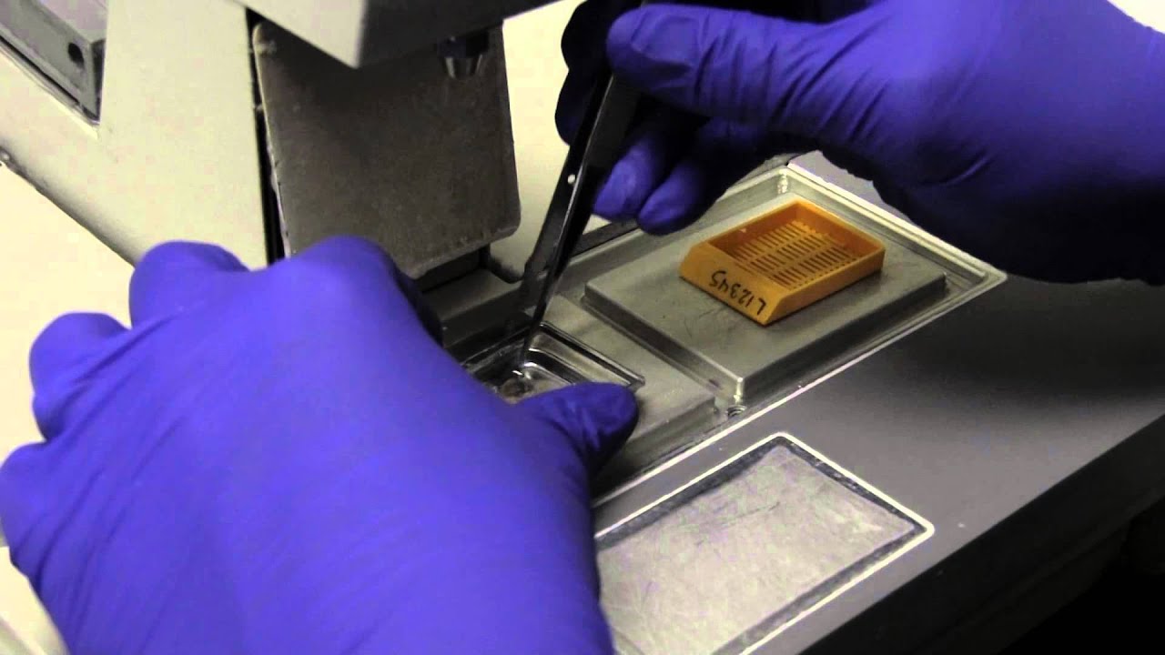

Video giving an overview of histology, slide preparation, histological stains, and types of microscopy. This video is a part of our Histology Video Course (https://youtube.com/playlist?l....ist=PLnr1l7WuQdDynxT
Specific topics: what is histology, general composition of tissues, histotechnology: how histology slides are prepared, histology stains, immunohistochemistry, light microscopy vs electron microscopy, and pro tips for learning histology
Additional YouTube Content
Anatomy Videos: https://youtube.com/playlist?l....ist=PLnr1l7WuQdDz2dK
Biochemistry videos: https://youtube.com/playlist?l....ist=PLnr1l7WuQdDzCUC
DaVinci Cases Videos: https://youtube.com/playlist?l....ist=PLnr1l7WuQdDyJUl
The DaVinci Hour Podcast: https://youtube.com/playlist?l....ist=PLnr1l7WuQdDwSm9
DaVinci Academy Website: https://www.dviacademy.com/

