- Physical Examination
- Surgical Examination
- Ophthalmology
- Clinical Skills
- Orthopedics
- Surgery Videos
- Laparoscopy
- Pediatrics
- Funny Videos
- Cardiothoracic Surgery
- Nursing Videos
- Plastic Surgery
- Otorhinolaryngology
- Histology and Histopathology
- Neurosurgery
- Dermatology
- Pediatric Surgery
- Urology
- Dentistry
- Oncology and Cancers
- Anatomy Videos
- Health and Fitness
- Radiology
- Anaesthesia
- Physical Therapy
- Pharmacology
- Interventional Radiology
- Cardiology
- Endocrinology
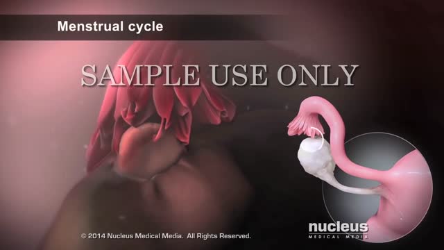
- Gynecology
- Emergency Medicine
- Psychiatry and Psychology
- Childbirth Videos
- General Medical Videos
- Nephrology
- Physiology
- Diet and Food Health
- Diabetes Mellitus
- Neurology
- Women Health
- Osteoporosis
- Gastroenterology
- Pulmonology
- Hematology
- Rheumatology
- Toxicology
- Nuclear Medicine
- Infectious Diseases
- Vascular Disease
- Reproductive Health
- Burns and Wound Healing
- Other
Top videos
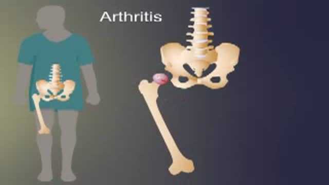
Arthritis of the hip causes severe pain, and sometimes requires surgical treatment, including hip replacement. This animated video show you what hip arthritis is, and how it causes symptoms.
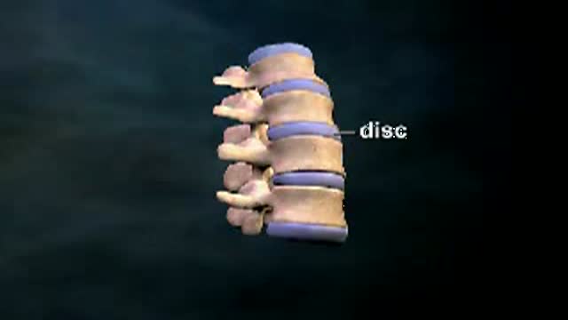
Herniated or Ruptured Disc: Between each of the vertebre of the spine is a disc which is filled with a gel type material to cushion the connection between the vertebre. With age or injury these intervertebral discs can rupture or herniate. This herniation causes them to push against the adjacent nerves which come from the spinal cord. This can frequently cause pain, numbenss and tingling. This animated video reviews herniated disc.
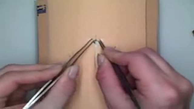
Demonstration of staple insertion and removal technique for laceration repair or wound closure in the operating room.

A parasitic twin (also known as an asymmetrical or unequal conjoined twin) is the result of the processes that produce vanishing twins and conjoined twins, and may represent a continuum between the two. Parasitic twins occur when a twin embryo begins developing in utero, but the pair does not fully separate, and one embryo maintains dominant development at the expense of the other. Unlike conjoined twins, one ceases development during gestation and is vestigial to a mostly fully-formed, otherwise healthy individual twin. The undeveloped twin is defined as parasitic, rather than conjoined, because it is incompletely formed or wholly dependent on the body functions of the complete fetus. The independent twin is called the autosite.
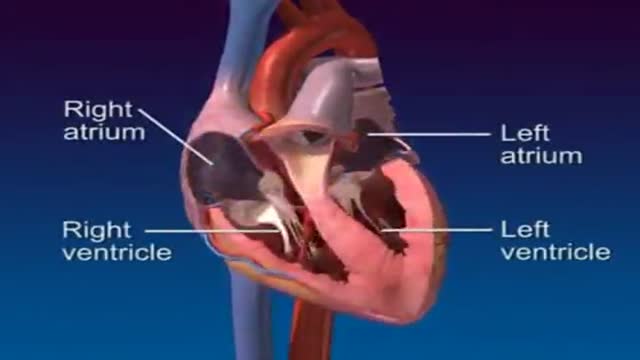
A 3D video clip showing anatomy and physiology of the heart
Intrauterine insemination (IUI) is a fertility treatment that involves placing sperm inside a woman's uterus to facilitate fertilization. The goal of IUI is to increase the number of sperm that reach the fallopian tubes and subsequently increase the chance of fertilization.
Patient 65-year-old of age who comes to the medical consultation with pain moderated pain in the right hypochondrium of “several years of evolution” but that it increased one week ago. Also, she shows pain in the umbilical region of “many years of evolution”, that is supported according to the patient - in a constant way.rnTo the examination, we observe an umbilical hernia, apparently divided into two parts. The hernia of the external region measures 25.1 centimeters x 18.0 centimeters and the one that occupies the average region measures 12.0 centimeters x 10.0 centimeters.rnPatient who comes to the medical consultation with moderated pain in the right hypochondrium of one year of evolution but it increased one week ago after eat duck.rnIn the ultrasound scan of the region of the right hypochondrium (patient came having breakfast, that is to say, without previous preparation ) we can observe the liver of 123.8 millimeters high, as well as the porta vein with a diameter of 7.3 millimeters.rnOn having observed the Gallbladder, we think that a side wall is increased in 2.7 mm (hyperechogenic) with several “echogenics points” in the interior (”Biliary Mud”).
The measurements of the gallbladder were: 39.0 x 17.4 millimeters.rnWe can appreciates an echogenic image in the interior that it would make think about stone. The stones are identified as echogenic foci casting acoustic shadowing but but this image did not appear and a re-evaluation is decided in 15 days.
Acalculous cholecystopathy which means disease or condition of the gallbladder without the presence of gallstones. You might also call it functional gallbladder disorder or impaired gallbladder emptying. Some causes may be chronic inflammation, a problem with the smooth muscles of the gallbladder or the muscle of the Sphincter of Oddi being too tight.
REMEMBER:
Umbilical hernia is a congenital malformation, especially common in infants of African descent, and more frequent in boys. An Acquired umbilical hernia directly results from increased intra-abdominal pressure and are most commonly seen in obese individuals.
Presentation:A hernia is present at the site of the umbilicus (commonly called a navel, or belly button) in the newborn; although sometimes quite large, these hernias tend to resolve without any treatment by around the age of 5 years. Obstruction and strangulation of the hernia is rare because the underlying defect in the abdominal wall is larger than in an inguinal hernia of the newborn. The size of the base of the herniated tissued is inversely correlated with risk of strangulation (i.e. narrow base is more likely to strangulate).
Babies are prone to this malformation because of the process during fetal development by which the abdominal organs form outside the abdominal cavity, later returning into it through an opening which will become the umbilicus.
Differential diagnosisrnImportantly this type of hernia must be distinguished from a paraumbilical hernia which occurs in adults and involves a defect in the midline near to the umbilicus, and from omphalocele.
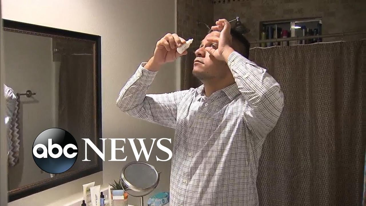
One man is speaking out about the potential risks of laser eye surgery, after he says the procedure left his vision permanently impaired.
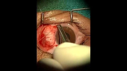
An eye web is a noncancerous, triangular growth that may occur on one or both eyes. It's more common in people who spend a lot of time in the sun, such as those who work outdoors. The painless growth may be slightly raised and contain obvious blood vessels. It may cause irritation and possibly affect vision. Treatment usually isn't necessary. Eyedrops or surgery may help in severe cases.
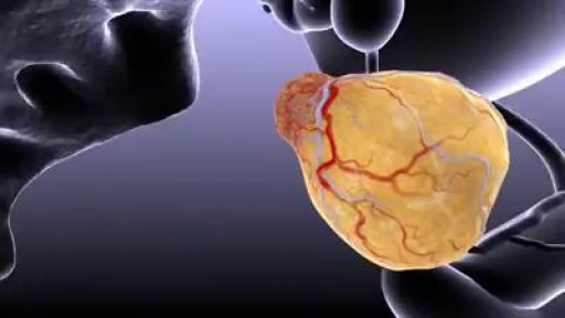
Prostate cancer is the most common cancer among men (after skin cancer), but it can often be treated successfully. If you have prostate cancer or are close to someone who does, knowing what to expect can help you cope. Here you can find out all about prostate cancer, including risk factors, symptoms, how it is found, and how it is treated.

Each year, thousands of babies in the U.S. are born addicted to opiates. And the problem is getting worse.

AZT Mechanism of Antiviral Activity
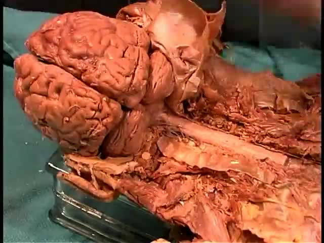
Anatomy of The Brain

Cervical cerclage can be placed via transvaginal, open transabdominal, or laparoscopic transabdominal approach, preferably before pregnancy. Recurrent late miscarriages may be due to a weak (sometimes called an incompetent) cervix that shortens or opens too early in pregnancy. Cervical cerclage involves placing a stitch around the upper part of the cervix to keep it closed; the operation may be carried out through the vagina, or through the abdomen, as an open or laparoscopic ('keyhole') procedure.
Parasitic twins: boy carrying dead twin inside him, giant tumor removed - tumors compilation
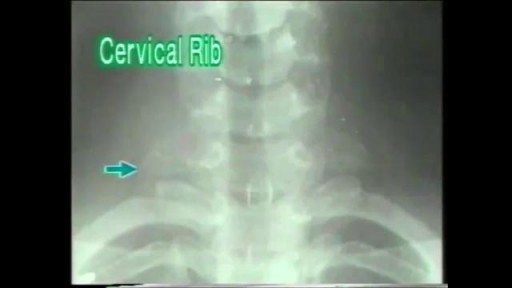
A cervical rib in humans is an extra rib which arises from the seventh cervical vertebra. Sometimes known as "neck ribs", their presence is a congenital abnormality located above the normal first rib. A cervical rib is estimated to occur in 0.2% (1 in 500 people) to 0.5% of the population.

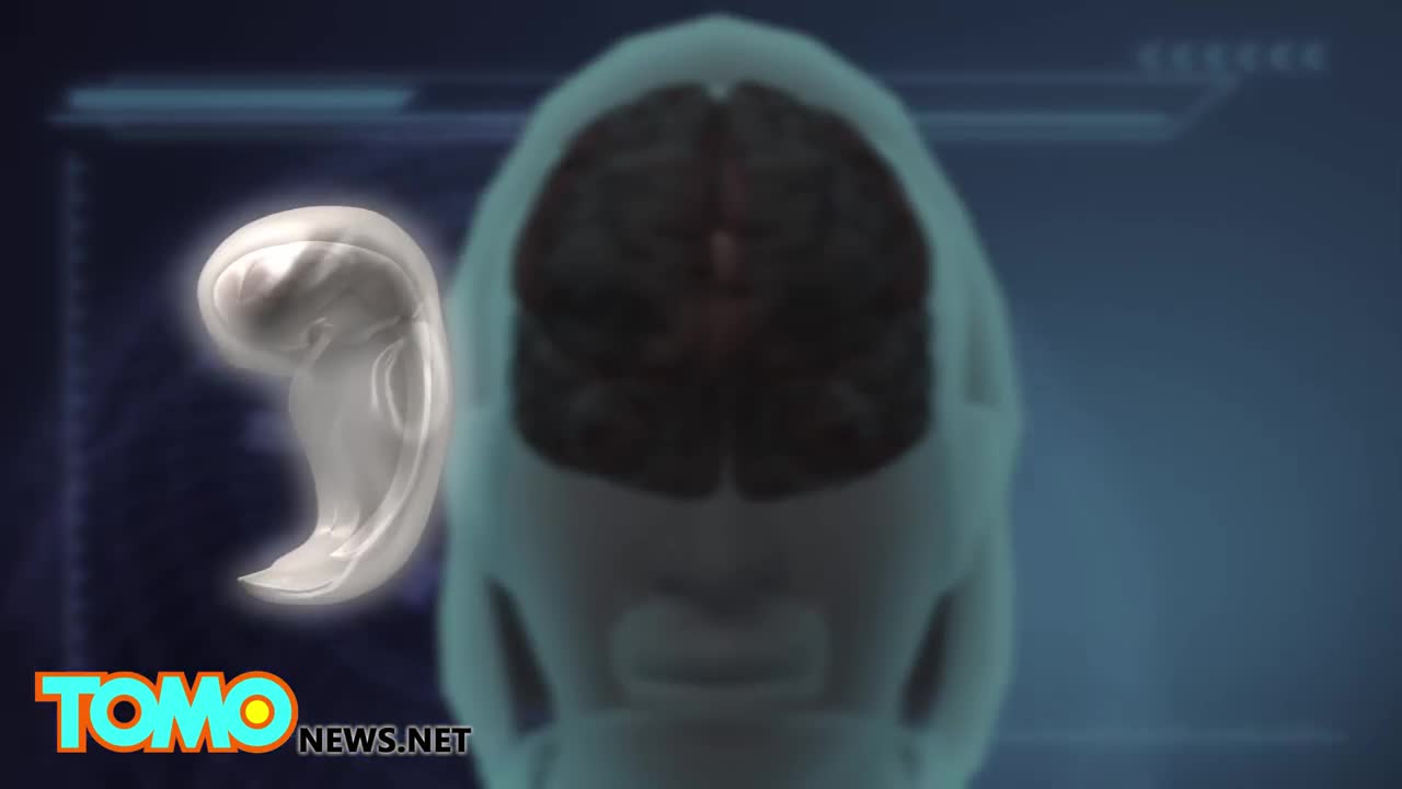
U.S. biotech firm Bioquark recently got approval to move forward with its ReAnima Project, in which it will try to reverse brain death in patients on life support

The preferred route of access for temporary transvenous pacing is the internal jugular vein followed by subclavian and femoral veins. However, all the major venous access sites (internal and external jugular, subclavian, brachial, femoral) have been used and each is associated with particular problems.

new study about the size of penis
Fremale to male gender reassignment surgery
