
- Physical Examination
- Surgical Examination
- Ophthalmology
- Clinical Skills
- Orthopedics
- Surgery Videos
- Laparoscopy
- Pediatrics
- Funny Videos
- Cardiothoracic Surgery
- Nursing Videos
- Plastic Surgery
- Otorhinolaryngology
- Histology and Histopathology
- Neurosurgery
- Dermatology
- Pediatric Surgery
- Urology
- Dentistry
- Oncology and Cancers
- Anatomy Videos
- Health and Fitness
- Radiology
- Anaesthesia
- Physical Therapy
- Pharmacology
- Interventional Radiology
- Cardiology
- Endocrinology
- Gynecology
- Emergency Medicine
- Psychiatry and Psychology
- Childbirth Videos
- General Medical Videos
- Nephrology
- Physiology
- Diet and Food Health
- Diabetes Mellitus
- Neurology
- Women Health
- Osteoporosis
- Gastroenterology
- Pulmonology
- Hematology
- Rheumatology
- Toxicology
- Nuclear Medicine
- Infectious Diseases
- Vascular Disease
- Reproductive Health
- Burns and Wound Healing
- Other
Top videos


Knee replacement involves replacing a knee joint that has been damaged or worn away, usually by arthritis or injury. Find out more here: https://www.bupa.co.uk/health-....information/knee-cli

Marcus Greatens, M.D., an orthopedic surgeon at Mayo Clinic Health System provides insight into a few of the things patients can expect to experience during knee replacement surgery.
LIKE, SUBSCRIBE & HIT THE 🔔 http://bit.ly/2VW2yV1
Mayo Clinic Health System offers outstanding care close to home. Part of Mayo Clinic, we meet most of your health care needs locally.
#mayoclinichealthcare #mayoclinicexperience #mayoclinichealthsystem #mayoclinic
Follow Mayo Clinic Health System on
Facebook:
http://bit.ly/2VXSYRv
On Instagram at:
http://bit.ly/30obB03
And also on Twitter:
http://bit.ly/2JgsHI5

Orthopedic surgeon Donald Polakoff, MD describes recovery time from knee replacement surgery.
SUBSCRIBE FOR MORE VIDEOS AND NEWS!
http://www.youtube.com/subscri....ption_center?add_use
CONNECT WITH US!
Google+ ► http://gplus.to/spuh
Facebook ► http://www.fb.com/SaintPetersUniversityHospital
Twitter ► http://twitter.com/sphcs_news
Pinterest ► http://www.pinterest.com/sphcs
Saint Peter's Healthcare System ► http://www.saintpetershcs.com

#clean #leg #surgery #knee
✍️Dr. Matthew Harb talk about knee replacement surgery
https://www.MatthewHarbMD.com/links
👨⚕️Orthopedic Hip and Knee Surgeon
📍Located in Washington DC, and Maryland
📚Education and Insight
🛠Minimally invasive, outpatient, hip and knee replacement surgery
👉Visit me Online: https://www.MatthewHarbMD.com
☎️Schedule a virtual or in office appointment: https://www.MatthewHarbMD.com/links
📲Follow me on Social Media:
💥TIKTOK: https://www.tiktok.com/@matthewharbmd
💥INSTAGRAM: https://www.instagram.com/thebonesurgeon
📲Please SUBSCRIBE for new #shorts #doctor videos!
https://www.youtube.com/channel/UCbte-7tl7kaAm1xfWgz6xvQ?app=desktop&sub_confirmation=1
✍️QUESTION — Have a question or comment about medicine, orthopedic surgery, or musculoskeletal conditions. Post in the comment sections and let me know!
Dr. Matthew Harb specializes in minimally invasive, muscle sparing, hip and knee replacement surgery. Minimally invasive surgery allows patients to recover faster and have less pain post operatively. Implants are tailored and custom fit to each patient to allow for improved performance. Dr. Harb’s expertise in rapid recovery protocols allow for quick recovery after surgery and excellent outcomes in patients with hip and knee arthritis. With minimally invasive, muscle sparing surgery patients can return to their lifestyles and get back to doing the things they love sooner. Dr. Harb performs outpatient joint replacement surgery with many of his patients walking independently and going home the day of surgery.
“My focus is excellence in patient care, expedited recovery after surgery, and getting people back to the normal activities they love. Our team focused approach is committed to superb outcomes, improving lives, and returning patients to living pain free.”

In this video, we have explained the procedure of total #knee #replacement #surgery in patient in 3D animation.
Learn more: https://ecgkid.com
_____________________________________________________________________
Knee replacement, commonly known as complete knee replacement or knee arthroplasty, is a surgical treatment that resurfaces a knee that has been destroyed by arthritis. The extremities of the bones that make up the knee joint, as well as the kneecap, are capped with metal and plastic pieces. Someone with severe arthritis or a major knee injury may benefit from this procedure.
The knee joint can be affected by a variety of arthritis forms. The degradation of joint cartilage and neighboring bone in the knees can be caused by osteoarthritis, a degenerative joint disease that primarily affects middle-aged and older persons. Rheumatoid arthritis produces pain and stiffness by inflaming the synovial membrane and resulting in an excess of synovial fluid. Traumatic arthritis, or arthritis caused by an injury, can harm the joints.
The purpose of knee replacement surgery is to resurface damaged areas of the knee joint and cure knee discomfort that has not responded to prior therapies.

Knee pain can happen at any age, but some doctors say they're seeing more people with osteoarthritis who are still young and active.
Subscribe to WCVB on YouTube for more: http://bit.ly/2526UpS
Get more Boston news: http://www.wcvb.com
Like us: https://www.facebook.com/wcvb5
Follow us: https://twitter.com/WCVB
Google+: https://plus.google.com/+wcvb

A growing number of patients having total knee replacement surgery are 55 or younger. Surgeons at Sunnybrook's Holland Centre perform more than 1,000 total knee replacements each year. Read more: http://sunnyview.sunnybrook.ca..../2011/11/snap-crackl
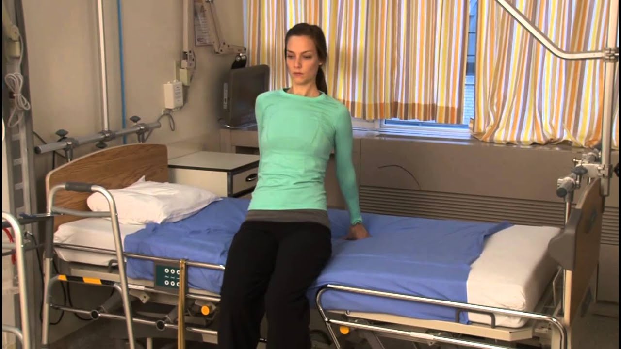
Patient information from Sunnybrook's Holland Musculoskeletal Program. For more, visit: http://sunnybrook.ca/holland

BioDigital Systems created this 3D animation of a knee replacement surgery.
---
BioDigital is happy to share helpful health information, but we do not offer medical advice. For medical advice, please contact your healthcare provider directly.
For more information on the content of this video, you can:
Access these 3D visuals: human.biodigital.com
Learn more about BioDigital: www.biodigital.com
Subscribe: www.youtube.com/c/BioDigital?sub_confirmation=1
Facebook: www.facebook.com/BioDigitalHuman
Instagram: www.instagram.com/biodigital.human
Twitter: twitter.com/biodigitalhuman
LinkedIn: www.linkedin.com/company/biodigital/ Medical disclaimer:
BioDigital, Inc is not a health care provider and we do not provide medical advice. You should not rely on the information provided on our sites or services as a substitute for, nor does it replace, professional medical advice, diagnosis, or treatment. The services are not intended to be used by consumers or clinicians in making treatment decisions. You are encouraged to seek professional medical diagnosis and treatment for any medical condition, and to discuss information from the sites and services with your healthcare provider. Information provided on the sites and media is provided for informational purposes and is in no way intended to substitute consulting a medical professional. Nothing stated or posted by BioDigital is intended to be, and must not be taken to be, the practice of medicine, the provision of medical care, or a tool relied on by patients or clinicians. If you rely on any of the information provided by BioDigital, you do so solely at your own risk.

The OrthoIllustrated® animation for total knee replacement is an educational tool to help patients better understand the diagnosis and treatment of arthritis.
- - - - -
Why Work Arthrex https://www.arthrex.com/job-seeker
Find an Arthrex Surgeon: https://doctorfinder.orthoillustrated.com
- - - - -
Join the Community:
LinkedIn: https://www.linkedin.com/company/arthrex
Facebook: https://www.facebook.com/Arthrex
Instagram: https://www.instagram.com/arthrex_inc/
Twitter: https://twitter.com/Arthrex
TikTok: https://www.tiktok.com/@arthrex
- - - - -
Arthrex Inc., headquartered in Naples, Florida, is a global leader in orthopedic surgical device design, research, manufacturing, and medical education. Arthrex develops and releases more than 1,000 new products and procedures every year to advance minimally invasive orthopedics worldwide.
For more information, visit https://www.arthrex.com
- - - - -
OrthoPedia is an innovative educational website that was created for anyone interested in learning about orthopedics from the first-year student to the experienced orthopedic surgeon.
Visit https://www.orthopedia.com to experience the future of Medical Education.

A new bionic body part that talks to your phone is the next frontier in knee replacements. It's called a smart knee, a new technology designed to improve recovery after surgery. Stephanie Stahl reports.

Outpatient -- or same-day -- knee replacement surgery is more convenient than traditional knee replacement surgery and often can help you recover faster.
Outpatient -- or same-day -- knee replacement surgery is more convenient than traditional knee replacement surgery and often can help you recover faster. At Duke Ambulatory Surgery Center Arringdon, your knee replacement will be followed immediately by physical therapy to get you moving and start your recovery process right away. Our expert joint replacement team ensures your knee replacement surgery is safe and effective so you can return to the comfort of your home as soon as possible.

Dr. Eric Janssen of SportsMED Orthopaedic Surgery & Spine Center in Huntsville, Alabama demonstrates a total knee replacement using dry bones model. In this demonstration he uses the Wright Medical Evolution Knee implant. This demonstrations does not include soft tissue.

Warren Pettaway of Detroit knew he needed to have his left knee checked out when he could no longer keep up while officiating basketball. The pain got so bad that running up and down the court or quickly changing directions was too much for him to continue doing what he loved.Only three weeks post-surgery, Warren is ready to get back in the game. He is able to move without issue and is getting things done around the house. He makes sure to do his therapy as directed and is exercising on his own in the gym. To learn more visit https://www.henryford.com/serv....ices/joint-replaceme
Carpal tunnel release (part 1). Skin incision and retraction. Procedure performed by Deepak Kapila, MD, Broward Health, Fort Lauderdale, FL. Courtesy of BroadcastMed (http://ortho.broadcastmed.com/....4229/videos/carpal-t
There are hundreds more procedural videos as well as news, features, resources and references on Medscape.com. Join today for free.
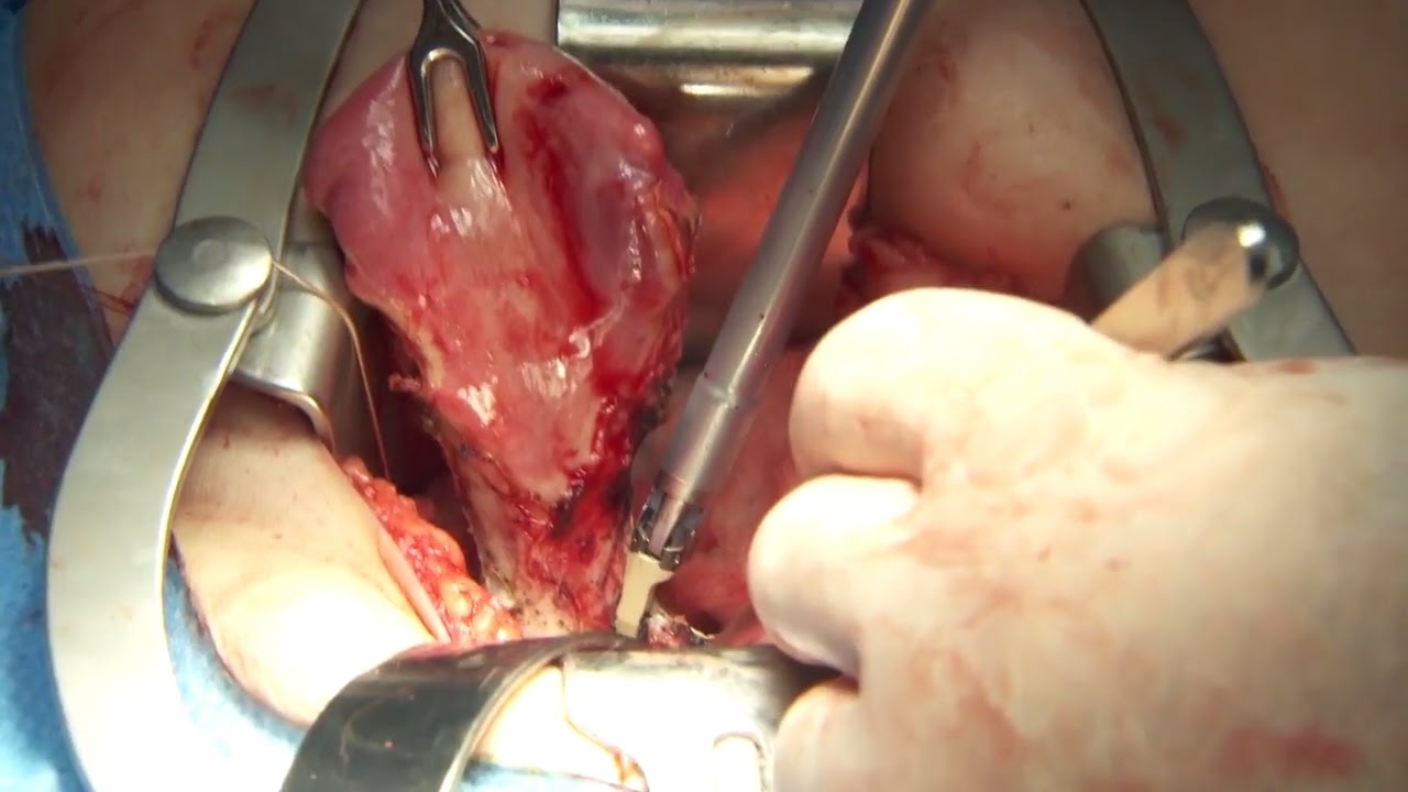
Olympus has extended the value of its award-winning combined surgical energy device, THUNDERBEAT, to open surgical procedures. Watch Dr. Francois Blaudeau master use of THUNDERBEAT Open Extended Jaw (OEJ) in a total abdominal hysterectomy.
http://medical.olympusamerica.com/products/thunderbeat?utm_source=youtube&utm_campaign=Total%20Abdominal%20Hysterectomy%20Surgery%20-%20THUNDERBEAT&utm_medium=description&utm_term=energy&utm_content=surgical

From UW Health's Neurosurgery Program: Learn more about the individual steps in the DBS surgery procedure. Visit uwhealth.org/dbs

A brain surgery called a craniectomy is performed to remove a part of your skull in order to relieve pressure in an area when your brain swells from a traumatic brain injury. It is also performed to treat medical conditions that cause your brain to swell or bleed that can be caused by an aneurysm, brain tumor or other cancer.
This 3d animation shows how the surgical procedure decreases intracranial pressure (ICP), intracranial hypertension (ICHT), or heavy bleeding (also called hemorrhaging) inside your skull. If left untreated, pressure or bleeding can compress your brain and push it down onto the brain stem. This can be fatal or cause permanent brain damage.
Brain surgery is a very serious procedure under any circumstances, but a craniectomy is done when there is an immediate risk to the brain and neurological function due to severe brain injury or stroke.
For more information about custom 3D animation depicting surgery, please visit https://www.amerra.com/.
Watch additional medical animations:
Accessing an implantable port training - 3D animation: https://youtu.be/xSTpxjyv4O4
Open Suctioning with a Tracheostomy Tube - 3D animation: https://youtu.be/wamB7jpWCiQ
Ventriculostomy Brain Surgery - 3d animation: https://youtu.be/pUy0YDzVNzs
Suctioning the endotracheal tube - medical animation: https://youtu.be/pN6-EYoeh3g
Functional endoscopic sinus surgery (FESS) - 3D animation: https://youtu.be/qKTRyowwaLA
How to insert a nasogastric tube for NG intubation - 3d animation: https://youtu.be/Abf3Gd6AaZQ
Oral airway insertion - oropharyngeal airway technique - 3D animation: https://youtu.be/caxUdNwjt34
Nasotracheal suctioning (NTS) - 3D animation: https://youtu.be/979jWMsF62c
Learn about hemorrhoids with #3d #animation: https://youtu.be/R6NqlMpsiiY
LASIK eye surgery - 3D animation: https://youtu.be/Bb8bnjnEM00
CPR cardiopulmonary resuscitation - 3D animation: https://youtu.be/G87knTZnhks
What are warts (HPV)? - 3D animation: https://youtu.be/guJ1J7rRs1w
How Macular Degeneration Affects Your Vision - 3D animation: https://youtu.be/ozZQIZ_52YY
NeoGraft hair transplant procedure – animation: https://youtu.be/C-eTdH2UPXI
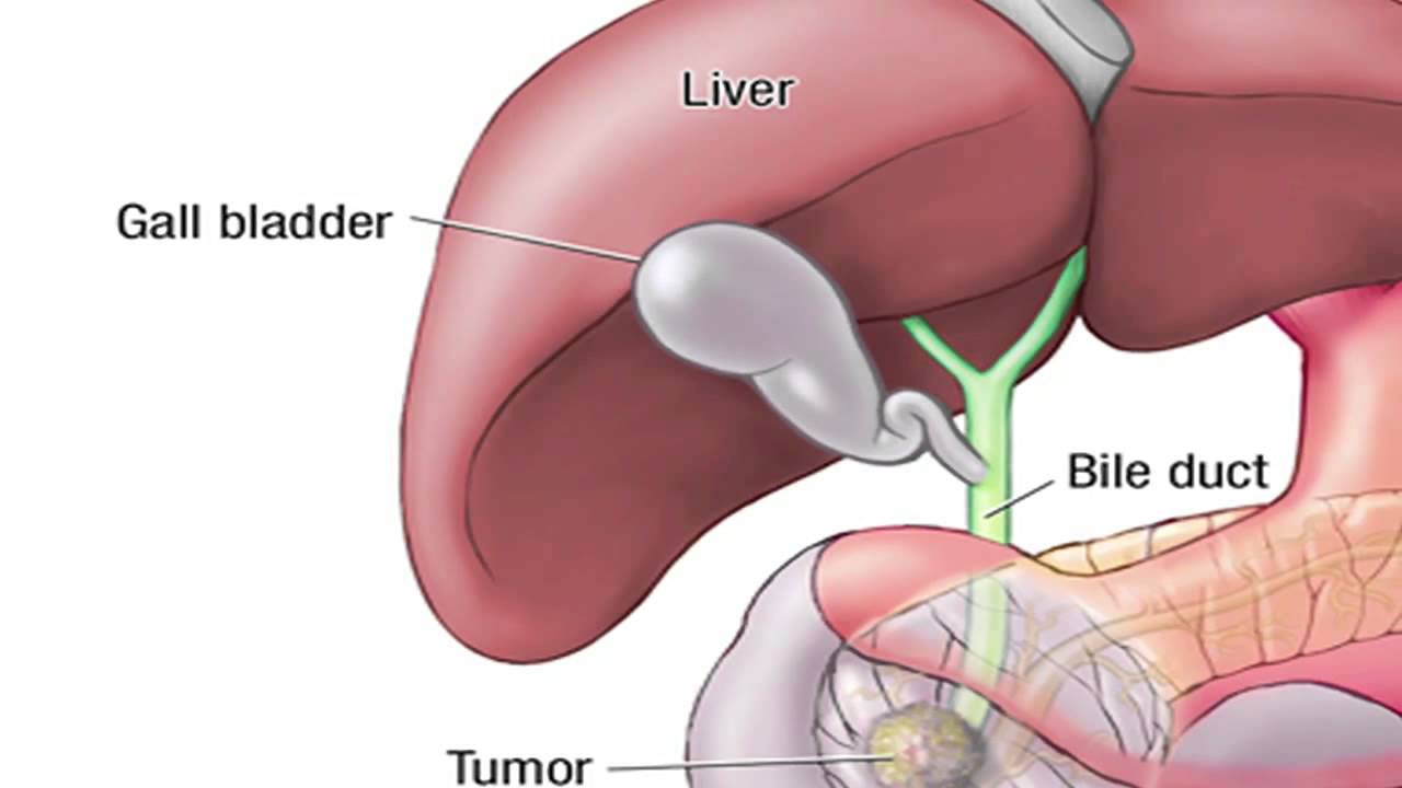
Dr. Horacio Asbun, Mayo Clinic in Florida, explains the Whipple procedure using this animated graphic of a pancreas. Cancer of the pancreas affects 45,000 people every year in the U.S., and it is the fourth leading cause of cancer-related deaths. The five-year overall survival rate if a tumor is detected early and surgically removed is 22 percent, versus 6 percent without early detection and surgery. To learn more, visit http://mayocl.in/2zk7FDi.
This video in Spanish/español: https://www.youtube.com/watch?v=N_zWboNMKWk
