- Physical Examination
- Surgical Examination
- Ophthalmology
- Clinical Skills
- Orthopedics
- Surgery Videos
- Laparoscopy
- Pediatrics
- Funny Videos
- Cardiothoracic Surgery
- Nursing Videos
- Plastic Surgery
- Otorhinolaryngology
- Histology and Histopathology
- Neurosurgery
- Dermatology
- Pediatric Surgery
- Urology
- Dentistry
- Oncology and Cancers
- Anatomy Videos
- Health and Fitness
- Radiology
- Anaesthesia
- Physical Therapy
- Pharmacology
- Interventional Radiology
- Cardiology
- Endocrinology
- Gynecology
- Emergency Medicine
- Psychiatry and Psychology
- Childbirth Videos
- General Medical Videos
- Nephrology
- Physiology
- Diet and Food Health
- Diabetes Mellitus
- Neurology
- Women Health
- Osteoporosis
- Gastroenterology
- Pulmonology
- Hematology
- Rheumatology
- Toxicology
- Nuclear Medicine
- Infectious Diseases
- Vascular Disease
- Reproductive Health
- Burns and Wound Healing
- Other
Top videos
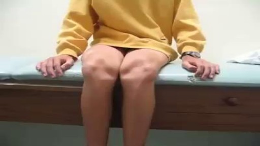
Kneecap dislocation Email this page to a friend Print Facebook Twitter Google+ Kneecap dislocation occurs when the triangle-shaped bone covering the knee (patella) moves or slides out of place. The problem usually occurs toward the outside of the leg. Causes Kneecap (patella) dislocation is often seen in women. It usually occurs after a sudden change in direction when your leg is planted. This puts your kneecap under stress.
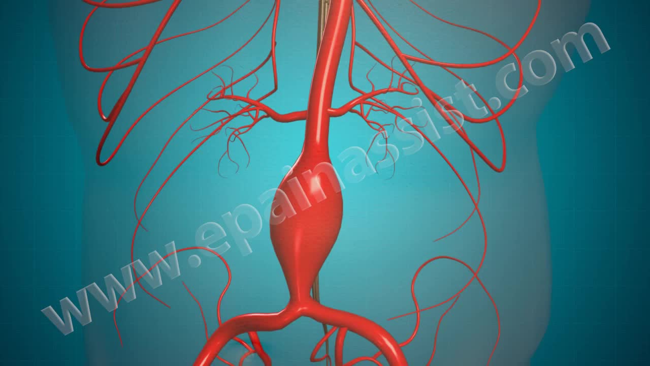
Open Abdominal Aortic and Endovascular Aneurysm Repair Surgery
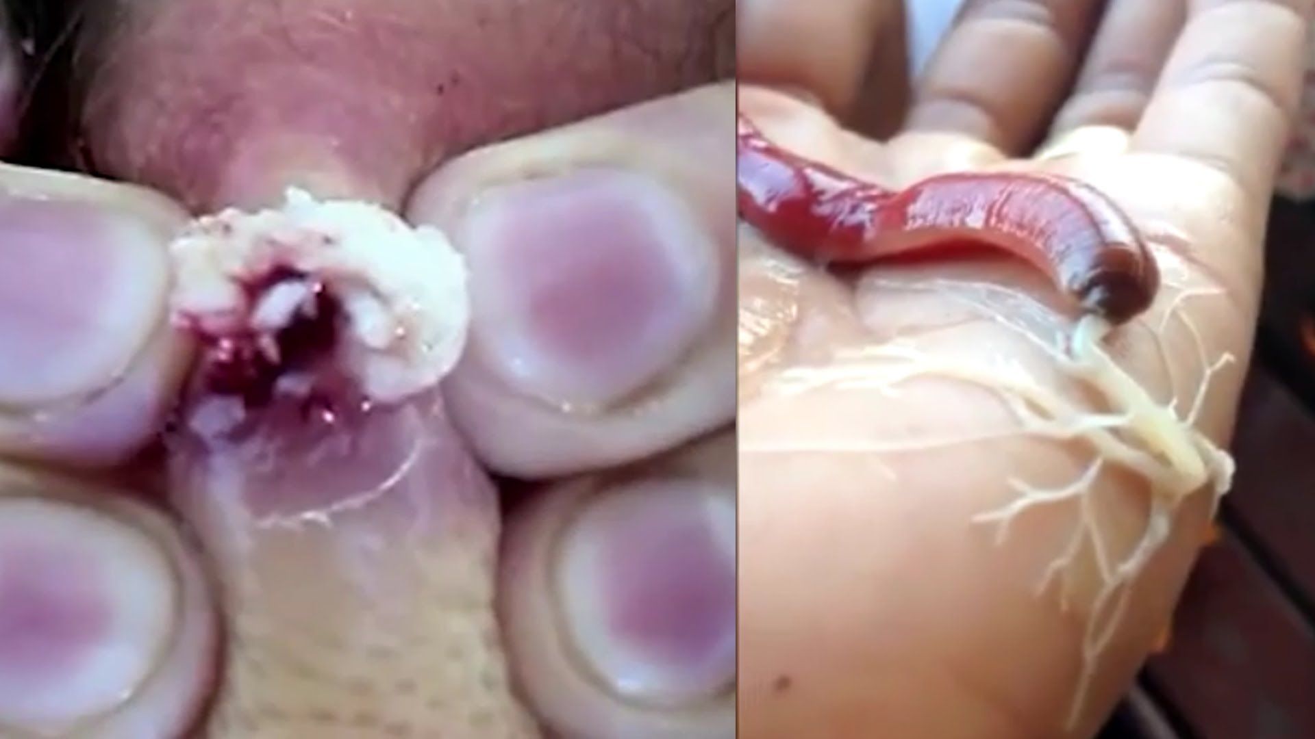
Watch that video to know about The Most Disgusting Parasites That Can Infect The Human Body
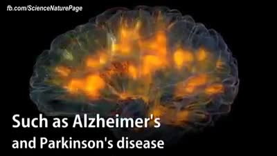
Researchers have received approval to bring 20 brain-dead humans back to life.

Tourette syndrome (also called Tourette's disorder or simply, "Tourette's") is an abnormal neurological condition characterized by motor and vocal tics. Tics are involuntary, rapid, sudden repetitive movements or sounds. Tics can be classified in a variety of ways. Motor tics can affect any part of the body including the head, neck, face, arms, shoulders, hands, feet, or legs. Facial tics, especially eye blinking, are usually the first symptoms of TS. Vocal tics are sounds that are made involuntarily. Vocal tics can include clearing the throat, coughing, sniffing, grunting, yelping, or shouting. In a few cases, vocal tics can include strange, inappropriate, or obscene words and phrases (called coprolalia). Vocal tics can also appear as constantly repeating the words of others (echolalia).

Thousands of Whiteheads! Why Do I Get Them?
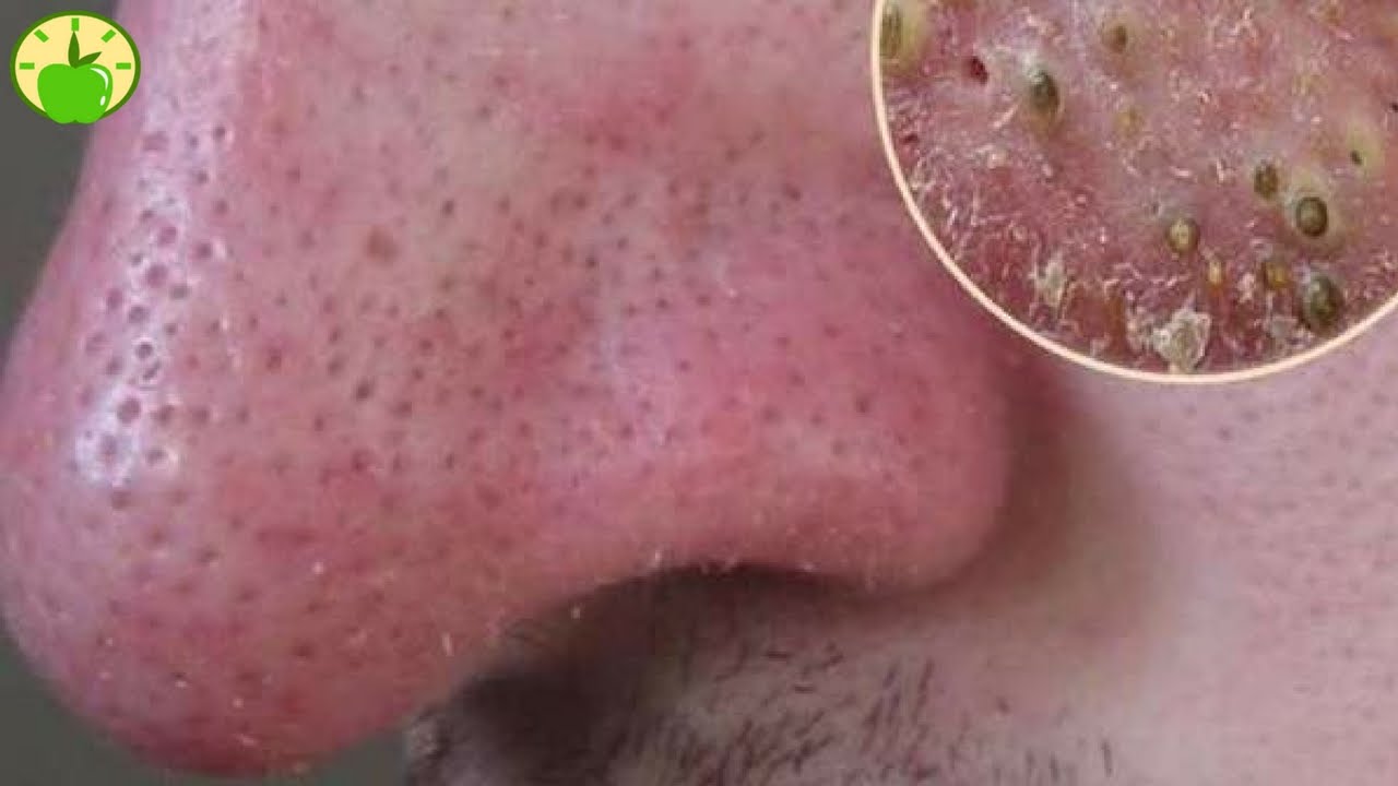
Watch that video to know How to Get Rid of Blackheads on Your Nose Naturally
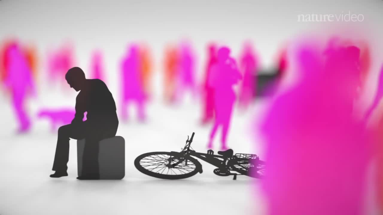
When you’re depressed, it can feel like you’ll never get out from under a dark shadow. However, even the most severe depression is treatable. So, if your depression is keeping you from living the life you want to, don’t hesitate to seek help. Learning about your depression treatment options will help you decide what approach is right for you. From therapy to medication to healthy lifestyle changes, there are many effective treatments that can help you overcome depression and reclaim your life.
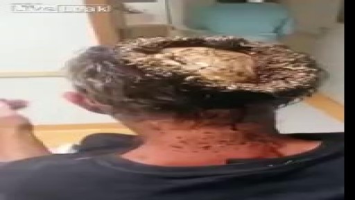
Watch that video of Unbelievable Head Infection Exposing a Man's Skull
Pneumonia is an infection that inflames the air sacs in one or both lungs. The air sacs may fill with fluid or pus (purulent material), causing cough with phlegm or pus, fever, chills, and difficulty breathing. A variety of organisms, including bacteria, viruses and fungi, can cause pneumonia.
The brain is the most complex organ in our body. It controls everything we do, from simple things such as breathing, to complex things such as co-ordinating our movements. The brain stores our memories, allows us to think and speak, and controls how we behave
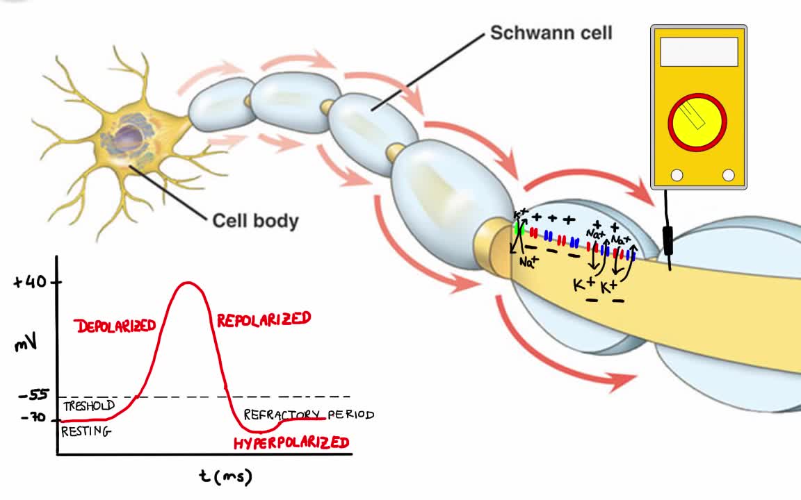
Your body has nerves that connect your brain to the rest of your organs and muscles, just like telephone wires connect homes all around the world. When you want your hand to move, your brain sends signals through your nerves to your hand telling the muscles to contract. But your nerves don’t just say “hand, move.” Instead your nerves send lots of electrical impulses (called action potentials) to different muscles in your hand, allowing you to move your hand with extreme precision.
Watch that video to know how to treat premature ejaculation naturally
Candidiase Tratamento, Remédio Para Unhas Com Fungos, Fungos Na Pele Como Tratar, Candidíase. http://candidiase-cura.plus101.com/ A intensa existência de substâncias químicas prejudiciais e metais tóxicos em nosso ambiente, nos medicamentos que tomamos, nos alimentos que comemos e até mesmo nos recheios dentários, cria um grande desafio para o nosso corpo se livrar efetivamente dessas toxinas resultando em uma ciclo vicioso que se manifesta em uma variedade de sintomas e problemas de saúde, entre eles é o crescimento excessivo de candida albicans que faz com que os sintomas da infecção por fungos aparecem. A acumulação de metais químicos e tóxicos no interior do corpo também pode levar a desequilíbrios hormonais, alterações genéticas, falhas no sistema imunológico, baixa eliminação, processo de cicatrização mais lenta, problemas de pele, alergias e danos no nervo e no cérebro. A presença de metais pesados ??no corpo (led, prata, mercúrio) proveniente de alimentos, o ar que respiramos, remédios e recheios dentários (contém 50% de amálgama), criam um ambiente ácido e anaeróbio (falta oxigênio) que incentiva a candida sobrecrescimento de fermento. Quando há sobrecarga de metal tóxico no intestino, o revestimento intestinal produz muco extra para impedir que os metais sejam absorvidos na corrente sanguínea. O problema é que esse muco cria um ambiente, que não possui oxigênio, incentivando bactérias e fungos, como organismos como o fermento Candida, a ficarem fora de controle. Além disso, a candida se liga a metais pesados ??(mesmo em seus enchimentos de amálgama) e cresce porque o corpo realiza uma tentativa desesperada de se proteger contra o envenenamento por metais pesados. Uma desintoxicação de metal profundo combinada com a remoção gradual do enchimento dentário de amalgama e substituindo-os por enchimentos brancos mais seguros é uma das etapas mais importantes e fundamentais na luta contra a infecção por levedura de Candida e restabelecendo o equilíbrio do corpo. O Único sistema holístico existente que vai lhe ensinar como curar Permanentemente sua Infecção fúngica, reequilibrar o seu corpo e conseguir a liberdade DURADOURA da Infecção do tipo candidíase! http://candidiase-cura.plus101.com/
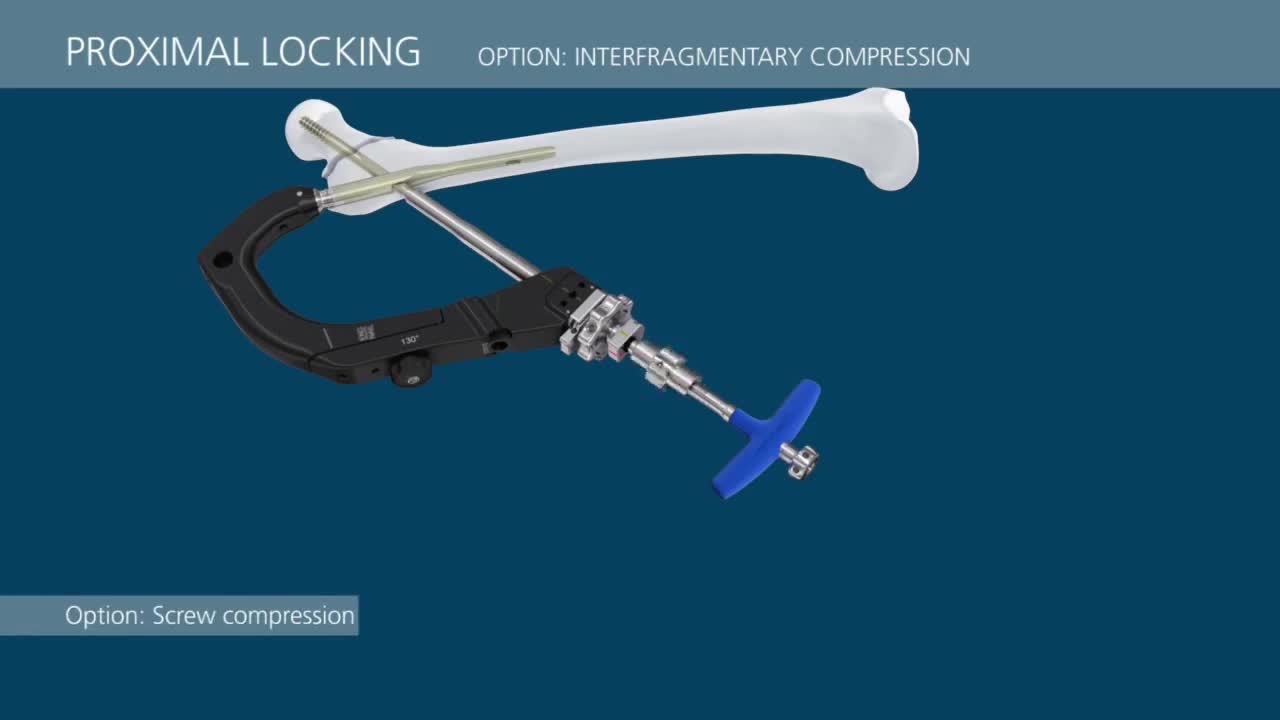
This video demonstrates a step-by-step technique for using the TFN-Advanced™ Proximal Femoral Nailing System (TFNA).
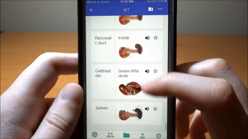
Watch that video to know How to Learn Human Anatomy Quickly and Efficiently?
Watch that video of a Bodybuilder's Colon Contains 10 lbs Meat Worms
Learn a simple way to tell if you might be suffering from an undiagnosed autoimmune condition. Examples of autoimmune conditions include: • alopecia areata
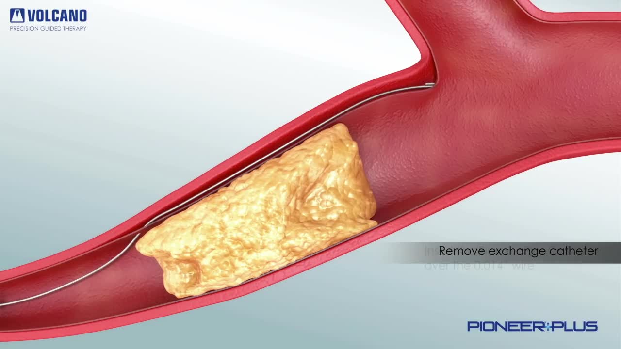
Pioneer Plus IVUS Re-Entry Catheter plaque removal

Percutaneous transluminal coronary angioplasty (PTCA) is a minimally invasive procedure to open up blocked coronary arteries, allowing blood to circulate unobstructed to the heart muscle.
