- Physical Examination
- Surgical Examination
- Ophthalmology
- Clinical Skills
- Orthopedics
- Surgery Videos
- Laparoscopy
- Pediatrics
- Funny Videos
- Cardiothoracic Surgery
- Nursing Videos
- Plastic Surgery
- Otorhinolaryngology
- Histology and Histopathology
- Neurosurgery
- Dermatology
- Pediatric Surgery
- Urology
- Dentistry
- Oncology and Cancers
- Anatomy Videos
- Health and Fitness
- Radiology
- Anaesthesia
- Physical Therapy
- Pharmacology
- Interventional Radiology
- Cardiology
- Endocrinology
- Gynecology
- Emergency Medicine
- Psychiatry and Psychology
- Childbirth Videos
- General Medical Videos
- Nephrology
- Physiology
- Diet and Food Health
- Diabetes Mellitus
- Neurology
- Women Health
- Osteoporosis
- Gastroenterology
- Pulmonology
- Hematology
- Rheumatology
- Toxicology
- Nuclear Medicine
- Infectious Diseases
- Vascular Disease
- Reproductive Health
- Burns and Wound Healing
- Other
Top videos
Thousands of Whiteheads! Why Do I Get Them?
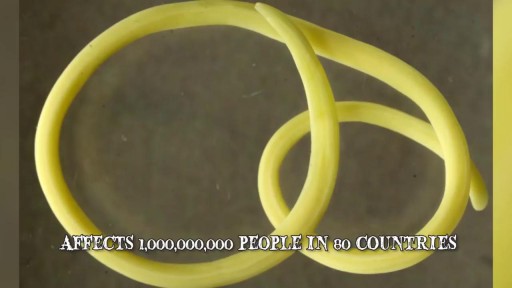
Watch that video of The 10 Most Invasive Parasites in the World

Nursing skills lab procedure for medication validation administration.

Watch that video of Amputated Hand Reattachment Surgery
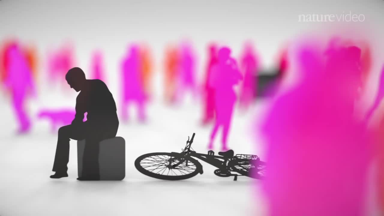
When you’re depressed, it can feel like you’ll never get out from under a dark shadow. However, even the most severe depression is treatable. So, if your depression is keeping you from living the life you want to, don’t hesitate to seek help. Learning about your depression treatment options will help you decide what approach is right for you. From therapy to medication to healthy lifestyle changes, there are many effective treatments that can help you overcome depression and reclaim your life.

Pneumonia is an infection that inflames the air sacs in one or both lungs. The air sacs may fill with fluid or pus (purulent material), causing cough with phlegm or pus, fever, chills, and difficulty breathing. A variety of organisms, including bacteria, viruses and fungi, can cause pneumonia.

Watch that video to know how to treat premature ejaculation naturally
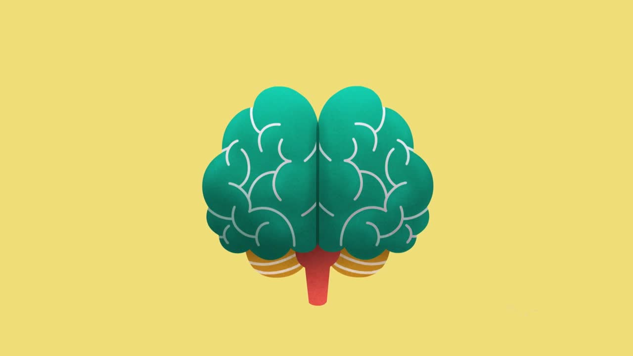
The brain is the most complex organ in our body. It controls everything we do, from simple things such as breathing, to complex things such as co-ordinating our movements. The brain stores our memories, allows us to think and speak, and controls how we behave

Watch that video of A man with one inch-wide hole in his face
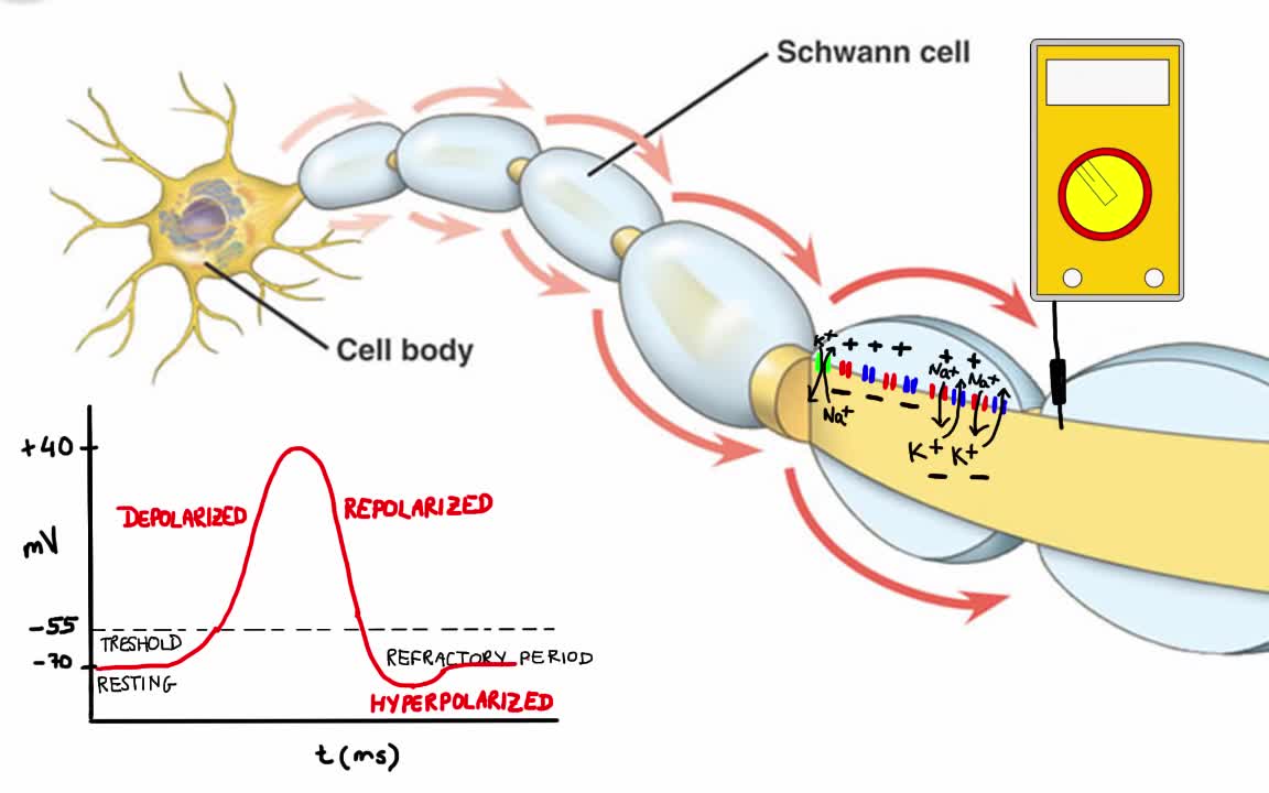
Your body has nerves that connect your brain to the rest of your organs and muscles, just like telephone wires connect homes all around the world. When you want your hand to move, your brain sends signals through your nerves to your hand telling the muscles to contract. But your nerves don’t just say “hand, move.” Instead your nerves send lots of electrical impulses (called action potentials) to different muscles in your hand, allowing you to move your hand with extreme precision.
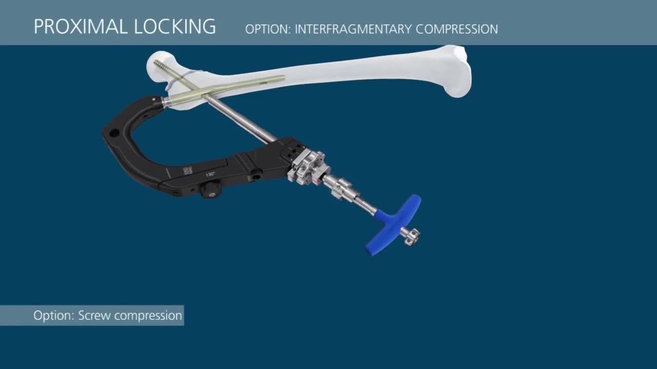
This video demonstrates a step-by-step technique for using the TFN-Advanced™ Proximal Femoral Nailing System (TFNA).

Aumento De Gluteos, Metacrilato En Gluteos, Aumento De Gluteos Natural, Operacion De Nalgas.--- http://aumente-gluteos.plus101.com/ --- Con una combinación de dieta, ejercicio y mejoras artificiales, puedes cambiar la forma de los glúteos rápidamente, sin importar tu tipo de cuerpo. Aunque no verás un cambio significativo en una semana, si dedicas un tiempo y haces ejercicios enfocados en los tres músculos principales de los glúteos: el glúteo mayor, el glúteo medio y el glúteo menor, tendrás unos glúteos más grandes. Enfócate en consumir muchas proteínas. Las proteínas son esenciales para el crecimiento y el desarrollo de los músculos, por lo que es importante comer el tipo correcto de proteínas. La proteína combinada con el ejercicio correcto aumentará definitivamente el tamaño de los glúteos. Algunas fuentes saludables de proteínas incluyen los huevos, las pechugas de pollo sin piel, el salmón, el atún, el queso cottage, el pavo, los frijoles, las legumbres, la carne de res magra y las nueces de soya. En cuanto a la carne, busca una que sea magra y sin procesar. Cuando compres el pescado, trata de hornearlo en lugar de freírlo. Elige el tipo correcto de carbohidratos y grasas. Existen muchas dietas que dicen que eliminan por completo los carbohidratos y las grasas, pero lo mejor no es eliminar los alimentos de la dieta, sino sustituirlos por opciones más saludables. Evita el exceso de calorías y la mala alimentación, alejándote de los carbohidratos procesados, como las papas fritas y la pasta. Los carbohidratos saludables incluyen la quinua, el camote, el arroz integral, los granos de avena enteros y los panes integrales. Las fuentes de grasas saludables que pueden ayudarte a perder peso y a tonificar los glúteos son los aceites de pescado, el aceite de oliva extra virgen, la mantequilla de almendras y las nueces. Abastécete de vegetales. Los vegetales suelen ser una parte olvidada de la dieta para agrandar los músculos. Al agregar vegetales a cada comida te darás cuenta de que tus niveles de energía serán más constantes y por lo tanto, podrás hacer un entrenamiento más fuerte ya que no sentirás demasiado cansancio. Descubre por qué las cirugías y los implantes no son la solución más efectiva. Olvídate del quirófano y ahorra tu dinero, porque con mi método resolverás el problema de “Síndrome de los glúteos planos” rápidamente. ingresa ahora a: http://aumente-gluteos.plus101.com/

Learn a simple way to tell if you might be suffering from an undiagnosed autoimmune condition. Examples of autoimmune conditions include: • alopecia areata

Como Aumentar Los Gluteos, Como Aumentar Las Pompas, Masajes Para Aumentar Gluteos--- http://aumente-gluteos.plus101.com/ --- Para conseguir un cuerpo perfecto hay que tomar en cuenta todo lo que consumimos día a día, razón por la cual te mencionaremos a continuación los mejores alimentos que debes comer frecuentemente para levantar los glúteos y piernas rápidamente: 1. Las Proteínas Las proteínas deben estar presentes dentro de tu dieta de manera constante, ya que ellas son las responsables del aumento del volumen de la masa muscular, te ayudarán a aumentar rápidamente los glúteos y piernas, para luego darle la firmeza y tonificación adecuada con los ejercicios. Proteínas de origen animal: Pollo sin piel, carne de res, pescado y ternera, considerando siempre que sean bajas en grasas, puedes encontrar proteínas además en los huevos y en los productos lácteos descremados. Proteínas de origen vegetal: Las puedes encontrar en las legumbres como lentejas, frijoles o garbanzos, en las semillas, las verduras y los cereales. 2. Carbohidratos Las mayorías de las personas cometen el error de eliminar de su dieta de manera completa los carbohidratos, los cuales son necesarios para el normal funcionamiento del organismo. Te recomendamos comer carbohidratos luego de una fuerte rutina de ejercicio, ya que es el momento ideal para que sean absorbidos completamente por las células musculares aumentando por lo tanto el tamaño de los glúteos y las piernas, entre los carbohidratos fundamentales para una buena alimentación tenemos: avena, granola, pan, arroz y pastas siempre que sean integrales. 3. Grasas Saludables No todas las grasas son malas para nuestra salud, existen algunas que son incluso indispensables para nuestros cuerpos, entre ellas tenemos: aceite de oliva, aceite de canola, nueces, aguacate, omega 3, aceite de almendra y de girasol. 4. Frutas y vegetales Las frutas y vegetales te ayudarán a hacer crecer los glúteos y piernas rápidamente gracias a sus altos contenidos en vitaminas y minerales que favorecen la regeneración de los músculos, dándote la energía y resistencia necesaria al momento de realizar tu rutina de ejercicios, así que inclúyelas dentro de tu dieta saludable para conseguir óptimos resultados. Alguna vez has deseado tener una cola más atractiva, pero sin invertir demasiado tiempo ni trabajo, Ingresa aqui: http://aumente-gluteos.plus101.com/
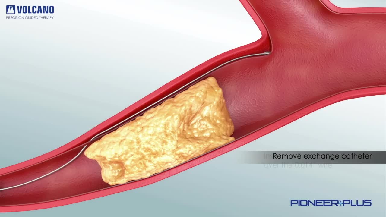
Pioneer Plus IVUS Re-Entry Catheter plaque removal

Percutaneous transluminal coronary angioplasty (PTCA) is a minimally invasive procedure to open up blocked coronary arteries, allowing blood to circulate unobstructed to the heart muscle.
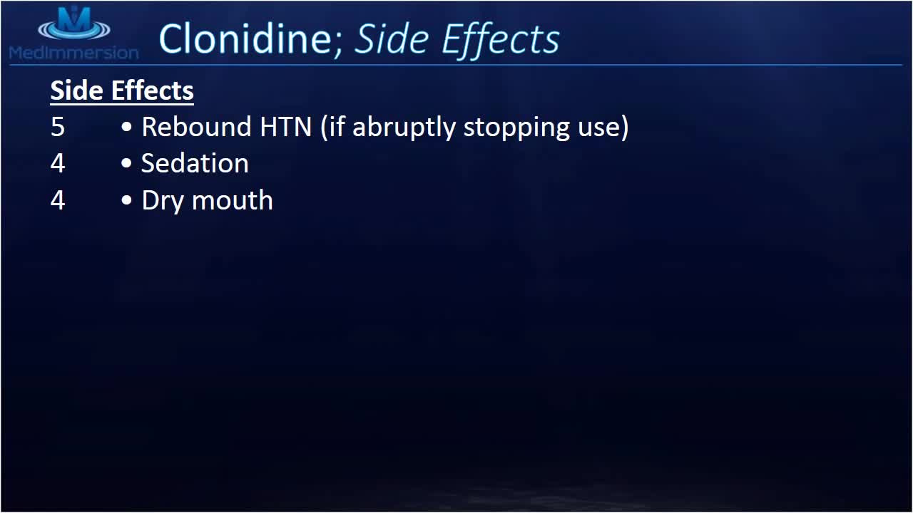
Clonidine lowers blood pressure by decreasing the levels of certain chemicals in your blood. This allows your blood vessels to relax and your heart to beat more slowly and easily. The Catapres brand of clonidine is used to treat hypertension (high blood pressure). The Kapvay brand is used to treat attention deficit hyperactivity disorder (ADHD). Clonidine is sometimes given with other medications

Broken or Dislocated Ankle Joint

Heart failure, sometimes known as congestive heart failure, occurs when your heart muscle doesn't pump blood as well as it should. Certain conditions, such as narrowed arteries in your heart (coronary artery disease) or high blood pressure, gradually leave your heart too weak or stiff to fill and pump efficiently.
