- Physical Examination
- Surgical Examination
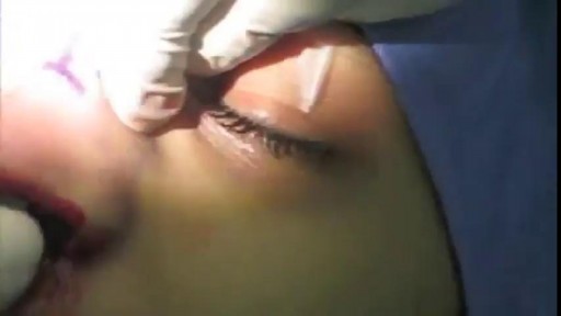
- Ophthalmology
- Clinical Skills
- Orthopedics
- Surgery Videos
- Laparoscopy
- Pediatrics
- Funny Videos
- Cardiothoracic Surgery
- Nursing Videos
- Plastic Surgery
- Otorhinolaryngology
- Histology and Histopathology
- Neurosurgery
- Dermatology
- Pediatric Surgery
- Urology
- Dentistry
- Oncology and Cancers
- Anatomy Videos
- Health and Fitness
- Radiology
- Anaesthesia
- Physical Therapy
- Pharmacology
- Interventional Radiology
- Cardiology
- Endocrinology
- Gynecology
- Emergency Medicine
- Psychiatry and Psychology
- Childbirth Videos
- General Medical Videos
- Nephrology
- Physiology
- Diet and Food Health
- Diabetes Mellitus
- Neurology
- Women Health
- Osteoporosis
- Gastroenterology
- Pulmonology
- Hematology
- Rheumatology
- Toxicology
- Nuclear Medicine
- Infectious Diseases
- Vascular Disease
- Reproductive Health
- Burns and Wound Healing
- Other
Top videos
What Causes Ulcers? No single cause has been found for ulcers. However, it is now clear that an ulcer is the end result of an imbalance between digestive fluids in the stomach and duodenum. Most ulcers are caused by an infection with a type of bacteria called Helicobacter pylori (H. pylori). Factors that can increase your risk for ulcers include: Use of painkillers called nonsteroidal anti-inflammatory drugs (NSAIDs), such as aspirin, naproxen (Aleve, Anaprox, Naprosyn, and others), ibuprofen (Motrin, Advil, some types of Midol, and others), and many others available by prescription; even safety-coated aspirin and aspirin in powered form can frequently cause ulcers. Excess acid production from gastrinomas, tumors of the acid producing cells of the stomach that increases acid output (seen in Zollinger-Ellison syndrome) Excessive drinking of alcohol Smoking or chewing tobacco Serious illness Radiation treatment to the area What Are the Symptoms of an Ulcer? An ulcer may or may not have symptoms. When symptoms occur, they may include: A gnawing or burning pain in the middle or upper stomach between meals or at night Bloating Heartburn Nausea or vomiting In severe cases, symptoms can include: Dark or black stool (due to bleeding) Vomiting blood (that can look like "coffee-grounds") Weight loss Severe pain in the mid to upper abdomen
Minimally invasive surgery has been shown to be feasible and safe in pediatric patients since 1975 when laparoscopic surgery was first used to treat a small bowel obstruction. Laparoscopy is an option for surgical repair of inguinal hernias in addition to the traditional open approach.
Function. Vitamin A helps form and maintain healthy skin, teeth, skeletal and soft tissue, mucus membranes, and skin. It is also known as retinol because it produces the pigments in the retina of the eye. Vitamin A promotes good vision, especially in low light. Vitamin deficiency anemia occurs when your body doesn't have enough of the vitamins needed to produce adequate numbers of healthy red blood cells. Red blood cells carry oxygen from your lungs throughout your body. If your diet is lacking in certain vitamins, vitamin deficiency anemia can develop.
Kegel exercises strengthen the pelvic floor muscles, which support the uterus, bladder, small intestine and rectum. You can do Kegel exercises, also known as pelvic floor muscle training, just about anytime. Start by understanding what Kegel exercises can do for you — then follow step-by-step instructions for contracting and relaxing your pelvic floor muscles.
This video: Multiple myeloma is a cancer that forms in a type of white blood cell called a plasma cell. Plasma cells help you fight infections by making antibodies that recognize and attack germs. Multiple myeloma causes cancer cells to accumulate in the bone marrow, where they crowd out healthy blood cells. Rather than produce helpful antibodies, the cancer cells produce abnormal proteins that can cause kidney problems. Treatment for multiple myeloma isn't always necessary. If you're not experiencing signs and symptoms, you may not require treatment. If signs and symptoms develop, a number of treatments can help control your multiple myeloma.


Less than a decade ago, corneal transplantation took a big leap forward with the introduction of Descemet’s stripping endothelial keratoplasty (DSEK), which removes only Descemet’s membrane and the diseased endothelium and replaces them with a thin, tripartite donor graft of posterior corneal stroma, Descemet’s membrane, and healthy endothelium. Then came DSAEK, in which the donor graft is prepared with an automated microkeratome, allowing for easier donor preparation and reproducible results by surgeons and eye bank technicians.1 DSAEK has proved to have many advantages over penetrating keratoplasty (PK) and its endothelial predecessors (see “A Brief History of Endothelial Keratoplasty”). Now DSAEK is being compared with a newer technique, Descemet’s membrane endothelial keratoplasty (DMEK), which has emerged as a promising alternative—grafting only Descemet’s membrane and endothelium, allowing for a pure anatomical replacement of only what was removed and the possibility of even better vision with quicker healing.1 Although indications for these procedures are similar, each has unique benefits and drawbacks. Five cornea surgeons offer their perspectives on the procedures and their thoughts on whether it may be time to move to the newer surgery.
The removal of a clot is called an embolectomy. An embolectomy might be done during a surgery. Or it might be done with a minimally invasive procedure that uses a catheter (a thin tube that is guided through a blood vessel). This type of treatment for pulmonary embolism is used only in rare cases.

http://sin-celulitis.good-info.co/ Como Quitar La Celulitis, Celulitis En La Pierna, Drenaje Linfatico Celulitis, Reducir Celulitis. Imagina Que En Tan Solo 8 Semenas Pudieras... Usar la ropa que quisieras sin preocuparte. Mirarte al espejo con total satisfacción. Usar ese traje de baño que tanto te gusta. Tener relaciones con tu pareja y volverlo loco con tu piel suave y lisa. Sentirte llena de confianza y optimismo. Olvidarte de la celulitis para siempre. ¿Has deseado alguna vez lucir una piel bella y sin los desagradables hoyuelos causados por la celulitis? Haz Clic Aqui. http://sin-celulitis.good-info.co
Repair of post-infarction ventricular septal defect (VSD) remains a challenging procedure with a high risk of VSD recurrence. In order to reduce this risk, a double patch and glue technique was introduced in the department in 1986. This surgical technique is hereunder presented. Since 1971, ninety-three patients have been operated on early (≪15 days) after the occurrence of a post-infarction VSD. This retrospective study allows to compare the results of this double patch and glue technique to those obtained with the conventional one, in terms of hospital death and VSD recurrence. The double patch and glue technique avoids recurrence of VSD and plays a part in reducing hospital mortality.

Shaking violently in a hospital crib, two tiny legs twitch and shudder uncontrollably. Wailing, muscle clenching and gasping for breath accompany the disturbing fit-like seizure. But, what makes the image all the more heartbreaking, is the newborn baby's diagnosis. Despite being just weeks old, the tiny baby is suffering the effects of drug withdrawal, having been born addicted to opioids. Every 19 minutes a child in the US is born with an opioid addiction - a devastating affliction inherited from their drug-addict mothers. While for most newborns the first precious weeks of life are full of love, care and adoration, for babies born addicted to drugs their first weeks are long, agonizing and distressing as they battle neonatal abstinence syndrome.
What is inside A Cyst? Watch it now

Hemodialysis is a process that uses a membrane (dialyzer) to: Remove wastes, such as urea, from the blood. Restore the proper balance of electrolytes in the blood. Eliminate extra fluid from the body.

Friedreich's ataxia is an inherited disease that damages your nervous system. The damage affects your spinal cord and the nerves that control muscle movement in your arms and legs. Symptoms usually begin between the ages of 5 and 15. The main symptom is ataxia, which means trouble coordinating movements. Specific symptoms include Difficulty walking Muscle weakness Speech problems Involuntary eye movements Scoliosis (curving of the spine to one side) Heart palpitations, from the heart disease which can happen along with Friedreich's ataxia People with Friedreich's ataxia usually need a wheelchair 15 to 20 years after symptoms first appear. In severe cases, people become incapacitated. There is no cure. You can treat symptoms with medicines, braces, surgery, and physical therapy.
A fractured rib is usually a result of a fall or accident. Prolonged coughing and sports with repetitive movement, such as golf, also can cause a rib fracture. Symptoms include pain when taking a deep breath, pressing on the injured area, or bending or twisting the body. In most cases, fractured ribs usually heal on their own in one or two months. Pain relievers can make it easier to breathe deeply.
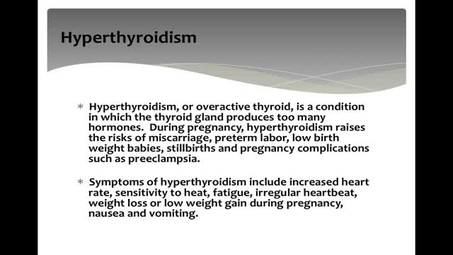
Uncontrolled hyperthyroidism during pregnancy can lead to serious health problems in the mother and the unborn baby. During pregnancy, mild hyperthyroidism does not require treatment. More severe hyperthyroidism is treated with antithyroid medications, which act by interfering with thyroid hormone production.

Primary infection with herpes simplex viruses (HSVs) is clinically more severe than recurrent outbreaks. However, most primary HSV-1 and HSV-2 infections are subclinical and may never be clinically diagnosed. Orolabial herpes Herpes labialis (eg, cold sores, fever blisters) is most commonly associated with HSV-1 infection. Oral lesions caused by HSV-2 have been identified, usually secondary to orogenital contact. Primary HSV-1 infection often occurs in childhood and is usually asymptomatic. Primary infection Symptoms of primary herpes labialis may include a prodrome of fever, followed by a sore throat and mouth and submandibular or cervical lymphadenopathy. In children, gingivostomatitis and odynophagia are also observed. Painful vesicles develop on the lips, the gingiva, the palate, or the tongue and are often associated with erythema and edema. The lesions ulcerate and heal within 2-3 weeks. Recurrences The disease remains dormant for a variable amount of time. HSV-1 reactivation in the trigeminal sensory ganglia leads to recurrences in the face and the oral, labial, and ocular mucosae. Pain, burning, itching, or paresthesia usually precedes recurrent vesicular lesions that eventually ulcerate or form a crust. The lesions most commonly occur in the vermillion border, and symptoms of untreated recurrences last approximately 1 week. Recurrent erythema multiforme lesions have been associated with orolabial HSV-1 recurrences. A recent study reported that HSV-1 viral shedding had a median duration of 48-60 hours from the onset of herpes labialis symptoms. They did not detect any virus beyond 96 hours of symptom onset.[7] Genital herpes HSV-2 is identified as the most common cause of herpes genitalis. However, HSV-1 has been increasingly identified as the causative agent in as many as 30% of cases of primary genital herpes infections likely secondary to orogenital contact. Recurrent genital herpes infections are almost exclusively caused by HSV-2. Primary infection Primary herpes genitalis occurs within 2 days to 2 weeks after exposure to the virus and has the most severe clinical manifestations. Symptoms of the primary episode typically last 2-3 weeks. In men, painful, erythematous, vesicular lesions that ulcerate most commonly occur on the penis, but they can also occur on the anus and the perineum. In women, primary herpes genitalis presents as vesicular/ulcerated lesions on the cervix and as painful vesicles on the external genitalia bilaterally. They can also occur on the vagina, the perineum, the buttocks, and, at times, the legs in a sacral nerve distribution. Associated symptoms include fever, malaise, edema, inguinal lymphadenopathy, dysuria, and vaginal or penile discharge. Females may also have lumbosacral radiculopathy, and as many as 25% of women with primary HSV-2 infections may have associated aseptic meningitis. Recurrences After primary infection, the virus may be latent for months to years until a recurrence is triggered. Reactivation of HSV-2 in the lumbosacral ganglia leads to recurrences below the waist. Recurrent clinical outbreaks are milder and often preceded by a prodrome of pain, itching, tingling, burning, or paresthesia. Individuals who are exposed to HSV and have asymptomatic primary infections may experience an initial clinical episode of genital herpes months to years after becoming infected. Such an episode is not as severe as a true primary outbreak. More than one half of individuals who are HSV-2 seropositive do not experience clinically apparent outbreaks. However, these individuals still have episodes of viral shedding and can transmit the virus to their sexual partners. Other HSV infections Localized or disseminated eczema herpeticum is also known as Kaposi varicelliform eruption. Caused by HSV-1, eczema herpeticum is a variant of HSV infection that commonly develops in patients with atopic dermatitis, burns, or other inflammatory skin conditions. Children are most commonly affected. Herpes whitlow, vesicular outbreaks on the hands and the digits, was most commonly due to infection with HSV-1. It usually occurred in children who sucked their thumbs and, prior to the widespread use of gloves, in dental and medical health care workers. The occurrence of herpes whitlow due to HSV-2 is increasingly recognized, probably due to digital-genital contact. Herpes gladiatorum is caused by HSV-1 and is seen as papular or vesicular eruptions on the face, arms, or torsos of athletes in sports involving close physical contact (classically wrestling). Disseminated HSV infection can occur in females who are pregnant and in individuals who are immunocompromised. These patients may present with atypical signs and symptoms of HSV, and the condition may be difficult to diagnose. Herpetic sycosis, a follicular infection with HSV, may present as a vesiculopustular eruption on the beard area. This infection often results from autoinoculation after shaving through a recurrent herpetic outbreak. Classically caused by HSV-1, there have been rare reports of relapsing beard folliculitis caused by type 2 HSV.[8] Neonatal HSV HSV-2 infection in pregnancy can have devastating effects on the fetus. Neonatal HSV usually manifests within the first 2 weeks of life and clinically ranges from localized skin, mucosal, or eye infections to encephalitis, pneumonitis, disseminated infection, and demise. Most women who deliver infants with neonatal HSV had no prior history, signs, or symptoms of HSV infection. Risk of transmission is highest in pregnant women who are seronegative for both HSV-1 and HSV-2 and acquire a new HSV infection in the third trimester of pregnancy. Factors that increase the risk of transmission from mother to baby include the type of genital infection at the time of delivery (higher risk with active primary infection), active lesions, prolonged rupture of membranes, vaginal delivery, and an absence of transplacental antibodies. The mortality rate for neonates is extremely high (>80%) if untreated.
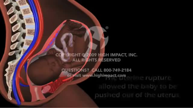
Uterine rupture is usually when the scar from your previous caesarean section tears open. Though it's uncommon, you should be aware of this risk, particularly if you're thinking about giving birth vaginally next time. It's possible for your scar to gape slightly while you're pregnant (scar dehiscence).
Nose Plastic Surgery: Open Rhinoplasty
