- Physical Examination
- Surgical Examination
- Ophthalmology
- Clinical Skills
- Orthopedics
- Surgery Videos
- Laparoscopy
- Pediatrics
- Funny Videos
- Cardiothoracic Surgery
- Nursing Videos
- Plastic Surgery
- Otorhinolaryngology
- Histology and Histopathology
- Neurosurgery
- Dermatology
- Pediatric Surgery
- Urology
- Dentistry
- Oncology and Cancers
- Anatomy Videos
- Health and Fitness
- Radiology
- Anaesthesia
- Physical Therapy
- Pharmacology
- Interventional Radiology
- Cardiology
- Endocrinology
- Gynecology
- Emergency Medicine
- Psychiatry and Psychology
- Childbirth Videos
- General Medical Videos
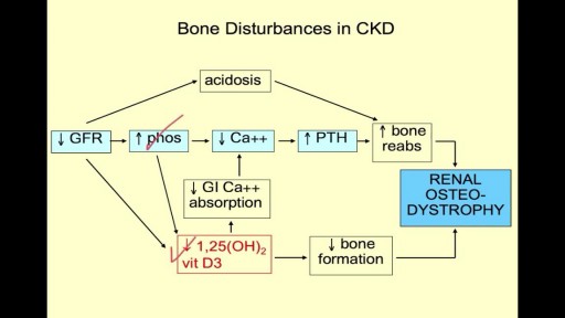
- Nephrology
- Physiology
- Diet and Food Health
- Diabetes Mellitus
- Neurology
- Women Health
- Osteoporosis
- Gastroenterology
- Pulmonology
- Hematology
- Rheumatology
- Toxicology
- Nuclear Medicine
- Infectious Diseases
- Vascular Disease
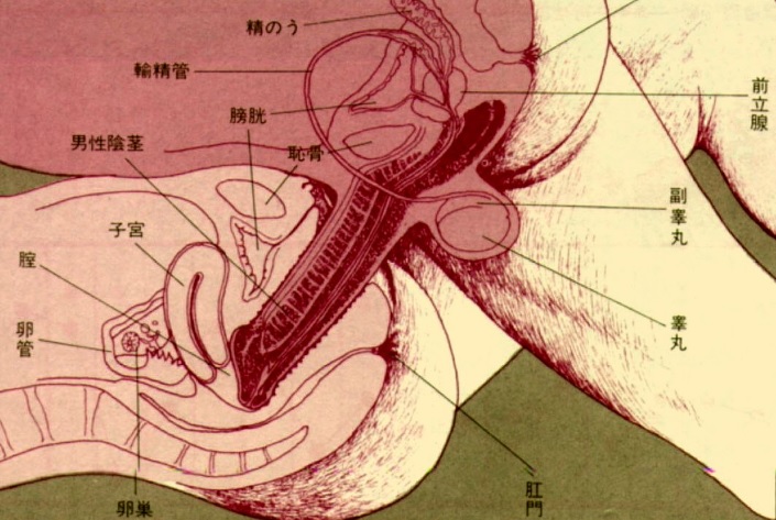
- Reproductive Health
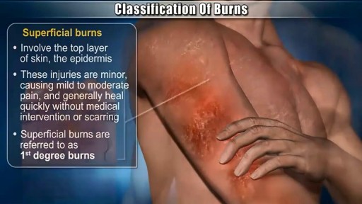
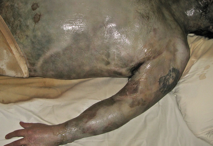
- Burns and Wound Healing
- Other
Top videos
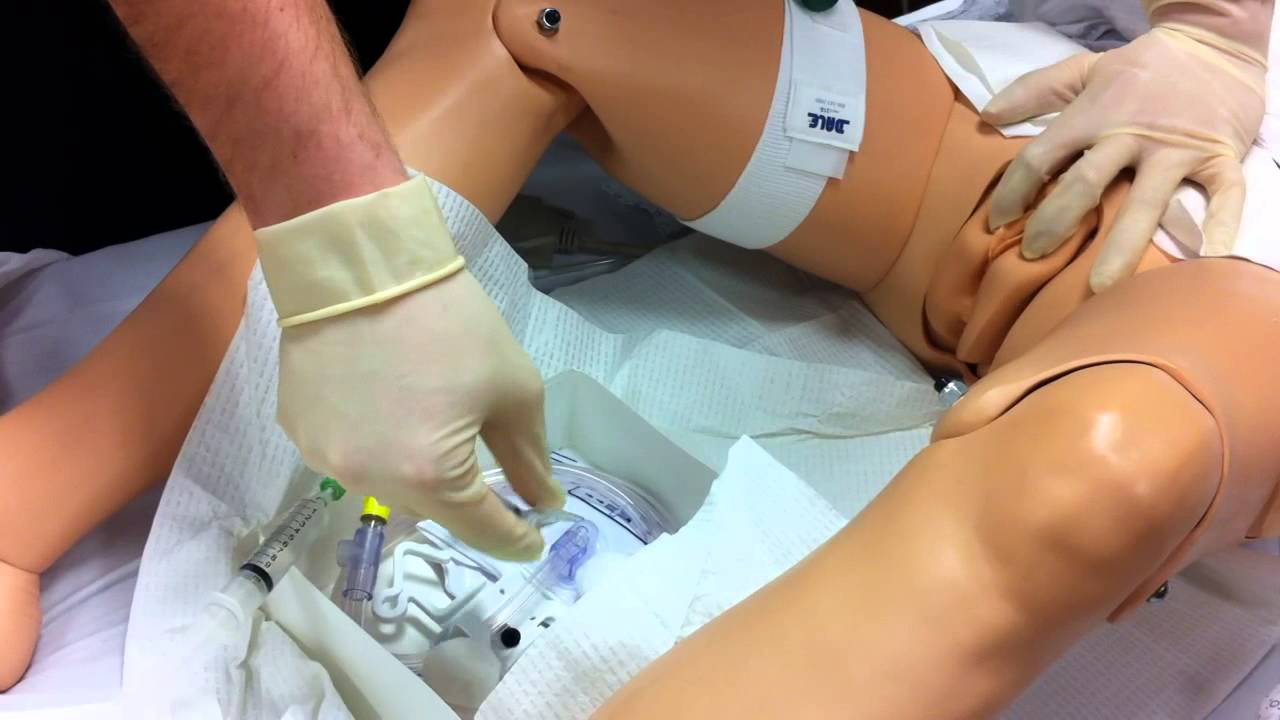
Watch that Female Foley Genital Catheter Insertion Procedure
Watch that Full Human Body Decay Process Video
Watch that video to know How To Increase Your Testosterone Levels, Naturally
Watch that video to learn How to Study The Human Anatomy

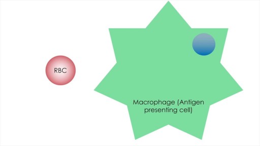
Hemophagocytic lymphohistiocytosis is a rare but life threatening condition characterised by activation of macrophages which result in phagocytosis of RBCs and cytokine mediated tissue damage. This presentation aims to discuss the genetic basis, clinical features, diagnostic criteria and management options in this serious condition. The management options in HLH include Etoposide, Dexamethasone, Cyclosorine, Tacrolimus, Alemtuzumab and stem cell transplant.
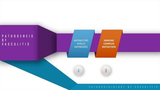
A step wise approach to the pathogenesis, types, disease entities and diagnosis of vasculitis. This discussion also includes the management options of vasculitis and their adverse drug reactions. In essence, vasculitis is a clfinicopathologic process characterised by inflammation and damage of blood vessels. This may be mainly due to three pathological processes which include immune complex deposition, anti-neutrophillic antibody formation and pathological T lymphocyte response and granuloma formation. The disease entities include Wegner's granulomatosis, Churg Strauss and many others. These present with palpable purpura, unexplained renal dysfunction etc which can be diagnosed based on biopsy and angiogram.
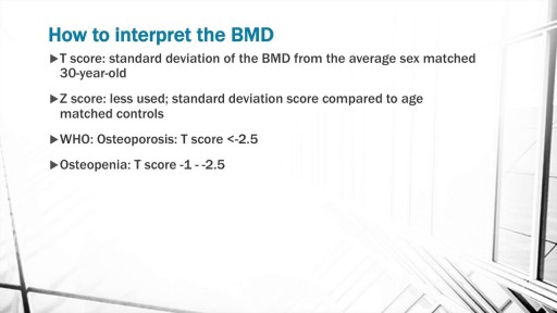
The discussion begins with a basic explanation of Bone biology taking into consideration the osteoblast and osteoclast balance. Concepts of RANK, RANK ligand and Osteoprotegerin are included. Risk factors for Osteoporosis such as Age, alcohol, smoking, sedentary lifestyle are also discussed.
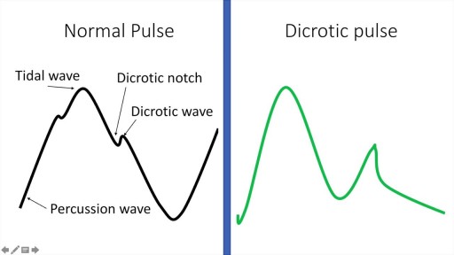
A detailed description of the Arterial Pulse including its waveform and pathological subtypes. Also discussed are the abnormal rates (tachycardia and bradycardia) and their causes, abnormal rhythm (including regularly regular and irregularly irregular pulses) and abnormal character (including pulses bisferiens, pulses parvus et tarsus, pulsus alternans, pulses paradoxus and others.) Description of pulse in various pathological states including Aortic stenosis and aortic regurgitation is also included. Finally there is also a description of the peripheral signs of aortic regurgitation.

Cigarette contain tobacco that is very harmful but vaporizers does not contain tobacco. ... The most basic difference between vaping and cigarette usage is that cigarettes require combustion. You need fire to light a cigarette. On the other hand, vaping requires electricity and creates vapor.

ESCLEROTERAPIA

Popping Cyst in the Ear Lobe
