- Physical Examination
- Surgical Examination
- Ophthalmology
- Clinical Skills
- Orthopedics
- Surgery Videos
- Laparoscopy
- Pediatrics
- Funny Videos
- Cardiothoracic Surgery
- Nursing Videos
- Plastic Surgery
- Otorhinolaryngology
- Histology and Histopathology
- Neurosurgery
- Dermatology
- Pediatric Surgery
- Urology
- Dentistry
- Oncology and Cancers
- Anatomy Videos
- Health and Fitness
- Radiology
- Anaesthesia
- Physical Therapy
- Pharmacology
- Interventional Radiology
- Cardiology
- Endocrinology
- Gynecology
- Emergency Medicine
- Psychiatry and Psychology
- Childbirth Videos
- General Medical Videos
- Nephrology
- Physiology
- Diet and Food Health
- Diabetes Mellitus
- Neurology
- Women Health
- Osteoporosis
- Gastroenterology
- Pulmonology
- Hematology
- Rheumatology
- Toxicology
- Nuclear Medicine
- Infectious Diseases
- Vascular Disease
- Reproductive Health
- Burns and Wound Healing
- Other
Top videos
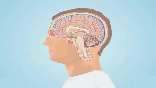
Subarachnoid hemorrhage is bleeding in the space between your brain and the surrounding membrane (subarachnoid space). Bleeding usually results from the rupture of an abnormal bulge in a blood vessel in your brain (brain aneurysm). Sometimes an abnormal tangle of blood vessels in your brain (arteriovenous malformation), trauma or other events cause bleeding. A subarachnoid hemorrhage may lead to permanent brain damage or death if not treated.
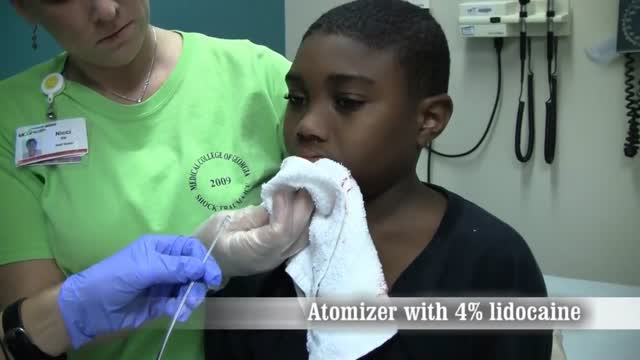
Nosebleeds common. Most often they are a nuisance and not a true medical problem. But they can be both. Nosebleed care Sit upright and lean forward. By remaining upright, you reduce blood pressure in the veins of your nose. This discourages further bleeding. Sitting forward will help you avoid swallowing blood, which can irritate your stomach. Pinch your nose. Use your thumb and index finger to pinch your nostrils shut. Breathe through your mouth. Continue to pinch for five to 10 minutes. Pinching sends pressure to the bleeding point on the nasal septum and often stops the flow of blood. To prevent re-bleeding, don't pick or blow your nose and don't bend down for several hours after the bleeding episode. During this time remember to keep your head higher than the level of your heart. If re-bleeding occurs, blow out forcefully to clear your nose of blood clots and spray both sides of your nose with a decongestant nasal spray containing oxymetazoline (Afrin, Mucinex Moisture Smart, others). Pinch your nose again as described above and call your doctor. When to seek emergency care The bleeding lasts for more than 20 minutes The nosebleed follows an accident, a fall or an injury to your head, including a punch in the face that may have broken your nose
Hypertensive emergencies encompass a spectrum of clinical presentations in which uncontrolled blood pressures (BPs) lead to progressive or impending end-organ dysfunction. In these conditions, the BP should be lowered aggressively over minutes to hours. Neurologic end-organ damage due to uncontrolled BP may include hypertensive encephalopathy, cerebral vascular accident/cerebral infarction, subarachnoid hemorrhage, and/or intracranial hemorrhage.[1] Cardiovascular end-organ damage may include myocardial ischemia/infarction, acute left ventricular dysfunction, acute pulmonary edema, and/or aortic dissection. Other organ systems may also be affected by uncontrolled hypertension, which may lead to acute renal failure/insufficiency, retinopathy, eclampsia, or microangiopathic hemolytic anemia.[1] With the advent of antihypertensives, the incidence of hypertensive emergencies has declined from 7% to approximately 1% of patients with hypertension.[2] In addition, the 1-year survival rate associated with this condition has increased from only 20% (prior to 1950) to a survival rate of more than 90% with appropriate medical treatment
Diabetes, often referred to by doctors as diabetes mellitus, describes a group of metabolic diseases in which the person has high blood glucose (blood sugar), either because insulin production is inadequate, or because the body's cells do not respond properly to insulin, or both. Patients with high blood sugar will typically experience polyuria (frequent urination), they will become increasingly thirsty (polydipsia) and hungry (polyphagia).
Catheter ablation is a minimally invasive procedure to treat atrial fibrillation. It can relieve symptoms and improve quality of life. During an ablation, the doctor destroys tiny areas in the heart that are firing off abnormal electrical impulses and causing atrial fibrillation. You will be given medicine to help you relax. A local anesthetic will numb the site where the catheter is inserted. Sometimes, general anesthesia is used. The procedure is done in a hospital where you can be watched carefully. Thin, flexible wires called catheters are inserted into a vein, typically in the groin or neck, and threaded up into the heart. There is an electrode at the tip of the wires. The electrode sends out radio waves that create heat. This heat destroys the heart tissue that causes atrial fibrillation or the heart tissue that keeps it happening. Another option is to use freezing cold to destroy the heart tissue. Sometimes, abnormal impulses come from inside a pulmonary vein and cause atrial fibrillation. (The pulmonary veins bring blood back from the lungs to the heart.) Catheter ablation in a pulmonary vein can block these impulses and keep atrial fibrillation from happening. View a slideshow of catheter ablation to see how the heart's electrical system works, how atrial fibrillation happens, and how ablation is done. Atrial Fibrillation: Should I Have Catheter Ablation? AV node ablation AV node ablation is a slightly different type of ablation procedure for atrial fibrillation. AV node ablation can control symptoms of atrial fibrillation in some people. It might be right for you if medicine has not worked, catheter ablation did not stop your atrial fibrillation, or you cannot have catheter ablation. With AV node ablation, the entire atrioventricular (AV) node is destroyed. After the AV node is destroyed, it can no longer send impulses to the lower chambers of the heart (ventricles). This controls atrial fibrillation symptoms. After AV node ablation, a permanent pacemaker is needed to regulate your heart rhythm. Nodal ablation can control your heart rate and reduce your symptoms, but it does not prevent or cure atrial fibrillation. AV node ablation helps about 9 out of 10 people.1 The procedure has a low risk of serious problems.2 View a slideshow of AV node ablation to see how the heart's electrical system works, how atrial fibrillation happens, and how AV node ablation is performed.
Tendon repair can be performed using: Local anesthesia (the immediate area of the surgery is pain-free) Regional anesthesia (the local and surrounding areas are pain-free) General anesthesia (the patient is asleep and pain-free) The surgeon makes a cut on the skin over the injured tendon. The damaged or torn ends of the tendon are sewn together. If the tendon has been severely injured, a tendon graft may be needed. In this case, a piece of tendon from the foot, toe, or another part of the body is often used. If needed, tendons are reattached to the surrounding tissue. The surgeon examines the area to see if there are any injuries to nerves and blood vessels. When the repair is complete, the wound is closed. If the tendon damage is too severe, the repair and reconstruction may have to be done at different times. The surgeon will perform one surgery to repair part of the injury, and then allow the hand to heal for a few weeks. Another surgery will be done later to complete the reconstruction and repair the tendon.
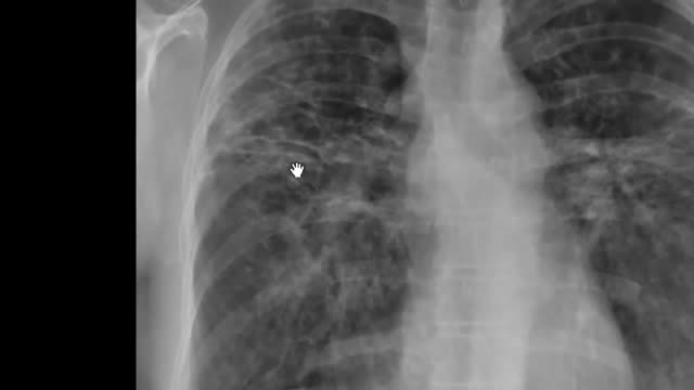
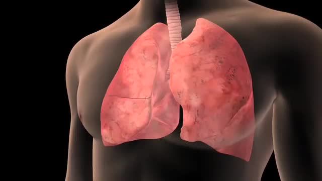
Pulmonary fibrosis is a lung disease that occurs when lung tissue becomes damaged and scarred. This thickened, stiff tissue makes it more difficult for your lungs to work properly. As pulmonary fibrosis worsens, you become progressively more short of breath. The scarring associated with pulmonary fibrosis can be caused by a multitude of factors. But in most cases, doctors can't pinpoint what's causing the problem. When a cause can't be found, the condition is termed idiopathic pulmonary fibrosis. The lung damage caused by pulmonary fibrosis can't be repaired, but medications and therapies can sometimes help ease symptoms and improve quality of life. For some people, a lung transplant might be appropriate.

First Aid for a suspected Fracture
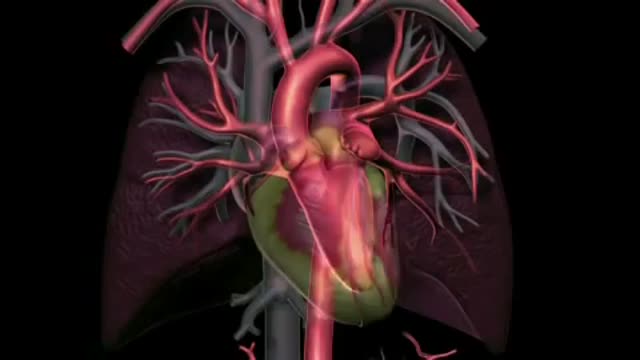
When the arteries in your heart become blocked, the condition is called coronary artery disease. It can be a serious condition if not treated. Coronary artery disease puts you at risk for a heart attack. Be sure you pay attention to your symptoms and manage your heart health risks.

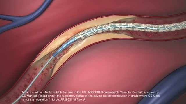
If you have a blocked artery, your doctor may need to open the blockage and restore blood flow using a small mesh tube called a stent. The stent is inserted in your artery during an angioplasty procedure. Until now, stents were permanent. Now there is a fully dissolving stent available to treat blockages.
An arteriovenous fistula is an abnormal connection or passageway between an artery and a vein. It may be congenital, surgically created for hemodialysis treatments, or acquired due to pathologic process, such as trauma or erosion of an arterial aneurysm.
Prostatitis is an infection or inflammation of the prostate gland that presents as several syndromes with varying clinical features. The term prostatitis is defined as microscopic inflammation of the tissue of the prostate gland and is a diagnosis that spans a broad range of clinical conditions. The National Institutes of Health (NIH) has recognized and defined a classification system for prostatitis in 1999.[1] The 4 syndromes of prostatitis are as follows: I - Acute bacterial prostatitis II - Chronic bacterial prostatitis III - Chronic prostatitis and chronic pelvic pain syndrome (CPPS; further classified as inflammatory or noninflammatory) IV - Asymptomatic inflammatory prostatitis
Lung cancer is a type of cancer that begins in the lungs. Your lungs are two spongy organs in your chest that take in oxygen when you inhale and release carbon dioxide when you exhale. Lung cancer is the leading cause of cancer deaths in the United States, among both men and women. Lung cancer claims more lives each year than do colon, prostate, ovarian and breast cancers combined. People who smoke have the greatest risk of lung cancer. The risk of lung cancer increases with the length of time and number of cigarettes you've smoked. If you quit smoking, even after smoking for many years, you can significantly reduce your chances of developing lung cancer.

can i get pregnant right after my period
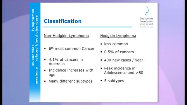
For most cancers, researchers are still trying to understand how they are caused. The same is true for lymphoma - doctors do not know what causes it, although it is more likely to occur in certain people.5,7,8 Medical researchers have identified certain risk factors that make lymphoma more likely, although they often do not understand why:5,8 Non-Hodgkin's lymphoma Age - most non-Hodgkin lymphomas are in people 60 years of age and over Sex - there are different rates of different types of non-Hodgkin's lymphoma across the sexes Ethnicity and location - in the US, African-Americans and Asian-Americans are less prone than white Americans, and the disease is more common in developed nations of the world Chemicals and radiation - some chemicals used in agriculture have been linked, as has nuclear radiation exposure Immune deficiency - for example, caused by HIV infection or in organ transplantation Autoimmune disease, in which the immune system attacks the body's own cells Infection - certain viral and bacterial infections increase the risk. The Helicobacter Infection has been implicated, as has the Epstein Barr Virus (the virus that causes glandular fever)13 See the American Cancer Society's page for more detail on risk factors for non-Hodgkin's lymphoma. Hodgkin's lymphoma Infectious mononucleosis - infection with Epstein-Barr virus Age - two specific groups are most affected: typically people in their 20s, and people over the age of 55 years Sex - slightly more common in men Location - most common in the US, Canada and northern Europe; least common in Asia Family - if a sibling has the condition, the risk is slightly higher, and very high if there is an identical twin Affluence - people from higher socioeconomic status are at greater risk HIV infection
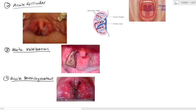
Tonsillitis is inflammation of the tonsils, two oval-shaped pads of tissue at the back of the throat — one tonsil on each side. Signs and symptoms of tonsillitis include swollen tonsils, sore throat, difficulty swallowing and tender lymph nodes on the sides of the neck. Most cases of tonsillitis are caused by infection with a common virus, but bacterial infections also may cause tonsillitis. Because appropriate treatment for tonsillitis depends on the cause, it's important to get a prompt and accurate diagnosis. Surgery to remove tonsils, once a common procedure to treat tonsillitis, is usually performed only when bacterial tonsillitis occurs frequently, doesn't respond to other treatments or causes serious complications.
An increased prevalence of cardiovascular disease (CVD) has been found in women of childbearing age,[1] with the presence of CVD in pregnant women posing a difficult clinical scenario in which the responsibility of the treating physician extends to the unborn fetus. Profound changes occur in the maternal circulation that have the potential to adversely affect maternal and fetal health, especially in the presence of underlying heart conditions. Up to 4% of pregnancies may have cardiovascular complications despite no known prior disease. The European Society of Cardiology has published guidelines on the management of cardiovascular disease during pregnancy.[
Honeymoon palsy from another individual sleeping on and compressing one's arm overnight. Saturday night palsy from falling asleep with one's arm hanging over the arm rest of a chair, compressing the radial nerve

Foot drop is a gait abnormality in which the dropping of the forefoot happens due to weakness, irritation or damage to the common fibular nerve including the sciatic nerve, or paralysis of the muscles in the anterior portion of the lower leg. It is usually a symptom of a greater problem, not a disease in itself.

Acute kidney failure occurs when your kidneys suddenly become unable to filter waste products from your blood. When your kidneys lose their filtering ability, dangerous levels of wastes may accumulate, and your blood's chemical makeup may get out of balance. Acute kidney failure — also called acute renal failure or acute kidney injury — develops rapidly over a few hours or a few days. Acute kidney failure is most common in people who are already hospitalized, particularly in critically ill people who need intensive care. Acute kidney failure can be fatal and requires intensive treatment. However, acute kidney failure may be reversible. If you're otherwise in good health, you may recover normal or nearly normal kidney function.
