- Physical Examination
- Surgical Examination
- Ophthalmology
- Clinical Skills
- Orthopedics
- Surgery Videos
- Laparoscopy
- Pediatrics
- Funny Videos
- Cardiothoracic Surgery
- Nursing Videos
- Plastic Surgery
- Otorhinolaryngology
- Histology and Histopathology
- Neurosurgery
- Dermatology
- Pediatric Surgery
- Urology
- Dentistry
- Oncology and Cancers
- Anatomy Videos
- Health and Fitness
- Radiology
- Anaesthesia
- Physical Therapy
- Pharmacology
- Interventional Radiology
- Cardiology
- Endocrinology
- Gynecology
- Emergency Medicine
- Psychiatry and Psychology
- Childbirth Videos
- General Medical Videos
- Nephrology
- Physiology
- Diet and Food Health
- Diabetes Mellitus
- Neurology
- Women Health
- Osteoporosis
- Gastroenterology
- Pulmonology
- Hematology
- Rheumatology
- Toxicology
- Nuclear Medicine
- Infectious Diseases
- Vascular Disease
- Reproductive Health
- Burns and Wound Healing
- Other
Top videos

If you are tired of dealing with glasses or squinting to read signs in the distance, then you should consider LASIK Eye Surgery. In this outpatient refractive procedure, lasers are used to correct vision issues by changing the structure of the cornea. This may entirely eliminate reliance upon glasses or contacts. In this interactive LASIK Eye Surgery, you will assist in numbing the patient’s eye and cleaning the area for the procedure. With a speculum, you will hold the eye open, mark the cornea using a water-soluble ink, then attach a suction ring to it. After that, a specialized blade device is used to cut into the corneal flap and peel it back so that the laser can clear away corneal tissue underneath. This process corrects the shape of the cornea in less than a minute before putting the corneal flap back in place. After the procedure, we will go over LASIK Eye Surgery recovery instructions. Scrub in and let’s get started!

The most reliable clinical sign to detect ascites is checking for bilateral flank dullness. If a patient with ascites is lying supine, fluid accumulates in the flank regions, leading to dullness on percussion. At the same time, the air-filled bowel loops are forced upwards by the free fluid due to buoyancy, resulting in tympanitic percussion. To locate specifically where dullness shifts to tympany, or the air-fluid level, percussion should be performed from the sides towards the middle. To confirm that the dullness is caused by ascites, ask the patient to switch to a lateral decubitus position. If ascites is present, the air-filled bowel loops will shift accordingly and remain at the surface of the fluid. As a result, the air-fluid level will shift as well. This is known as shifting dullness.
Subscribe to AMBOSS YouTube for the latest clinical examination videos, medical student interviews, study tips and tricks, and live webinars!
Free 5 Day Trial: https://go.amboss.com/amboss-YT
Instagram: https://www.instagram.com/amboss_med/
Facebook: https://www.facebook.com/AMBOSS.Med/
Twitter: https://twitter.com/ambossmed
Blog: https://blog.amboss.com/us
#AMBOSSMed #ClinicalExamination
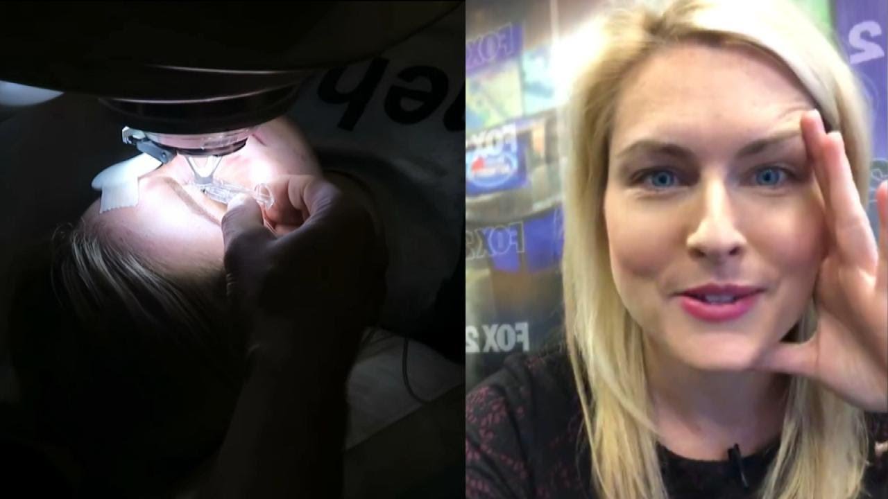
Detroit TV meteorologist Jessica Starr posted a heart-wrenching video on social media a month before dying by suicide this week. She had told viewers she was struggling in the aftermath of undergoing Lasik surgery. After learning of her death, her heartbroken colleagues on WJBK fought back tears live on TV. Twelve people have died by suicide after suffering pain and even blindness after the operation. Inside Edition also spoke to a doctor who wants the surgery banned. #InsideEdition
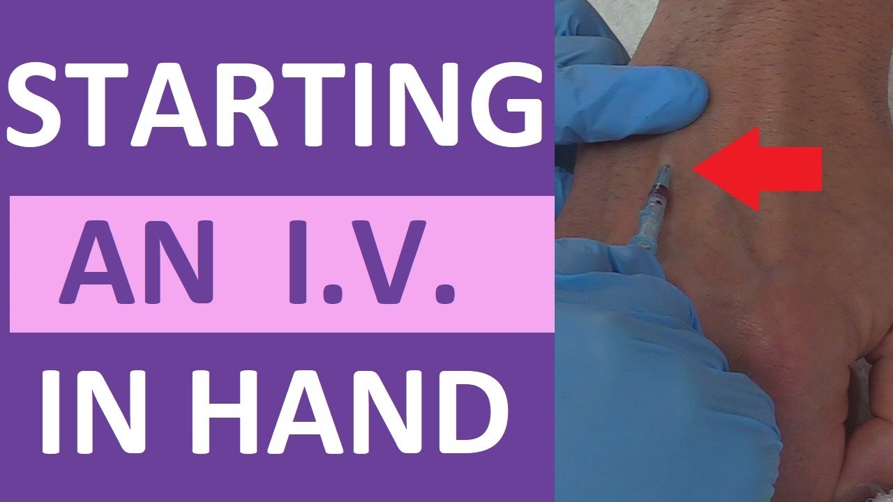
How to start a peripheral IV in the dorsum of the hand: clinical nursing skill technique.
Starting an IV (intravenous catheter) can be an intimidating experience for nurses, especially nursing students and new nurses. However, nurses will perform IV insertions often, so this is an important nursing skill to learn.
Before starting an IV, always follow the protocols of your facility, as well as manufacturer's instructions for any supplies used.
In this video, Nurse Sarah demonstrates how to start a peripheral IV in the dorsum of the hand. Prior to inserting the IV, you'll want to do the following:
-Gather supplies
-Perform hand hygiene
-Prepare supplies (including priming the saline flush, removing air from extension tubing, opening packages, completing labels, and any other steps required by your facility.
-Locate a suitable vein
-Perform hand hygiene
-Don gloves
If the patient has a lot of hair, you might want to use clippers to trim the hairs prior to starting the IV. You may also apply a tourniquet to help veins move near the surface of the skin.
Next, you'll want to clean the site using the cleaner that came in the IV start kit, such as ChloraPrep.
Once the site has dried completely, you can insert the IV. Stabilize the vein with your non-dominant hand, and insert the IV's needle into the vein, watching carefully for blood return (or a blood flash) in the chamber. Advance the IV around 2mm more to ensure the plastic cannula is in the vein, then thread the cannula into the vein and press the needle safety button.
Notes: https://www.registerednursern.....com/how-to-start-an-
IV Video Series: https://www.youtube.com/watch?v=MbG_1-_mnoo&list=PLQrdx7rRsKfXr6kruqEpIovf66sxo0gxh
This video also demonstrates how to flush the IV using the push-pause method, how to secure the IV using the Tegaderm dressing that came with the IV start kit, considerations of the different cap types and the clamp sequence, and more.
For more information, watch the complete tutorial.
#nurse #nursing #iv #startiv #ivtherapy
Website: https://www.registerednursern.com/
More Videos: https://www.youtube.com/watch?v=R2XMro13dD0&list=UUPyMN8DzkFl2__xnTEiGZ1w
Nursing Gear: https://teespring.com/stores/registerednursern
Instagram: https://www.instagram.com/registerednursern_com/
Facebook: https://www.facebook.com/RegisteredNurseRNs
Twitter: https://twitter.com/NursesRN
Popular Playlists:
NCLEX Reviews: https://www.youtube.com/playli....st?list=PLQrdx7rRsKf
Fluid & Electrolytes: https://www.youtube.com/playli....st?list=PLQrdx7rRsKf
Nursing Skills: https://www.youtube.com/playli....st?list=PLQrdx7rRsKf

http://www.amerra.com In this patient education video from Colorectal Surgical Associates in Houston, Texas, learn more about the single incision laparoscopic colectomy procedure. This minimally invasive procedure uses a mini incision that
results in less pain, fewer complications, earlier recovery, and a smaller scar. Colorectal cancer is the second leading cause of cancer death in the United States. For more information please visit our website: www.csamd.com or call (713)-790-0600.

#dialysis #uvahealth
If your kidney function is declining and medications and other treatments aren’t working, dialysis can offer life-saving care. UVA has one of the largest dialysis programs in the country. Nephrologist Daphne Knicely, MD, explains the types of home dialysis and how they can work to fit your life.
Find out more at: https://uvahealth.com/services/dialysis
Transcript
Dialysis is just a way to replace the kidneys when they're not working anymore. So when the kidneys stop working, they stop getting rid of water, stop balancing the chemistry, stop getting rid of the toxins. Then dialysis does its job by balancing the chemistries, getting rid of the toxins, and help remove fluid. It doesn't fix the kidneys. It just replaces them.
I usually think of dialysis as two components. There's hemodialysis and peritoneal dialysis. So peritoneal dialysis can only be done at home. Hemodialysis can be done in a center, or it can be done at home.
Hemodialysis is where you have some sort of access to the blood. Either some sort of shunt in the arm that connects an artery and vein, or a catheter. And it allows for blood to leave you, go through a machine, get cleaned, chemistries balanced, and then comes back to you.
For home hemodialysis, the patient actually learns how to do that treatment. It's a very simple machine, very user-friendly. Training is usually about anywhere from four weeks up to eight weeks, and you work one-on-one with a nurse. You still see the physician. You come in about once a month, maybe twice a month, to get labs. You'll see a social worker, and a nutritionist at the same time.
Peritoneal dialysis takes place by putting a tube into your abdomen. And we take dialysis fluid that's chemically balanced. When we put it into the abdomen, it uses those little blood vessels to pull toxins out, to balance chemistries, kind of like little filters. Now, after it sits in there for several hours, we drain it out.
Anyone that needs dialysis is a candidate for home dialysis. There's not one type of dialysis that's going to make you live longer. They're all equal. The goal is to pick the type of dialysis that fits with your life.
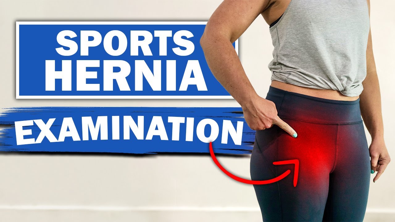
We will show you what a sports hernia examination (aka athletic pubalgia, gilmore's groin, lower abdominal pain) and rule out a diagnosis of hip impingement. Rehab exercises are suggested based on the results.
If you're experiencing any of these symptoms, don't hesitate to schedule a sports hernia examination. I can help you determine the best treatment plan to promote your recovery and avoid future injury. Subscribe to my channel to stay updated on the latest medical news and tips!
If you would like to know more about sports hernias and other diagnoses for front of hip, groin, adductor and lower abdominal strain, watch our detailed webinar here: https://bit.ly/37thtNF
For treatment, come visit us or schedule a virtual session. www.p2sportscare.com
Costa Mesa CA 715-502-4243
#sportshernia #abdominal #hippain
Sports Hernia Diagnosis
What Is A Sports Hernia?
A sports hernia is tearing of the transversalis fascia of the lower abdominal or groin region. A common misconception is that a sports hernia is the same as a traditional hernia. The mechanism of injury is rapid twisting and change of direction within sports, such as football, basketball, soccer and hockey.
The term “sports hernia” is becoming mainstream with more professional athletes being diagnosed. The following are just to name a few:
Torii Hunter
Tom Brady
Ryan Getzlaf
Julio Jones
Jeremy Shockey
If you follow any of these professional athletes, they all seem to have the same thing in common: Lingering groin pain. If you play fantasy sports, this is a major headache since it seems so minor, but it can land a player on Injury Reserve on a moments notice. In real life, it is a very frustrating condition to say the least. It is hard to pin point, goes away with rest and comes back after activity, but is hardly painful enough to make you want to stop. It lingers and is always on your mind. And if you’re looking for my step-by-step sports hernia rehab video course here it is.
One the best definitions of Sport hernias is the following by Harmon:
The phenomena of chronic activity–related groin pain that it is unresponsive to conservative therapy and significantly improves with surgical repair.”
This is truly how sports hernias behave in a clinical setting. It is not uncommon for a sports hernia to be unrecognized for months and even years. Unlike your typical sports injury, most sports medicine offices have only seen a handful of cases. It’s just not on most doctors’ radar. The purpose of this article is not only to bring awareness about sports hernias, but also to educate.
Will you find quick fixes in this article for sports hernia rehab?
Nope. There is no quick fix for this condition, and if someone is trying to sell you one, they are blowing smoke up your you-know-what.
Is there a way to decrease the pain related to sports hernias?
Yes. Proper rehab and avoidance of activity for a certain period of time will assist greatly, but this will not always stop it from coming back. Pain is the first thing to go and last thing to come. Do not be fooled when you become pain-free by resting it. Pain is only one measure of improvement in your rehab. Strength, change of direction, balance and power (just to name a few) are important, since you obviously desire to play your sport again. If you wanted to be a couch potato, you would be feeling better in no time. Watching Sports Center doesn’t require any movement.
Why is this article so long?
There is a lot of information on sports hernias available to you on the web. However, much of the information is spread out all over the internet and hard for athletes to digest due to complicated terminology. This article lays out the foundational terminology you will need to understand what options you have with your injury. We will go over anatomy, biomechanics, rehab, surgery, and even the fun facts. The information I am using is from the last ten years of medical research, up until 2016. We will be making updates overtime when something new is found as well. So link to this page and share with friends. This is the best source for information on sports hernias you will find.
Common Names (or Aliases?) for Sports Hernias
Sportsman’s Hernia
Athletic Pubalgia
Gilmore’s Groin
How Do You Know If You Have A Sports Hernia?
Typical athlete characteristics:
Male, age mid-20s
Common sports: soccer, hockey, tennis, football, field hockey
Motions involved: cutting, pivoting, kicking and sharp turns
Gradual onset
How A Sports Hernia Develops
Chronic groin pain typically happens over time, which is why with sports hernias, we do not hear many stories of feeling a “pop” or a specific moment of injury. It is the result of “overuse” mechanics stemming from a combination of inadequate strength and endurance, lack of dynamic control, movement pattern abnormalities, and discoordination of motion in the groin area.
#SPORTSHERNIAEXAM #california

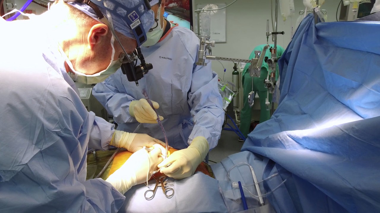
Dr. Ailawadi, M.D., the Chair of Cardiac Surgery at Michigan Medicine, specializes in minimally invasive valve surgery as well as complex cardiac operations. This video shows step by step footage of a Coronary Artery Bypass Graft (CABG) in a complex patient. In this case, CABG was performed through a sternotomy (through the breast bone) using the internal thoracic artery and saphenous leg veins to bypass obstructed coronary arteries. In this video, Dr. Ailawadi will perform a triple vessel bypass (CABG) which has been shown to minimize the risk of future heart attack and help patients live longer in the setting of complex coronary artery disease.
To learn more about cardiac surgery at Michigan Medicine, visit: https://medicine.umich.edu/dept/cardiac-surgery
To learn more about Frankel Cardiovascular Center, visit: https://www.umcvc.org/
To watch the full playlist, visit: https://www.youtube.com/playli....st?list=PLNxqP-XbH8B
-------------------------------------------------------
Subscribe to Michigan Medicine’s YouTube channel for upcoming videos and future live streams featuring our experts answering your questions.
-------------------------------------------------------
Follow Michigan Medicine on Social:
Twitter: https://twitter.com/umichmedicine
Instagram: https://www.instagram.com/umichmedicine/
Facebook: https://www.facebook.com/MichiganMedicine/
Follow the U-M Frankel Cardiovascular Center on Social:
Twitter: https://twitter.com/umichcvc
Facebook: https://www.facebook.com/Unive....rsityofMichiganCardi
#MichiganMedicine #MedEd #CardiacSurgery #UniversityOfMichiganHealth #FrankelCardiovascularCenter #Cardiology #CardiacSurgeon

Ellis demonstrates how to clean a reusable inner cannula, care for a tracheostomy site, and suction a tracheostomy.
Our Critical Nursing Skills video tutorial series is taught by Ellis Parker MSN, RN-BC, CNE, CHS and intended to help RN and PN nursing students study for your nursing school exams, including the ATI, HESI and NCLEX.
#ClinicalSkills #NCLEX #tracheostomy #patientcare #ATI #Kaplan #LVN #PN #RN #nurseeducator #nurse #nursingstudent #murse #clinicals #clinicalnursingskills
00:00 What to expect Tracheostomy Care and Suctioning
0:33 Explaining the process Tracheostomy Care and Suctioning
1:10 Positioning patient for a Tracheostomy Care and Suctioning
1:33 Opening tray
1:46 Pouring saline
1:58 Removing inner cannula
2:14 Removing clean gloves
2:25 Donning sterile gloves
3:16 Showing tray contents
3:53 Removing previous dressing
4:06 Pouring saline
4:27 Cleaning stoma
5:10 Cleaning faceplate
5:20 Drying site
5:30 Cleaning inner cannula
6:00 Drying inner cannula
6:20 Reinserting inner cannula
6:40 Placing new gauze
7:00 Replacing ties
8:00 Replacing oxygen
8:13 Preparing for suction
8:58 Checking suction
9:30 Opening saline
9:42 Opening kit
9:58 Donning sterile gloves
11:04 Setting up saline container
11:20 Pouring saline
11:52 Connecting catheter to suction
12:46 Inserting catheter
13:10 Removing catheter
13:24 Rinsing catheter
13:40 Reoxyginating
14:05 Reinserting catheter
14:17 Removing catheter
14:29 Rinsing catheter
14:44 Reoxyginating
14:55 Cleaning up
15:09 Chatting about sterility
17:00 Checking a tie
🚨 Reminder: shipping deadlines are looming 👀
🎁 Regular Shipping: Order by Friday, December 15
🚀 Expedited Shipping: Order by Monday, December 18
🔍 Still searching for last-minute gifts? Consider a Level Up RN Gift Card! 💌 It’s not only a thoughtful present but also the perfect way to share treasures like Pharmacology Flashcards OR digital treasures like Flashables Digital Nursing Flashcards & the Level Up RN membership. Give the gift of knowledge this holiday season! 🧠⚡️💖 bit.ly/LevelUpRNGC
🚪 Access our Cram Courses, Quizzes and Videos all in one ad free space with Level Up RN Membership https://bit.ly/LevelUpRNMembership
Want more ways to MASTER Clinical Skills? Check out our flashcards & videos!
👇👇👇👇👇👇👇👇👇👇
👉 https://bit.ly/clinicalnursingskills 👈
☝️👆☝️👆☝️👆☝️👆☝️👆
This is your one-stop-shop for materials to help you LEARN & REVIEW so you can PASS Nursing School.
🤔🤔🤔 DO YOU WANT TO PASS your classes, proctored exams and the NCLEX? 🤔🤔🤔 Our resources are the best you can buy. They are built with a single goal: help you pass with no fluff. Everything you need, and nothing you don’t. Don’t take our word for it, though! Check out our hundreds of ⭐️⭐️⭐️⭐️⭐️ reviews from nurses who passed their exams and the NCLEX with Level Up RN.
🗂️ Our Ultimate Nursing School Survival kit is your number 1 resource to get through nursing school and to pass the NCLEX. Whether you're just starting school or you’re already prepping for the NCLEX, this bundle of flashcards is the best you can buy. It covers all the information you need to know to pass all your exams and it has FREE shipping!
➡️ https://bit.ly/TUNSSK ⬅️
L👀king for EVEN MORE resources to survive Nursing School? Make your Nursing School experience your own! Life’s difficult enough—learning shouldn’t be.
🪅 Games https://nursesquad.com
💻 Digital resources https://bit.ly/NursingStudyCourses
📅 Organizational tools https://bit.ly/OrganizingSchool
✨Want perks? Join our channel!
https://youtube.com/leveluprn/join
🏷 Head to https://leveluprn.com/specials for all our latest deals!🥳️
📧 LOOKING FOR FREE RESOURCES TO HELP WITH YOUR EXAMS? Get exclusive tips, latest video releases and more delivered to your email!
➡️ https://leveluprn.com/signup ⬅️
⚕ 👩 LEVEL UP NURSE SQUAD 👩⚕️
All of the nurses at Level Up RN are here to help! Cathy Parkes started helping her fellow classmates back when she was in nursing school, tutoring so they could pass their exams and graduate. After she got her BSN and started working as an RN at Scripps Encinitas Hospital, she started this YouTube channel to help nursing students around the world. Since then she has built a team of top-notch dedicated nurses and nurse educators who are focused on improving nursing education and supporting career advancement for nurses everywhere. With flashcards, videos, courses, organizational tools and more, we are singularly focused on helping students and nurses Level Up on their exams and nursing careers.

It’s called gamma knife surgery, but there’s no cutting involved.
It’s been used at Mayo Clinic for 30 years as an alternative to open brain surgery.
The patient’s head is held still during the procedure with a headframe, which also serves as a map for the radiation. Using 3D imaging — typically an MRI — as a guide, the gamma knife is targeted directly at the tumor.
And with no hospital stay and minimal side effects, it’s a procedure that is efficient and can be lifesaving.
More health and medical news on the Mayo Clinic News Network. https://newsnetwork.mayoclinic.org/
Journalists: Clean and nat sound versions of this pkg available for download at https://newsnetwork.mayoclinic.org/
Register (free) at https://newsnetwork.mayoclinic.org/request-account/

Jennifer Lawton, M.D., is professor and chief of the Johns Hopkins Division of Cardiac Surgery, as well as director of the Cardiac Surgery Research Laboratory and program director of the cardiothoracic fellowship training program at Johns Hopkins. Her areas of expertise include valve surgery, including minimally invasive surgery, coronary artery bypass grafting on- and off-pump, all arterial revascularization, as well as surgery for aortic dissection and ascending aneurysm. For more information about Dr. Lawton visit http://www.hopkinsmedicine.org..../heart_vascular_inst

A brain surgery called a craniectomy is performed to remove a part of your skull in order to relieve pressure in an area when your brain swells from a traumatic brain injury. It is also performed to treat medical conditions that cause your brain to swell or bleed that can be caused by an aneurysm, brain tumor or other cancer.
This 3d animation shows how the surgical procedure decreases intracranial pressure (ICP), intracranial hypertension (ICHT), or heavy bleeding (also called hemorrhaging) inside your skull. If left untreated, pressure or bleeding can compress your brain and push it down onto the brain stem. This can be fatal or cause permanent brain damage.
Brain surgery is a very serious procedure under any circumstances, but a craniectomy is done when there is an immediate risk to the brain and neurological function due to severe brain injury or stroke.
For more information about custom 3D animation depicting surgery, please visit https://www.amerra.com/.
Watch additional medical animations:
Accessing an implantable port training - 3D animation: https://youtu.be/xSTpxjyv4O4
Open Suctioning with a Tracheostomy Tube - 3D animation: https://youtu.be/wamB7jpWCiQ
Ventriculostomy Brain Surgery - 3d animation: https://youtu.be/pUy0YDzVNzs
Suctioning the endotracheal tube - medical animation: https://youtu.be/pN6-EYoeh3g
Functional endoscopic sinus surgery (FESS) - 3D animation: https://youtu.be/qKTRyowwaLA
How to insert a nasogastric tube for NG intubation - 3d animation: https://youtu.be/Abf3Gd6AaZQ
Oral airway insertion - oropharyngeal airway technique - 3D animation: https://youtu.be/caxUdNwjt34
Nasotracheal suctioning (NTS) - 3D animation: https://youtu.be/979jWMsF62c
Learn about hemorrhoids with #3d #animation: https://youtu.be/R6NqlMpsiiY
LASIK eye surgery - 3D animation: https://youtu.be/Bb8bnjnEM00
CPR cardiopulmonary resuscitation - 3D animation: https://youtu.be/G87knTZnhks
What are warts (HPV)? - 3D animation: https://youtu.be/guJ1J7rRs1w
How Macular Degeneration Affects Your Vision - 3D animation: https://youtu.be/ozZQIZ_52YY
NeoGraft hair transplant procedure – animation: https://youtu.be/C-eTdH2UPXI

http://www.highimpact.com - This brain surgery animation was used to demonstrate a young girl's craniotomy, cranioplasty, and reconstructive skull surgery after her vehicle was struck by a tractor-trailer. The procedures included the evacuation of a large epidural hematoma, the draining of the epidural space, and the reassembly of bone fragments to repair the skull.
More Brain Surgery Animations: https://tinyurl.com/y6m4lkdf
WHAT HAPPENED
A teenage girl was riding home with her parents and boyfriend from a Wednesday night church service when a tractor-trailer struck the back driver’s side of their car as they were traveling through an intersection. The impact sent the car spinning into oncoming traffic where it struck another vehicle. When paramedics arrived, the 17-year-old was unresponsive with bleeding from her left ear and a laceration from behind her left ear.
She was rushed to the hospital where she underwent a series of CT scans that showed a severely comminuted open skull fracture with an underlying 1.1 cm subdural hematoma. She was taken to the operating room where an emergency craniotomy was performed to evacuate the hematoma and reassemble the skull fragments. The patient gradually began to wake up and was discharged six days later, after she showed she could maneuver up and down the hallway.
The biggest challenge in a traumatic brain injury case like this - where most of the damages are deeply underlying and undetectable on the surface - is that the only visual evidence is in the form of 2D black-and-white radiographic films. This can look ambiguous to the typical juror because it’s often difficult to discern where these snapshots are located inside the person’s skull. Tony Seaton, Esq., and Robert Bates, Esq., needed to reinforce this 2D radiographic evidence with maximum 3D context.
We equipped them with a custom Diagnostic Slice Chooser: an interactive presentation that presents radiographic slides within a three-dimensional model of the patient’s head. We also designed the model accurately to the patient’s likeness and colorized the films to highlight key areas of damage. The attorneys could show the complete depth and magnitude of his client’s injuries at every level both before and after the surgery. After establishing the full extent of damages, we also created an animation to walk viewers through the surgical experience the patient would undergo as a result of her injuries.
The visual presentation helped jurors understand the destructive impact this collision had on this young teenager’s life, and Mr. Seaton and Mr. Bates, Esq., were able to acquire a $4.5M settlement for his client.
Read the Full Case Study: https://tinyurl.com/yy4v2dyh

http://www.nucleushealth.com/ - This 3D medical animation depicts two operations, called craniotomy and craniectomy, in which the skull is opened to access the brain. The normal anatomy of the skull and tissues surrounding the brain are shown, including arteries and veins. The animation lists the common reasons for these procedures, and briefly introduces intracranial pressure.
Video ID: ANH13109
Transcript:
Your doctor may recommend a craniotomy or a craniectomy procedure to treat a number of different brain diseases, injuries, or conditions.
Your skull is made of bone and serves as a hard, protective covering for your brain. Just inside your skull, three layers of tissue, called meninges, surround your brain. The thick, outermost layer is the dura mater. The middle tissue layer is the arachnoid mater and the innermost layer is the pia mater. Between the arachnoid mater and the pia mater is the subarachnoid space, which contains blood vessels and a clear fluid called cerebrospinal fluid. Blood vessels, called bridging veins, connect the surface of your brain with the dura mater. Other blood vessels, called cerebral arteries, bring blood to your brain.
Inside your skull, normal brain function requires a delicate balance of pressure between the blood in your blood vessels, the cerebrospinal fluid that surrounds your brain, and your brain tissue. This is called normal intracranial pressure. Increased intracranial pressure may result from: brain tumors, head injuries, problems with your blood vessels, or infections in your brain or spinal cord. These conditions put pressure on your brain and may cause it to swell or change shape inside your skull, which can lead to serious brain injury.
Your doctor may recommend a craniotomy to remove: abnormal brain tissue, such as a brain tumor, a sample of tissue by biopsy, a blood clot, called a hematoma, excess cerebrospinal fluid, or pus from an infection, called an abscess.
A craniotomy may also be done to: relieve brain swelling,
stop bleeding, called a hemorrhage, repair abnormal blood vessels, repair skull fractures, or repair damaged meninges.
Finally, a craniotomy may also be done to: treat brain conditions, such as epilepsy, deliver medication to your brain, or implant a medical device, such as a deep brain stimulator.
The most common reason for a craniotomy is to remove a brain tumor.
#Craniotomy #Craniectomy #BrainSurgery

For more than 25 years, The Children's Hospital of Philadelphia — the first Level 1 Pediatric Trauma Center in Pennsylvania — has provided unparalleled medical and surgical care for all injured children, including those with the most severe injuries.
Learn what makes the Trauma Center at CHOP a Level 1 Pediatric Trauma Center, and how our work toward trauma prevention, research advances and overall trauma awareness provides hope for reduced injuries in the future.
Learn more about the Trauma Center at CHOP: http://www.chop.edu/trauma.
Dr. Joseph McGinn explains minimally invasive bypass, the procedure he pioneered as an alternative to open heart surgery.

A tummy tuck is a surgical process that removes excess fat and skin. Learn more about the procedure by watching this video!
Looking to book a consultation? Call Zuri Plastic Surgery now at 786-804-1603 or DM us today to schedule a complimentary consultation with Dr. Z.
Un tummy tuck es un procedimiento quirúrgico que elimina el exceso de grasa y piel. ¡Aprenda más sobre este procedimiento viendo este video!
¿Quiere agendar una consulta? Llame a Zuri Plastic Surgery ahora al 786-804-1603 o envíenos un DM hoy para programar una consulta gratuita con el Dr. Z.

Neurosurgeon Sujit Prabhu, M.D., discusses what happens after surgery and how a patient recovers.
Learn more: http://www.mdanderson.org/educ....ation-and-research/d
Request an appointment at MD Anderson by calling 1-877-632-6789 or online: https://my.mdanderson.org/requestappointment

#abdomenliposuction #laserskintightening #drprashantyadav #cosmeticsurgery #plasticsurgery #dezireclinicindia #weightloss #shorts #360degreeabdomenliposuction #lowerbackliposuction
Weight Loss After 360° Abdomen liposuction result, Abdomen Liposuction, lower back liposuction, 360 degree abdomen liposuction
☎️ For more info:
WhatsApp Your Details to know the Cost
Delhi - 8956880644, 9717470550, Pune - 9222122122, Bangalore- 8971224700, Gurugram - 9272007896, Ahmedabad - 9711162746
Why choose Dezire Clinic For Your Cosmetic and plastic surgery treatment ?
Dezire Clinic is a top searched clinic surgical and nonsurgical cosmetic procedure in India when comes to “Cosmetic, Skin ,Laser and Hair transplantation”.
Like and Share the video if you find it useful. Do not forget to Subscribe to our channel to get more updates.
Subscribe on YouTube https://youtube.com/dezireclin....ic?sub_confirmation=
https://youtube.com/dezireplas....ticsurgerycenter?sub
🎦 https://www.youtube.com/dezireclinic
🎦 https://www.youtube.com/DezirePlasticSurgeryCenter
👍🏻 https://www.facebook.com/drprashantmch/
👍🏻 https://www.facebook.com/dezireclinic
📸 https://www.instagram.com/drprashantdezireclinic/
📸 https://www.instagram.com/dezireclinics/
🐥 https://twitter.com/drprashantmch
👍🏻 https://www.linkedin.com/in/drprashantyadav/
🌐 Website: https://www.dezireclinic.in/
📧 dezireclinicindia@gmail.com
📧 info@dezireclinic.in
Dr. Prashant Yadav (M.S., M.Ch. Plastic Surgery ) & Founder of Dezire Clinic
Disclaimer: The content of this channel is for informational and educational purposes only. This content should not be considered a substitute for advice provided by a certified plastic or cosmetic surgeon. Patients must be properly diagnosed by a healthcare professional on an individual basis in order to achieve the desired results. There is no guarantee of getting the results and outcomes shown in videos, as the results can vary at the end. We will not be held liable for any harm caused by someone misusing our name.
#plasticsurgery #cosmeticsurgery #dezireclinic #drprashantyadav
🔥 Multivitamins for Men: https://lynxshort.com/Multivitamins-for-Men
✨ Multivitamins for Women: https://lynxshort.com/Multivitamins-for-Women
⭐ Multivitamins for Kids: https://lynxshort.com/Multivitamins-for-Kids
📙 Book of the Day 📚 https://lynxshort.com/Book-of-the-Day
This is one of the most interesting medical topics to discuss. What are the responsibilities of a doctor? What are the basic skills a doctor needs to have? and what are the responsibilities of a doctor?
**** CONNECT ****
- " Medical Videos " Android application on Google Play store:
https://play.google.com/store/....apps/details?id=com.
https://healthusher.com
https://www.facebook.com/MedicalVideosAnimated
https://www.instagram.com/medical_videos1
👉 Support Us to Help Us Continue Making Videos.. Thanks in Advance :)
- Via PayPal: https://www.paypal.me/medicalvideos
- Via Patreon: https://www.patreon.com/medicalvideosanimated
- The creator:
Pharmacist. Alaa Nasr
#MedicalVideosAnimated
Affiliate Disclaimer: This video and description contains affiliate links, which means that if you click on one of the product links, I'll receive a small commission. This is at no extra cost to you and in many cases include exclusive discounts where applicable. This helps support the channel and allows me to continue to make free videos like this. Thank you for the support!
