
- Physical Examination
- Surgical Examination
- Ophthalmology
- Clinical Skills
- Orthopedics
- Surgery Videos
- Laparoscopy
- Pediatrics
- Funny Videos
- Cardiothoracic Surgery
- Nursing Videos
- Plastic Surgery
- Otorhinolaryngology
- Histology and Histopathology
- Neurosurgery
- Dermatology
- Pediatric Surgery
- Urology
- Dentistry
- Oncology and Cancers
- Anatomy Videos
- Health and Fitness
- Radiology
- Anaesthesia
- Physical Therapy
- Pharmacology
- Interventional Radiology
- Cardiology
- Endocrinology
- Gynecology
- Emergency Medicine
- Psychiatry and Psychology
- Childbirth Videos
- General Medical Videos
- Nephrology
- Physiology
- Diet and Food Health
- Diabetes Mellitus
- Neurology
- Women Health
- Osteoporosis
- Gastroenterology
- Pulmonology
- Hematology
- Rheumatology
- Toxicology
- Nuclear Medicine
- Infectious Diseases
- Vascular Disease
- Reproductive Health
- Burns and Wound Healing
- Other
Top videos
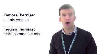

Try our MULTIPLE CHOICE QUESTIONS and WATCH MORE VIDEOS at www.boxmedicine.com!
Inguinal and femoral hernias need not be confusing. In this tutorial you will be presented with colourful diagrams and animations to cover important areas, such as the anatomy of what goes on in these two conditions, the examination of groin hernias and a simple explanation of the difference between incarceration, strangulation and obstruction, in and amongst a systematic look at the clinical topic. More tutorials at www.boxmedicine.com.

If you’ve lost a significant amount of weight, either after pregnancy or through exercise and dietary changes, excess skin and weakened abdominal muscles can leave you self-conscious about your appearance. In this video, Dr. Catherine Hannan and Dr. Lauren Patrick, two of our Board-Certified Plastic Surgeons, are performing a Tummy Tuck (Abdominoplasty) surgery. Tummy Tuck surgery gets rid of the excess skin, as well as tightens your abdominal muscles, resulting in a flatter and smoother abdomen. The results of the surgery are permanent except in cases of large weight gain or pregnancy after surgery.
We are so excited to have taken a part in our patient's body transformation journey!
Before & After Gallery:
https://www.westendplasticsurg....ery.com/surgical/bod
To learn more, visit our website or call (202) 785-4187
http://www.westendplasticsurgery.com
~~~~~~~~~~~~~~~~~~~
Social Media:
✨ Instagram: http://www.instagram.com/westendplasticsurgery
✨ Facebook: http://www.facebook.com/westendplasticsurgery
✨ Twitter: http://www.twitter.com/weplasticsurg
✨ Blog: https://www.westendplasticsurgery.com/blog
✨ Business Inquiries: info@westendplasticsurgery.com
~~~~~~~~~~~~~~~~~~~
#TummyTuck #Abdominoplasty

***************************************************************************
MEDICAL ANIMATION TRANSCRIPT:
Laparoscopic Ovarian Drilling (LOD)
A surgical treatment for women with PCOS
Women with PCOS usually have ovaries with a thick outer layer.
Ovarian drilling works by breaking through the thick outer surface and lowering the amount of testosterone made by the ovaries
A small incision is made in the abdomen.
Carbon dioxide gas is used to inflate the abdomen.
Very small holes are made in the ovaries.
Ovarian drilling can help restore ovulation and improve the chances of becoming pregnant.
***************************************************************************
*TimeStamps*
0:00 Introduction
0:15 Procedure of Laparoscopic Ovarian Drilling (LOD)
***************************************************************************
Let us watch this 3D video to understand what is Laparoscopic Ovarian Drilling for PCOS, why it is done, how well it works, and what to expect.
***************************************************************************
Get credible information on various health topics follow us on:
* Facebook: https://www.facebook.com/eremedium
* Instagram: https://www.instagram.com/eremedium/
* LinkedIn: https://www.linkedin.com/company/13197441/
* Twitter: https://twitter.com/eremedium
***************************************************************************
Disclaimer: Eremedium blogs and videos are for informational purposes only and should not be construed as advice or as a substitute for consulting a physician. It is not a substitute for medical advice or treatment from a healthcare professional.
#pcos #pcostreatment #laparascopicovariandrilling
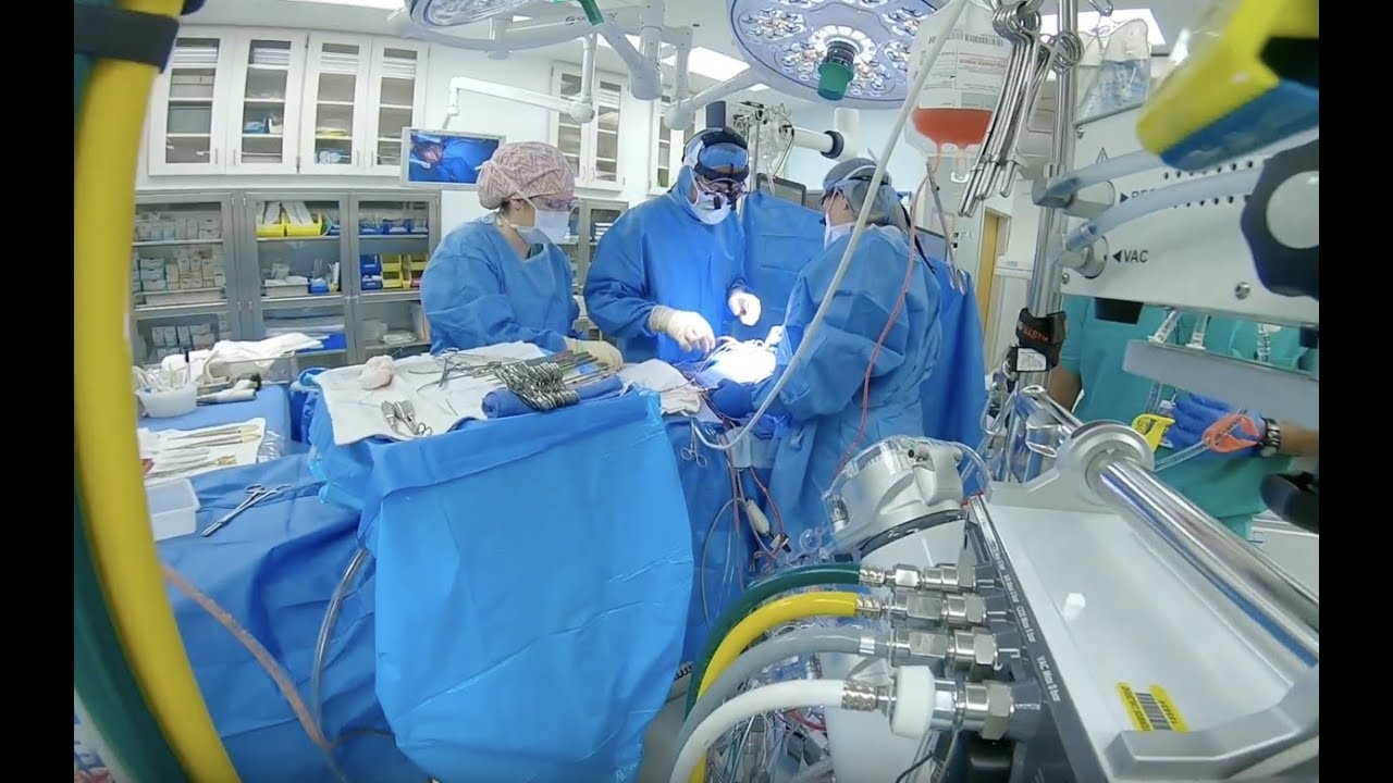
"I’m essentially taking care of the baby right now to give them 60 or 70 or 80 years of life so I have to perform my best every time. Every single time. That is a commitment that I have to the parents."
The highest standard. That’s what cardiothoracic surgeon Sergio Carrillo demands of himself every time he steps into the OR. Dr. Carrillo and his Heart Center team at Nationwide Children’s Hospital treat patients with congenital heart disease with the simplest to the most complex procedures.
Connect with a specialist: http://bit.ly/2LU2kJn
The Heart Center at Nationwide Children's: http://bit.ly/2LTQmPR
Advancing cardiac care through research: http://bit.ly/2LXFqAD
Tissue Engineering Research & Innovation: http://bit.ly/2LUD0Ts
Heart & Chest Surgery, What to Expect: http://bit.ly/2LVQr5J
Meet our Heart Center Team: http://bit.ly/2LUvdF9

Sports Hernia Self Test (TRY IT)
714-502-4243 | Costa Mesa, CA | http://www.p2sportscare.com
[FREE GIFT] Audio Download
#sportshernia #hernia #hippain
Sports Hernia Diagnosis
What Is A Sports Hernia?
A sports hernia is tearing of the transversalis fascia of the lower abdominal or groin region. A common misconception is that a sports hernia is the same as a traditional hernia. The mechanism of injury is rapid twisting and change of direction within sports, such as football, basketball, soccer and hockey.
The term “sports hernia” is becoming mainstream with more professional athletes being diagnosed. The following are just to name a few:
Torii Hunter
Tom Brady
Ryan Getzlaf
Julio Jones
Jeremy Shockey
If you follow any of these professional athletes, they all seem to have the same thing in common: Lingering groin pain. If you play fantasy sports, this is a major headache since it seems so minor, but it can land a player on Injury Reserve on a moments notice. In real life, it is a very frustrating condition to say the least. It is hard to pin point, goes away with rest and comes back after activity, but is hardly painful enough to make you want to stop. It lingers and is always on your mind. And if you’re looking for my step-by-step sports hernia rehab video course here it is.
One the best definitions of Sport hernias is the following by Harmon:
The phenomena of chronic activity–related groin pain that it is unresponsive to conservative therapy and significantly improves with surgical repair.”
This is truly how sports hernias behave in a clinical setting. It is not uncommon for a sports hernia to be unrecognized for months and even years. Unlike your typical sports injury, most sports medicine offices have only seen a handful of cases. It’s just not on most doctors’ radar. The purpose of this article is not only to bring awareness about sports hernias, but also to educate.
Will you find quick fixes in this article for sports hernia rehab?
Nope. There is no quick fix for this condition, and if someone is trying to sell you one, they are blowing smoke up your you-know-what.
Is there a way to decrease the pain related to sports hernias?
Yes. Proper rehab and avoidance of activity for a certain period of time will assist greatly, but this will not always stop it from coming back. Pain is the first thing to go and last thing to come. Do not be fooled when you become pain-free by resting it. Pain is only one measure of improvement in your rehab. Strength, change of direction, balance and power (just to name a few) are important, since you obviously desire to play your sport again. If you wanted to be a couch potato, you would be feeling better in no time. Watching Sports Center doesn’t require any movement.
Why is this article so long?
There is a lot of information on sports hernias available to you on the web. However, much of the information is spread out all over the internet and hard for athletes to digest due to complicated terminology. This article lays out the foundational terminology you will need to understand what options you have with your injury. We will go over anatomy, biomechanics, rehab, surgery, and even the fun facts. The information I am using is from the last ten years of medical research, up until 2016. We will be making updates overtime when something new is found as well. So link to this page and share with friends. This is the best source for information on sports hernias you will find.
Common Names (or Aliases?) for Sports Hernias
Sportsman’s Hernia
Athletic Pubalgia
Gilmore’s Groin
How Do You Know If You Have A Sports Hernia?
Typical athlete characteristics:
Male, age mid-20s
Common sports: soccer, hockey, tennis, football, field hockey
Motions involved: cutting, pivoting, kicking and sharp turns
Gradual onset
How A Sports Hernia Develops
Chronic groin pain typically happens over time, which is why with sports hernias, we do not hear many stories of feeling a “pop” or a specific moment of injury. It is the result of “overuse” mechanics stemming from a combination of inadequate strength and endurance, lack of dynamic control, movement pattern abnormalities, and discoordination of motion in the groin area.
There is a lot going on in the groin area. There are many muscles, tendons, and fascia pulling in different directions. These contracting structures need to coordinate together for any athletic motion. This perspective is also known as the injury prevention model.

Contact us to find out more http://www.londonvisionclinic.com/contact-us/ A video of an actual lasik eye surgery procedure taking place, using VisuMax and MEL 80 at the London Vision Clinic.
Visit http://www.londonvisionclinic.....com/are-you-suitable to find out more about the laser eye surgery process.

Histology of all the different types of cartilage. This video is a part of our Histology Video Course (https://youtube.com/playlist?l....ist=PLnr1l7WuQdDynxT
All Histology Videos: https://youtube.com/playlist?l....ist=PLnr1l7WuQdDynxT
Thank you to our sponsor Doc2Doc Lending, the Personal Lending platform designed for Doctors, by Doctors. Check out https://doc2doclending.com/davinci to learn more today.
Additional YouTube Content
Biochemistry videos: https://youtube.com/playlist?l....ist=PLnr1l7WuQdDzCUC
Anatomy Videos: https://youtube.com/playlist?l....ist=PLnr1l7WuQdDz2dK
DaVinci Cases Videos: https://youtube.com/playlist?l....ist=PLnr1l7WuQdDyJUl
The DaVinci Hour Podcast: https://youtube.com/playlist?l....ist=PLnr1l7WuQdDwSm9
DaVinci Academy Website: https://www.dviacademy.com/

http://www.laparoscopyhospital.com
For the surgeon to develop the same level of proficiency and dexterity in the endoscopic environment as he may possess in open surgery is not a simple matter. The use of proper Mishra's Knot, are essential. Participating in an in-depth, systematic training program in a laboratory setting is essential before applying endoscopic Mishra's Knot techniques to humans. Successful acquisition of these Mishra's Knot skill requires that the surgeon be motivated to succeed and willing to invest the time and effort necessary to do so. Succumbing to the temptation of mechanical devices in lieu of acquiring the manual skills results in a questionable dependence on disposable technology and reduces the cost effectiveness of the minimally invasive approach. It is the adoption of Mishra's Knotting skills by the surgeon that will expand the surgeon's capability of performing increasingly advanced endoscopic surgical procedures.
For more information please contact:
World Laparoscopy Hospital
Cyber City, DLF Phase II, Gurgaon
NCR Delhi, 122002, India
Phone & WhatsApp: +919811416838, + 91 9999677788
contact@laparoscopyhospital.com

Not sure what to expect with your child's upcoming surgery at Wesley Children's Hospital? This guided tour will walk you through the process to make both patients and families feel as comfortable as possible.

Watch Dr. Robert Thomas, of Panorama Orthopedics & Spine Center, perform a Mako Knee replacement. He narrates each step of the process.
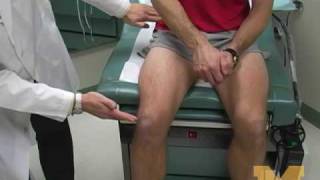
Dr. Yeong Kwok speaks about knee pain and demonstrates a stretch designed to treat tendonitis.

Watch as neurosurgeon Dr. Shawn Hervey-Jumper performs awake brain surgery on a 31-year-old woman with a brain tumor at UCSF Health.
Learn more here: https://magazine.ucsf.edu/awak....e-brain-cancer-surge
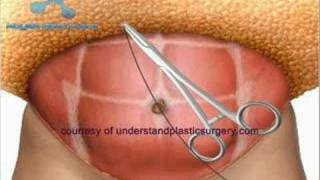
http://www.bodysculptor.com. Dr. Otto Placik, Board Certified Chicago based plastic surgeon demonstrates the results of a muscle separation(rectus diastasis) repair using 3 dimesional CAT scan and photographic images

For more information, visit https://ukhealthcare.uky.edu/doctors.

Linen Changes (with Patient in Bed)- Nursing Skills
FREE Nursing School Cheat Sheets at: http://www.NURSING.com
Get the full lesson on Patient Linen Changes here:
https://nursing.com/lesson/ski....lls-01-02-linen-chan
Get the full lesson on Bed Baths here:
https://nursing.com/lesson/skills-01-01-bed-bath/
Check out our new Nurse Care Plan Lessons here:
https://bit.ly/3BPRfPL
Get Access to Thousands of Lessons here:
https://nursing.com/courses/
Welcome to the NURSING Family, we call it the most supportive nursing cohort on the planet.
At NURSING.com, we want to help you remove the stress and overwhelm of nursing school so that you can focus on becoming an amazing nurse.
Check out our freebies and learn more at: (http://www.nursing.com)
Linen Changes (with Patient in Bed)- Nursing Skills
In this video, we’re going to show you how to change the linens with a patient in the bed. This might be after a bed bath or during incontinence care. So check out the bed bath video to see what got us up to this point. We love you guys! Go out and be your best selves today! And, as always, happy nursing!
Bookmarks:
0.05 Linen change introduction
0.16 Linen change supplies
0.30 Adjusting the patient/ sheet removal
1.00 Secure new fitted sheet
1.12 Pro tip
1.40 Roll patient back over
1.50 Repeat linen removal
2.02 Linen disposal
2.20 Wrinkle check
2.31 Reposition the patient for comfort
2.40 Covering the patient/ tuck-in
2.48 Pillowcase change (trick)
3.30 Making the patient comfortable
3.40 Linen change outro
Visit us at https://nursing.com/medical-disclaimer/ for disclaimer information.
NCLEX®, NCLEX-RN® are registered trademarks of the National Council of State Boards of Nursing, INC. and hold no affiliation with NURSING.com.

What is hemodialysis and how does it work? Who needs it? How do you prepare for it? In the United States, over 30 million Americans have kidney disease, and sometimes, kidney disease progresses to kidney failure or end-stage renal disease. When this happens, you cannot survive unless you have a kidney transplant or some form of dialysis. So today we're going to talk about hemodialysis.
Your kidneys are the two kidney bean-shaped organs that are located in your lower back, or in your flanks. And the kidneys are responsible for filtering out or cleaning your blood. They get rid of excess waste, excess toxins, and excess fluids. If your kidneys stop functioning, then you develop renal failure or end-stage renal disease.
What is Hemodialysis?
Hemodialysis, or blood dialysis, is the filtering of your blood outside of your body. So, if your kidneys stop working properly, the hemodialysis acts as a substitute kidney. Now it's important to note that hemodialysis does not actually correct your own kidney function. It does not fix or treat your kidneys.
#hemodialysis #drfrita
What is The Dialyzer?
The dialyzer is actually the filter. It's the main powerhouse of the hemodialysis system, and it is what actually acts as the substitute kidney. In the dialyzer, you have these hollow fibers that run through it, and these fibers are bathed in something called dialysates, or dialysis fluid.
How Often Are Patients Treated With Hemodialysis?
Most patients who are on hemodialysis are on it between three and six hours, about three days a week, especially if they go to a center.
How Does Hemodialysis Work?
So when you are on dialysis, how does your blood get from your body to the hemodialysis machine and then back to your body? Well, it does so through tubes, and those tubes are connected to your access, and we'll talk about access in just a moment. But as far as the tubing, the tubing is connected to your body.
Types Of Hemodialysis Access
Arteriovenous Fistula or AV Fistula
The AV fistula is the gold standard as far as hemodialysis access is concerned because it gives you the most efficient hemodialysis and it is the least likely to be infected.
Arteriovenous Graft or AV Graft
The AV graft is very similar to the AV fistula in that you still have a surgically connected artery and a vein, usually in the arm, but in the case where if you have veins that are rather thin or arteries that are thin and maybe too weak in order to really give you a properly functioning, substantial AV fistula, then the vascular surgeon may opt to add an artificial material in order to make that shunt a little stronger, or little more durable. And so, an AV graft is another option for dialysis access.
Catheter
If you're in a situation where you need temporary dialysis, or if you have acute kidney injury, then you may have a temporary Vascath placed, and it's usually placed in a vein of the neck, the internal jugular vein, or it can be placed in the groin, or in the femoral vein.
Who Needs Hemodialysis Treatment?
How do you know if you need hemodialysis, and when is it time to prepare? Well, if you follow up with your kidney doctor (nephrologist) regularly, he or she will be watching your labs. They'll be able to see those signs of your kidneys not functioning properly.

James Dunn, MD, PhD is a pediatric surgeon at Stanford Children's Health. He is board certified in General Surgery and Pediatric Surgery.
Learn more at https://www.stanfordchildrens.....org/en/service/gener

A recap of Mater Hospital patient Helen's story as she progressed from experiencing chronic knee pain due to osteoarthritis, through to knee replacement treatment and ultimately a new lease on life.
Dedicated to surgical excellence and patient-centred care, the Mater Hospital North Sydney is regarded as a leading orthopaedic hospital and the only Australian hospital to be accepted into the International Society of Orthopaedic Centres.
For more information, click here: https://bit.ly/3bvhY8G

Mini-Laparoscopic Cholecystectomy with Intraoperative Cholangiogram for Symptomatic Cholelithiasis (Gallstones) - Extended
Authors: Brunt LM1, Singh R1, Yee A2
Published: September 26, 2017
AUTHOR INFORMATION
1 Department of Surgery, Washington University, St. Louis, Missouri
2 Division of Plastic and Reconstructive Surgery, Washington University, St. Louis, Missouri
DISCLOSURE
No authors have a financial interest in any of the products, devices, or drugs mentioned in this production or publication.
ABSTRACT
Minimal invasive laparoscopic cholecystectomy is the typical surgical treatment for cholelithiasis (gallstones), where patients present with a history of upper abdominal pain and episodes of biliary colic. The classic technique for minimal invasive laparoscopic cholecystectomy involves four ports: one umbilicus port, two subcostal ports, and a single epigastric port. The Society of American Gastrointestinal and Endoscopic Surgeons (SAGES) has instituted a six-step strategy to foster a universal culture of safety for cholecystectomy and minimize risk of bile duct injury. The technical steps are documented within the context of the surgical video for (1) achieving a critical view of safety for identification of the cystic duct and artery, (2) intraoperative time-out prior to management of the ductal structures, (3) recognizing the zone of significant risk of injury, and (4) routine intraoperative cholangiography for imaging of the biliary tree. In this case, the patient presented with symptomatic biliary colic due to a gallstone seen on the ultrasound in the gallbladder. The patient was managed a mini-laparoscopic cholecystectomy using 3mm ports for the epigastric and subcostal port sites with intraoperative fluoroscopic cholangiogram. Specifically, the senior author encountered a tight cystic duct preventing the insertion of the cholangiocatheter and the surgical video describes how the author managed the cystic duct for achieving a cholangiogram, in addition to the entire technical details of laparoscopic cholecystectomy.

© 2023 Elsevier. All rights reserved. What are lymph nodes? Lymph nodes are small secondary lymphoid organs that are found along lymphatic vessels throughout the body.
Find our full video library only on Osmosis Prime: http://osms.it/more.
Join over 3 million current & future clinicians who learn by Osmosis, and over 130 universities around the world who partner with us to make medical and health education more engaging and efficient. We have unparalleled tools and materials to prepare you to succeed in school, on board exams, and as a future clinician. Sign up for a free trial at http://osms.it/more. If you're interested in exploring an institutional partnership, visit osmosis.org/educators to request a personalized demo.
Follow us on social:
Facebook: http://osms.it/facebook
Twitter: http://osms.it/twitter
Instagram for med: http://osms.it/instagram
Instagram for nursing: https://osms.it/ignursing
Linkedin: https://osms.it/linkedin
Our Vision: Everyone who cares for someone will learn by Osmosis.
Our Mission: To empower the world’s clinicians and caregivers with the best learning experience possible. Learn more here: http://osms.it/mission
Medical disclaimer: Knowledge Diffusion Inc (DBA Osmosis) does not provide medical advice. Osmosis and the content available on Osmosis's properties (Osmosis.org, YouTube, and other channels) do not provide a diagnosis or other recommendation for treatment and are not a substitute for the professional judgment of a healthcare professional in diagnosis and treatment of any person or animal. The determination of the need for medical services and the types of healthcare to be provided to a patient are decisions that should be made only by a physician or other licensed health care provider. Always seek the advice of a physician or other qualified healthcare provider with any questions you have regarding a medical condition. © 2023 Elsevier. All rights reserved.
