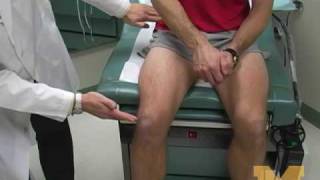
- Physical Examination
- Surgical Examination
- Ophthalmology
- Clinical Skills
- Orthopedics
- Surgery Videos
- Laparoscopy
- Pediatrics
- Funny Videos
- Cardiothoracic Surgery
- Nursing Videos
- Plastic Surgery
- Otorhinolaryngology
- Histology and Histopathology
- Neurosurgery
- Dermatology
- Pediatric Surgery
- Urology
- Dentistry
- Oncology and Cancers
- Anatomy Videos
- Health and Fitness
- Radiology
- Anaesthesia
- Physical Therapy
- Pharmacology
- Interventional Radiology
- Cardiology
- Endocrinology
- Gynecology
- Emergency Medicine
- Psychiatry and Psychology
- Childbirth Videos
- General Medical Videos
- Nephrology
- Physiology
- Diet and Food Health
- Diabetes Mellitus
- Neurology
- Women Health
- Osteoporosis
- Gastroenterology
- Pulmonology
- Hematology
- Rheumatology
- Toxicology
- Nuclear Medicine
- Infectious Diseases
- Vascular Disease
- Reproductive Health
- Burns and Wound Healing
- Other
Top videos

In this video, I am talking about the best histology resources available on the internet. All the links to the resources I talked are here -
1. Amit's lectures - https://www.youtube.com/channe....l/UCwdAyZnA6FEE0Iqsw
2. VIBS histology - https://www.youtube.com/c/VIBSHistology/featured
3. Dr. Eman Sadek Histology Queen - https://www.youtube.com/channe....l/UCHXGb5GphBKKN-xD3
4. BIOC 21 Histology lectures - https://www.youtube.com/playli....st?list=PLKnI3Jl97pW
5. https://medicalschoolpathology.com/
7. Udemy - https://clnk.in/qfEB
Buy this awsm book for Histology - https://amzn.to/3wSX1Oh

This Basic Laparoscopic Surgery: Abdominal Access and Trocar Introduction course will teach you the steps of Laparoscopic Surgery. View the full course for free by signing up on our website: https://www.incision.care/
What is Laparoscopic Surgery:
Laparoscopic surgery describes procedures performed using one or multiple small incisions in the abdominal wall in contrast to the larger, normally singular incision of laparotomy. The technique is based around principles of minimally invasive surgery (or minimal access surgery): a large group of modern surgical procedures carried out by entering the body with the smallest possible damage to tissues. In abdominopelvic surgery, minimally invasive surgery is generally treated as synonymous with laparoscopic surgery as are procedures not technically within the peritoneal cavity, such as totally extraperitoneal hernia repair, or extending beyond the abdomen, such as thoraco-laparoscopic esophagectomy. The term laparoscopy is sometimes used interchangeably, although this is often reserved to describe a visual examination of the peritoneal cavity or the purely scopic component of a laparoscopic procedure. The colloquial keyhole surgery is common in non-medical usage.
Surgical Objective of Laparoscopic Surgery:
The objective of a laparoscopic approach is to minimize surgical trauma when operating on abdominal or pelvic structures. When correctly indicated and performed, this can result in smaller scars, reduced postoperative morbidity, shorter inpatient durations, and a faster return to normal activity. For a number of abdominopelvic procedures, a laparoscopic approach is now generally considered to be the gold-standard treatment option.
Definitions
Developments of Laparoscopic Surgery:
Following a number of smaller-scale applications of minimally invasive techniques to abdominopelvic surgery, laparoscopic surgery became a major part of general surgical practice with the introduction of laparoscopic cholecystectomy in the 1980s and the subsequent pioneering of endoscopic camera technology. This led to the widespread adoption of the technique by the early- to mid-1990s. The portfolio of procedures that can be performed laparoscopically has rapidly expanded with improvements in instruments, imaging, techniques and training — forming a central component of modern surgical practice and cross-specialty curricula [2]. Techniques such as laparoscopically assisted surgery and hand-assisted laparoscopic surgery have allowed the application of laparoscopic techniques to a greater variety of pathology. Single-incision laparoscopic surgery, natural orifice transluminal endoscopic surgery, and minilaparoscopy-assisted natural orifice surgery continue to push forward the applications of minimally invasive abdominopelvic techniques; however, the widespread practice and specific indications for these remain to be fully established. More recently, robotic surgery has been able to build on laparoscopic principles through developments in visualization, ergonomics, and instrumentation.
This Basic Laparoscopic Surgery: Abdominal Access and Trocar Introduction course will teach you:
- How to access the abdomen using an open, closed, and direct optical-entry technique
- Principles underlying safe abdominal insufflation
- The vascular anatomy of the abdominal wall and its implications for trocar placement
- How to introduce trocars into the peritoneal cavity
- The principle of triangulation and how this can be applied to organizing a laparoscopic surgical field
Specific attention is given to these hazards you may encounter:
- Intravascular, intraluminal, or extraperitoneal needle position
- Limitations of a closed introduction technique
- Abdominal surgical history
- Limitations of an open introduction technique
- Optical trocar entry in thin individuals
- Visualization of non-midline structures
- Limitations of direct optical-entry techniques
- Limitations of clinical examination to confirm intraperitoneal insufflation
- Leakage of insufflation gas
These tips are designed to help you improve your understanding and performance:
- Alternative left upper quadrant approach
- Testing Veress needle before use
- Lifting the abdominal wall for Veress needle introduction
- "Hanging-drop test"
- Palmer's test
- Confirming intra-abdominal insufflation
- Subcutaneous tissue retraction
- Anatomy of the umbilicus
- Retraction of abdominal wall fascia
- Finger sweep of anterior abdominal wall
- Lifting the abdominal wall for optical trocar introduction
- Identification of venous bleeding at the end of a procedure
- Identification of inferior epigastric vessels by direct vision
- Peritoneal folds of the anterior abdominal wall
- Transillumination of superficial epigastric vessels
- Infiltration of local anesthetic at port sites
- Aiming of trocars
- Selection of trocar size
- Maintaining direct vision

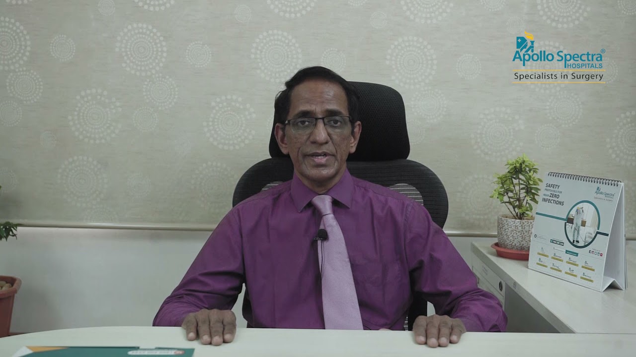
Dr. Mohan Rao, Senior General & Laparoscopic consultant at Apollo Spectra Hospitals, MRC Nagar explains How can one self-examination of Hernia be done

A drill. A mallet. A robot. Go inside the operating room to see how Northwestern Medicine Orthopaedic Surgeon Linda Idris Suleiman, MD, uses these tools for a total knee replacement.
#insidetheor

In this video, we have explained the procedure of total #knee #replacement #surgery in patient in 3D animation.
Learn more: https://ecgkid.com
_____________________________________________________________________
Knee replacement, commonly known as complete knee replacement or knee arthroplasty, is a surgical treatment that resurfaces a knee that has been destroyed by arthritis. The extremities of the bones that make up the knee joint, as well as the kneecap, are capped with metal and plastic pieces. Someone with severe arthritis or a major knee injury may benefit from this procedure.
The knee joint can be affected by a variety of arthritis forms. The degradation of joint cartilage and neighboring bone in the knees can be caused by osteoarthritis, a degenerative joint disease that primarily affects middle-aged and older persons. Rheumatoid arthritis produces pain and stiffness by inflaming the synovial membrane and resulting in an excess of synovial fluid. Traumatic arthritis, or arthritis caused by an injury, can harm the joints.
The purpose of knee replacement surgery is to resurface damaged areas of the knee joint and cure knee discomfort that has not responded to prior therapies.

The OrthoIllustrated® animation for total knee replacement is an educational tool to help patients better understand the diagnosis and treatment of arthritis.
- - - - -
Why Work Arthrex https://www.arthrex.com/job-seeker
Find an Arthrex Surgeon: https://doctorfinder.orthoillustrated.com
- - - - -
Join the Community:
LinkedIn: https://www.linkedin.com/company/arthrex
Facebook: https://www.facebook.com/Arthrex
Instagram: https://www.instagram.com/arthrex_inc/
Twitter: https://twitter.com/Arthrex
TikTok: https://www.tiktok.com/@arthrex
- - - - -
Arthrex Inc., headquartered in Naples, Florida, is a global leader in orthopedic surgical device design, research, manufacturing, and medical education. Arthrex develops and releases more than 1,000 new products and procedures every year to advance minimally invasive orthopedics worldwide.
For more information, visit https://www.arthrex.com
- - - - -
OrthoPedia is an innovative educational website that was created for anyone interested in learning about orthopedics from the first-year student to the experienced orthopedic surgeon.
Visit https://www.orthopedia.com to experience the future of Medical Education.
Tough to beat! Head #InsideTheOR with S. Christopher Malaisrie, MD, and witness open heart surgery by one of the best cardiology and heart surgery programs in the nation as ranked by US News and World Report.

If you’ve lost a significant amount of weight, either after pregnancy or through exercise and dietary changes, excess skin and weakened abdominal muscles can leave you self-conscious about your appearance. In this video, Dr. Catherine Hannan and Dr. Lauren Patrick, two of our Board-Certified Plastic Surgeons, are performing a Tummy Tuck (Abdominoplasty) surgery. Tummy Tuck surgery gets rid of the excess skin, as well as tightens your abdominal muscles, resulting in a flatter and smoother abdomen. The results of the surgery are permanent except in cases of large weight gain or pregnancy after surgery.
We are so excited to have taken a part in our patient's body transformation journey!
Before & After Gallery:
https://www.westendplasticsurg....ery.com/surgical/bod
To learn more, visit our website or call (202) 785-4187
http://www.westendplasticsurgery.com
~~~~~~~~~~~~~~~~~~~
Social Media:
✨ Instagram: http://www.instagram.com/westendplasticsurgery
✨ Facebook: http://www.facebook.com/westendplasticsurgery
✨ Twitter: http://www.twitter.com/weplasticsurg
✨ Blog: https://www.westendplasticsurgery.com/blog
✨ Business Inquiries: info@westendplasticsurgery.com
~~~~~~~~~~~~~~~~~~~
#TummyTuck #Abdominoplasty

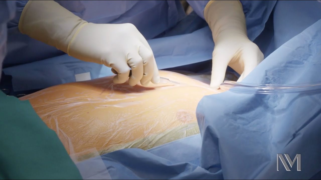
Dr. Neel Joshi, Clinical Chief, Department of Surgery at Cedars Sinai, describes his technique for trocar removal at the end of laparoscopic cholecystectomy.
#medicaleducation #laparoscopicsurgery
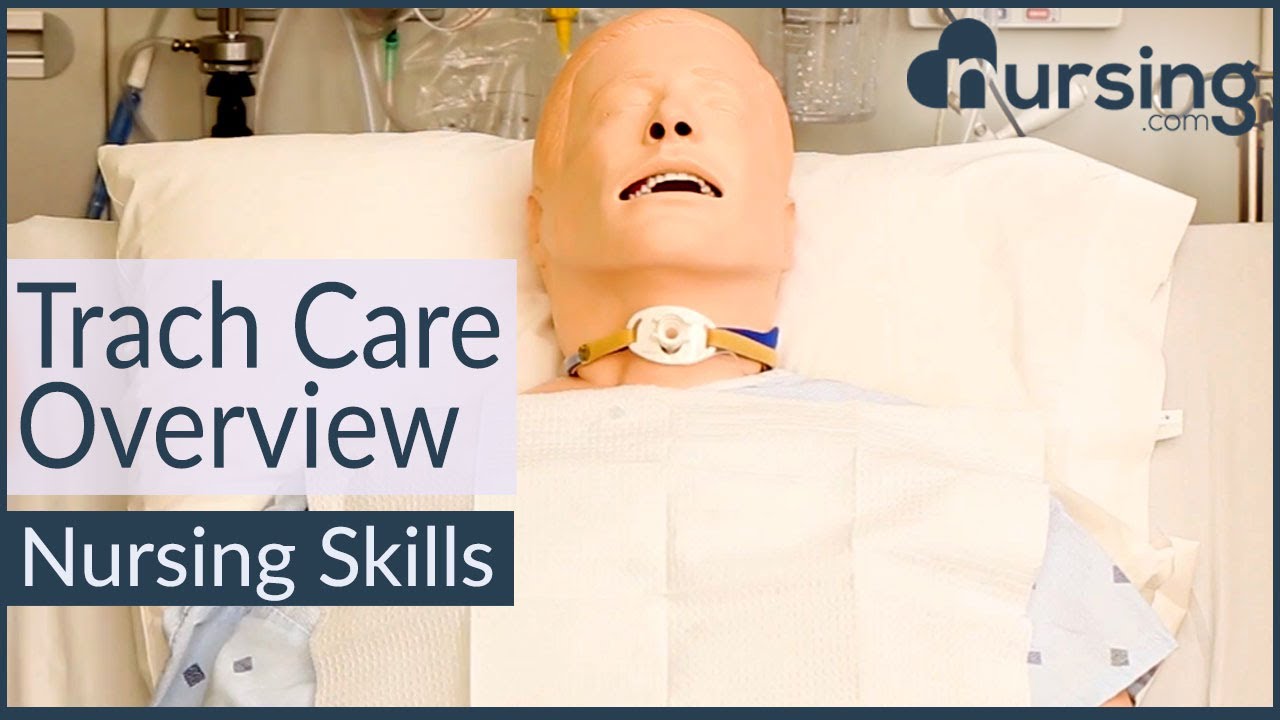
Learn what's working for other Nursing Students! Check out our Top 10 Most Popular Lessons Here: https://bit.ly/3nda5u3
Get the full lesson here: https://nursing.com/lesson/ski....lls-03-04-trach-care
Welcome to the NURSING Family, we call it the most supportive nursing cohort on the planet.
At NURSING.com, we want to help you remove the stress and overwhelm of nursing school so that you can focus on becoming an amazing nurse.
Check out our freebies and learn more at: (http://www.nursing.com)
Trach Care Overview (Nursing Skills):
In this video we’re going to look at trach care. Remember you should always suction the patient before trach care, so if you haven’t watched that skill video yet, make sure you watch it!
Click here: https://nursing.com/lesson/ski....lls-03-03-trach-suct
And remember as you’re doing this, you want to be assessing the stoma for signs of infection or skin breakdown.
Bookmarks:
0:00 Introduction
0:30 Set up sterile field
1:00 Apply gloves
1:12 Remove inner canula and dressing
1:30 Apply sterile gloves
2:05 Clean secretions
2:56 Clean stoma
3:48 Replace inner canula
4:14 Change trach ties
5:50 Apply dressing
Visit us at https://nursing.com/medical-disclaimer/ for disclaimer information.
NCLEX®, NCLEX-RN® are registered trademarks of the National Council of State Boards of Nursing, INC. and hold no affiliation with NURSING.com.
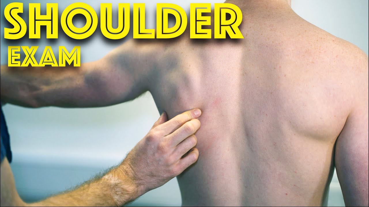
Shoulder Clinical Examination - Medical School Clinical Skills - Dr Gill
Personally, I find the shoulder examination the most complex examination possibly as there are so many variations and special tests. Some of which overlap and some will relate specifically to a patients presentation.
Often in a medical school syllabus, only select special tests will be used. In this shoulder exam demonstration, we include the Hawkins-Kennedy Test looking for impingement. This is dovetailed with examination for bicipital tendonitis as this is another possible cause of impingement type symptoms.
This shoulder upper limb exam follows the standard "Look, Feel, Move" orthopaedic exam approach, and overall order as set out in MacLeods Clinical Examination
Watch further orthopaedic examinations for your OSCE revision:
The Spine Examination:
https://youtu.be/pJxMHa6SCgU
Knee Examination
https://youtu.be/oyKH4EYfJDM
Hip Joint Clinical Examination
https://youtu.be/JC9GKq5nSdQ
________
Please note that there is no ABSOLUTE way to perform a clinical examination. Different institutions and even clinicians will have differing degrees of variations - the aim is the effectively identify medically relevant signs.
However during OSCE assessments. Different medical schools, nursing colleges, and other health professional courses will have their own preferred approach to a clinical assessment - you should concentrate on THEIR marks schemes for your assessments.
The examination demonstrated here is derived from Macleods Clinical Examination - a recognized standard textbook for clinical skills.
#ShoulderExamination #ClinicalSkills #DrGill

Watch Dr. Robert Thomas, of Panorama Orthopedics & Spine Center, perform a Mako Knee replacement. He narrates each step of the process.

Dr. Yeong Kwok speaks about knee pain and demonstrates a stretch designed to treat tendonitis.

Watch more clips of Dr. James Kelly - https://www.youtube.com/playli....st?list=PLe2Je5-cHxP And for more information about brain injury and PTSD, please visit us at https://www.brainline.org.
Watch more clips of Pat LaFontaine - https://www.youtube.com/playli....st?list=PL5F3273C3C8
© 2018 WETA All Rights Reserved
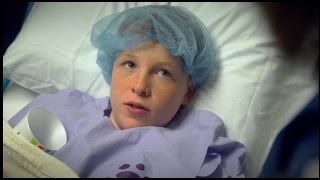
Our surgeons take a compassionate, family-centered approach to both inpatient and outpatient care. We’re committed to making sure both you and your child understand our process. Told through a kid's eyes, this video tour reveals our caring approach.
To learn more about pediatric surgery at Stamford Hospital, visit: https://www.stamfordhealth.org..../care-treatment/pedi

Devi Shetty, founder of Narayana Health in India, reflects on the remarkable fact that, after 26 years of operation, the cost of heart surgery at Narayana Health has come down dramatically, and shares some of the strategies used to maintain high quality with low patient cost.
Learn more about the Creating Emerging Markets Project and explore its many compelling interviews: https://www.hbs.edu/creating-e....merging-markets/Page


Linen Changes (with Patient in Bed)- Nursing Skills
FREE Nursing School Cheat Sheets at: http://www.NURSING.com
Get the full lesson on Patient Linen Changes here:
https://nursing.com/lesson/ski....lls-01-02-linen-chan
Get the full lesson on Bed Baths here:
https://nursing.com/lesson/skills-01-01-bed-bath/
Check out our new Nurse Care Plan Lessons here:
https://bit.ly/3BPRfPL
Get Access to Thousands of Lessons here:
https://nursing.com/courses/
Welcome to the NURSING Family, we call it the most supportive nursing cohort on the planet.
At NURSING.com, we want to help you remove the stress and overwhelm of nursing school so that you can focus on becoming an amazing nurse.
Check out our freebies and learn more at: (http://www.nursing.com)
Linen Changes (with Patient in Bed)- Nursing Skills
In this video, we’re going to show you how to change the linens with a patient in the bed. This might be after a bed bath or during incontinence care. So check out the bed bath video to see what got us up to this point. We love you guys! Go out and be your best selves today! And, as always, happy nursing!
Bookmarks:
0.05 Linen change introduction
0.16 Linen change supplies
0.30 Adjusting the patient/ sheet removal
1.00 Secure new fitted sheet
1.12 Pro tip
1.40 Roll patient back over
1.50 Repeat linen removal
2.02 Linen disposal
2.20 Wrinkle check
2.31 Reposition the patient for comfort
2.40 Covering the patient/ tuck-in
2.48 Pillowcase change (trick)
3.30 Making the patient comfortable
3.40 Linen change outro
Visit us at https://nursing.com/medical-disclaimer/ for disclaimer information.
NCLEX®, NCLEX-RN® are registered trademarks of the National Council of State Boards of Nursing, INC. and hold no affiliation with NURSING.com.
