- Physical Examination
- Surgical Examination
- Ophthalmology
- Clinical Skills
- Orthopedics
- Surgery Videos
- Laparoscopy
- Pediatrics
- Funny Videos
- Cardiothoracic Surgery
- Nursing Videos
- Plastic Surgery
- Otorhinolaryngology
- Histology and Histopathology
- Neurosurgery
- Dermatology
- Pediatric Surgery
- Urology
- Dentistry
- Oncology and Cancers
- Anatomy Videos
- Health and Fitness
- Radiology
- Anaesthesia
- Physical Therapy
- Pharmacology
- Interventional Radiology
- Cardiology
- Endocrinology
- Gynecology
- Emergency Medicine
- Psychiatry and Psychology
- Childbirth Videos
- General Medical Videos
- Nephrology
- Physiology
- Diet and Food Health
- Diabetes Mellitus
- Neurology
- Women Health
- Osteoporosis
- Gastroenterology
- Pulmonology
- Hematology
- Rheumatology
- Toxicology
- Nuclear Medicine
- Infectious Diseases
- Vascular Disease
- Reproductive Health
- Burns and Wound Healing
- Other
Top videos
Podalic version is an obstetric procedure wherein the fetus is turned within the womb such that one or both feet present through the cervix during childbirth. It is used most often in cases where the fetus lies transversely or in another abnormal position in the womb.
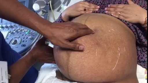
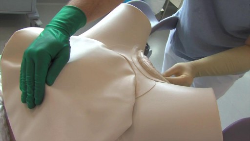
External cephalic version is a process by which a breech baby can sometimes be turned from buttocks or foot first to head first. External cephalic version (ECV) is a manual procedure that is advocated by national guidelines for breech presentation singleton pregnancy, in order to enable vaginal delivery.
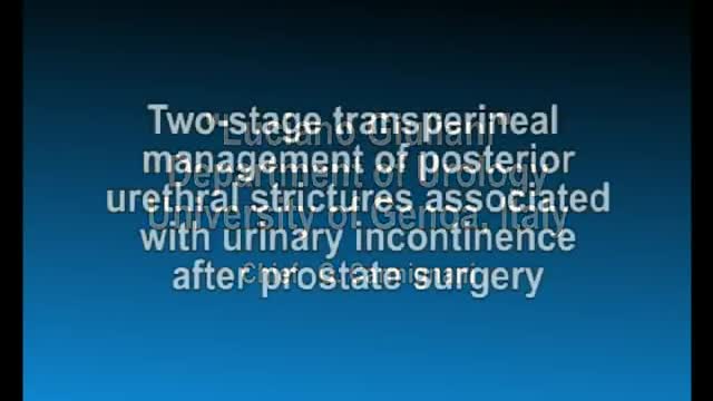
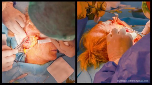
Posterior Urethral Strictures Associated with Urinary Incontinence after Prostatectomy Management
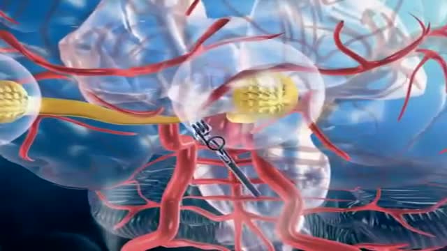
A brain (cerebral) aneurysm is a bulging, weak area in the wall of an artery that supplies blood to the brain. In most cases, a brain aneurysm causes no symptoms and goes unnoticed. In rare cases, the brain aneurysm ruptures, releasing blood into the skull and causing a stroke. When a brain aneurysm ruptures, the result is called a subarachnoid hemorrhage. Depending on the severity of the hemorrhage, brain damage or death may result. The most common location for brain aneurysms is in the network of blood vessels at the base of the brain called the circle of Willis. What causes a brain aneurysm? A person may inherit the tendency to form aneurysms, or aneurysms may develop because of hardening of the arteries (atherosclerosis) and aging. Some risk factors that can lead to brain aneurysms can be controlled, and others can't. The following risk factors may increase your risk for an aneurysm or, if you already have an aneurysm, may increase your risk of it rupturing: Family history. People who have a family history of brain aneurysms are more likely to have an aneurysm than those who don't. Previous aneurysm. People who have had a brain aneurysm are more likely to have another. Gender. Women are more likely to develop a brain aneurysm or to suffer a subarachnoid hemorrhage. Race. African Americans are more likely than whites to have a subarachnoid hemorrhage. High blood pressure. The risk of subarachnoid hemorrhage is greater in people who have a history of high blood pressure. Smoking. In addition to being a cause of high blood pressure, the use of cigarettes may greatly increase the chances of a brain aneurysm rupturing.
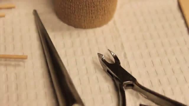
Ingrown Toenail Surgery HD
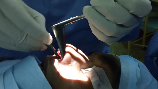
Initial treatment of a deviated septum may be directed at managing the symptoms of the tissues lining the nose, which may then contribute to symptoms of nasal obstruction and drainage. Your doctor may prescribe: Decongestants. Decongestants are medications that reduce nasal tissue swelling, helping to keep the airways on both sides of your nose open. Decongestants are available as a pill or as a nasal spray. Use nasal sprays with caution, however. Frequent and continued use can create dependency and cause symptoms to be worse (rebound) after you stop using them. Decongestants have a stimulant effect and may cause you to be jittery as well as elevate your blood pressure and heart rate. Antihistamines. Antihistamines are medications that help prevent allergy symptoms, including obstruction and runny nose. They can also sometimes help nonallergic conditions such as those occurring with a cold. Some antihistamines cause drowsiness and can affect your ability to perform tasks that require physical coordination, such as driving. Nasal steroid sprays. Prescription nasal corticosteroid sprays can reduce inflammation in your nasal passage and help with obstruction or drainage. It usually takes from one to three weeks for steroid sprays to reach their maximal effect, so it is important to follow your doctor's directions in using them. Medications only treat the swollen mucus membranes and won't correct a deviated septum.
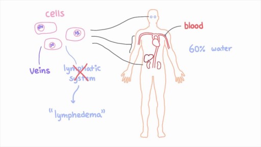
The lymphatic system is a network of specialized vessels (lymph vessels) throughout the body whose purpose is to collect excess lymph fluid with proteins, lipids, and waste products from the tissues. This fluid is then carried to the lymph nodes, which filter waste products and contain infection-fighting cells called lymphocytes. The excess fluid in the lymph vessels is eventually returned to the bloodstream. When the lymph vessels are blocked or unable to carry lymph fluid away from the tissues, localized swelling (lymphedema) is the result.
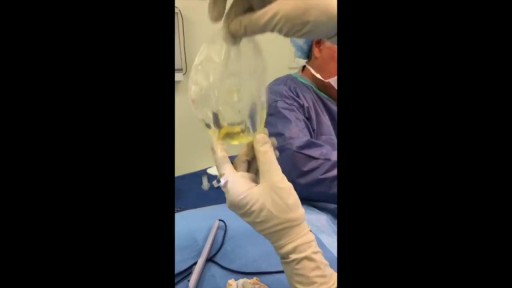
As you can see I access the left implant from the periareolar incisions which I made at the lower portion of the areola. As I entered the capsule and begin to remove the implant I noticed a lot of fluid surrounding the implant. Right away I know this is a rupture and that the mammogram was incorrect. Mammograms are very helpful in detecting cancer but often not ruptures. When implants rupture, it is important to have them replaced as soon as possible to avoid excessive scarring in the breasts. If too much scar tissue has accumulated around the deflated implant, it becomes difficult to create a normal breast shape in the future. Therefor know the signs of a ruptured implant such as, painful to touch, visible asymmetry or loss of integrity to the bag. For more information please visit: www.drlinder.com
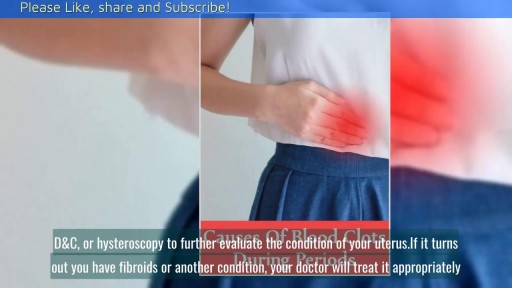
Heavy period blood can be especially alarming if it contains clots. In most cases, though, red, brown, or even black menstrual blood clots are normal—just bits of the endometrium (the lining of the uterus) that are shed during menstruation.
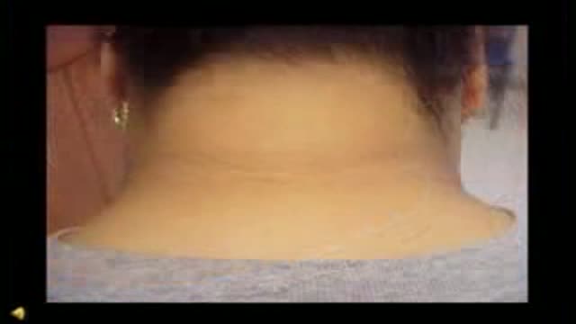
Acanthosis Nigricans Insulin Resistance
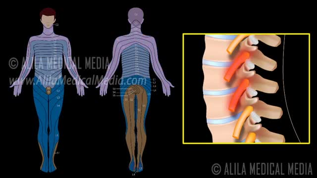
A nerve root block is an injection of local anesthetic (numbing medicine) and steroid injected under X-ray guidance into the area where the nerve exits the spinal column. A nerve root block is usually ordered by your doctor for pain in the arm or leg that follows the path of a single nerve. A nerve root block may be diagnostic (a test to determine the source of your pain) and/or therapeutic (to relieve your pain). If you get a period of sustained pain relief from the injection, the block may be repeated. Sometimes the block is done to help identify whether or not surgery might be helpful and at what level such surgery might be most helpful.
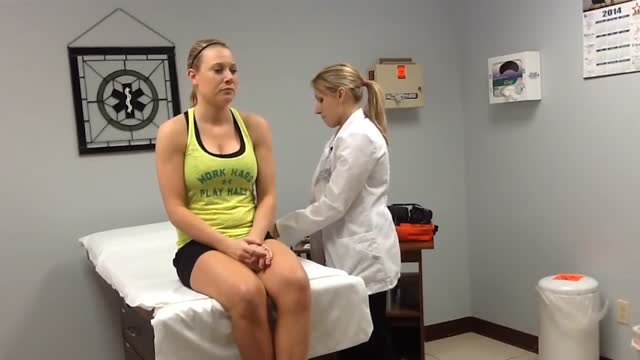
Head to Toe Assesment
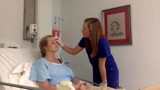
Physical assessment is taking an educated, systematic look at all aspects of an individual’s health status utilizing knowledge, skills and tools of health history and physical exam. To collect data- information about the client’s health, including physiological, psychological, sociocultural and spiritual aspects To establish actual and potential problems To establish the nurse-client relationship Method: The history is done first, then the physical examination focuses on finding data associated with the history. Health History- obtained through interview and record review. Physical exam- accomplished by tools and techniques ** A complete assessment is not necessarily carried out each time. A comprehensive assessment is part of a health screening examination. On admission, you will do an admission assessment (not necessarily including everything presented here) and document it on the admission form. You will do a daily shift assessment (patient systems review). And, if client has a specific problem, you may assess only that part of the body (focused). Data Collection: Information is organized into objective and subjective data: Subjective: Apparent only to person affected; includes client’s perceptions, feelings, thoughts, and expectations. It cannot be directly observed and can be discovered only asking questions. Objective: Detectable by an observer or can be tested against an acceptable standard; tangible, observable facts; includes observation of client behavior, medical records, lab and diagnostic tests, data collected by physical exam. ** To obtain data for the nursing health history, you must utilize good interview techniques and communications skills. Record accurately. DO NOT ASSUME. D. Frameworks for Health Assessment There are two main frameworks utilized in health assessment: Head to Toe- systematic collection of data starting with the head and working downward. Functional Health Assessment- Gordon’s 11 functional health patterns that address the behaviors a person uses to maintain health. PERSON is the ACC-ADN framework for assessment. It is similar to Gordon's functional health patterns.
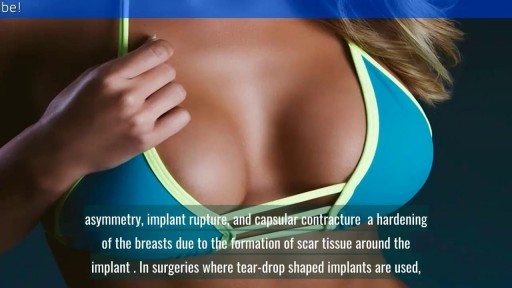
reast Augmentation: From Cost to Complications || Common gynaecological problems in women Breast augmentation (aka augmentation mammaplasty) is one of the most popular cosmetic procedures performed in the U.S. today. Despite controversy over the use of silicone breast implants, women have shown a continuing and growing eagerness to surgically enhance the size and shape of their breasts. If you are a healthy, non-smoking women who are at or near their ideal weight, with enough of their own breast tissue to cover and support an implant adequately, then you are a good candidate for breast augmentation surgery.

We are aware that the "official" way to use an ear candle is small end down into the ear, but for this video, we have elected to use it the way most "lay" public would (small end up). Ear candling is an alternative medicine practice that is thought to remove earwax. However, this video illustrates how ineffective this practice is in removing earwax... and can potentially be even harmful. And yes... It is still frequently practiced.

Integrative Physical Examination Lecture

Abdominal Physical Examination Lecture
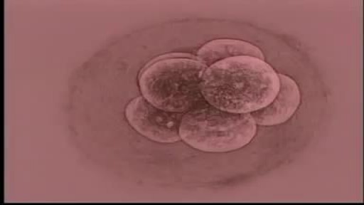
Almost all the cells in your body were produced by mitosis. The only exception is sperm or eggs which are produced by a different type of cell division called meiosis. During fertilization the sperm and egg unite to form a single cell called the zygote which contains chromosomes from both the sperm and egg.

Watch that video of an Ingrown hair turns into 140 Lbs tumor in man’s stomach