
- Physical Examination
- Surgical Examination
- Ophthalmology
- Clinical Skills
- Orthopedics
- Surgery Videos
- Laparoscopy
- Pediatrics
- Funny Videos
- Cardiothoracic Surgery
- Nursing Videos
- Plastic Surgery
- Otorhinolaryngology
- Histology and Histopathology
- Neurosurgery
- Dermatology
- Pediatric Surgery
- Urology
- Dentistry
- Oncology and Cancers
- Anatomy Videos
- Health and Fitness
- Radiology
- Anaesthesia
- Physical Therapy
- Pharmacology
- Interventional Radiology
- Cardiology
- Endocrinology
- Gynecology
- Emergency Medicine
- Psychiatry and Psychology
- Childbirth Videos
- General Medical Videos
- Nephrology
- Physiology
- Diet and Food Health
- Diabetes Mellitus
- Neurology
- Women Health
- Osteoporosis
- Gastroenterology
- Pulmonology
- Hematology
- Rheumatology
- Toxicology
- Nuclear Medicine
- Infectious Diseases
- Vascular Disease
- Reproductive Health
- Burns and Wound Healing
- Kidney Zone Podcast
- Other


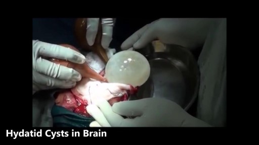
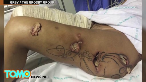



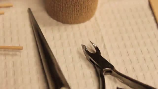
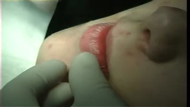

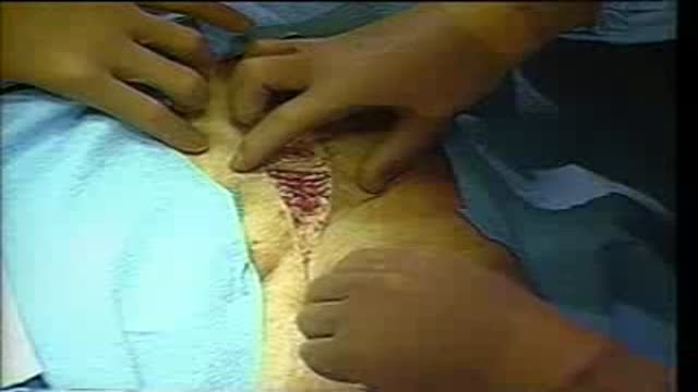
Full Swelling Examination

Educational only: videos on MedicalVideos.com are for general information and are not medical advice. Always consult your own doctor about your health.
ost of us come across this particular sign quite often. Of course, you can just jump to the numerous investigations and one after another, rule out the possible causes, finally getting to the diagnosis. For me, that’s no fun at all. Although I still don’t know whether I am going to become a surgeon or not (embarassing for me, since I’m going to be done with med-school this year), its pretty fascinating. If I were to work in a country whether investigations aren’t that expensive, I would definitely just perform a small examination and take a short history, sending off my patient to get a myriad of investigations, reporting to me after a while, with the diagnosis in his reports.
This video in 30 seconds
A classic surgical-teaching lecture sets out how to examine a swelling in a traditional order. It proceeds through site, number, shape and measured size, surface and skin changes, then palpation for temperature, tenderness, consistency and mobility. Special tests including fluctuation, transillumination, impulse on coughing and reducibility are demonstrated, with examples from everyday surgical practice.
Key points
- Inspection covers site, number, shape, size and surface changes
- Palpation begins with temperature, tenderness and confirmed measurements
- Fluctuation and transillumination tests are demonstrated for cystic swellings
- Impulse on coughing, reducibility and compressibility are tested
- Fixity to skin and to underlying muscle is tested last
Chapters
- 0:03 — Inspecting site, number and size
- 4:41 — Palpation: warmth, tenderness, consistency
- 8:19 — Special tests for cystic swellings
- 12:01 — Transillumination test demonstrated
- 13:18 — Impulse, reducibility and compressibility
- 16:48 — Fixity to skin and muscle
Video Transcript
A classic surgical-teaching lecture on how to examine a swelling. It proceeds in the traditional order — site, number, shape and measured size, surface and skin changes, then palpation for consistency and mobility, testing the swelling with the underlying muscle relaxed and then contracted. Examples are drawn from everyday surgical practice, from dermoid cysts and lipomatosis to melanoma and squamous cell carcinoma.
Read the full transcript
Examination of a swelling. On inspection, first note the site of the swelling, that is the exact anatomical location and the number, whether it is single or multiple. Some swellings have a typical position which is diagnostic of the swelling. For example, a post-autocular dermoid behind the ear, or a meningo seal over the back in the midline, or an external angular dermoid at the lateral end of the eyebrow. Then note the number. Most swellings will be solitary, but some are typically multiple. For example, multiple cutaneous swellings in multiple neurofibromatosis or von Reckling-Horsen's disease,
multiple subcutaneous swellings in multiple lipomatosis or Durkheim's disease. Now note the shape and size of the swelling. The shape may be a regular one like spherical, povoid or kidney shape, or it may be an irregular shaped swelling. Then measure the exact size in centimeters using a measuring tape. Measure in longitudinal and transverse axis and where possible the depth. The depth of the swelling can be better judged on palpation, so measurement with a scale may also be done during palpation. Now examine the surface of the swelling. Does it have any characteristic color?
For example, capillary hemangioma as seen on the forehead of this child is bright red in color, or malignant melanoma as seen on the heel of this patient is black in color. Next is the surface smooth or irregular. Note the typical philiform branches over a papilloma and cauliflower surface of a squamous cell carcinoma. Now note the skin over the swelling whether it is normal, inflamed or ulcerated. A tense shiny skin with prominent veins suggests a sarcoma. A black punctum on the skin over the swelling is diagnostic of a sebaceous cyst.
Red edymata skin over this sebaceous cyst indicates inflammation or an infected cyst. Note the redness and shininess of skin in this injection abscess of the right buttock. Presence of a scar of previous surgery suggests that the swelling is a recurrent one. An edymata skin with multiple pits like an orange peel termed as Oda orange appearance suggests an infiltrating malignant lump blocking cutaneous lymphatics. Now inspect the swelling for a few seconds for any visible pulsations that is a movement or increase in size synchronous with each heartbeat. Swelling arising from the arteries like aneurysms will be pulsatile
as seen in this aneurysm of the ascending orta bulging through the anterior chest wall. Lastly depending on the site and type of the swelling a few specific signs should be looked for. If the swelling is over the abdomen or chest or spinal canal, ask the patient to cuff and look for an impulse on cuffing. A visible increase in the size of the swelling synchronous with the cuff is termed as an impulse on cuffing as seen in this in guainal hernia. If the swelling is on the limbs inspect the distal limb for any pressure effects like edema or nore palsy.
Note the foot drop caused by a bone tumor in the upper tibia. Similarly if the swelling is on the front of the neck look for movement on the glutation. To revise on inspection note the site of the swelling and whether it is single or multiple. Note the shape and measure the size. Inspect the surface and inspect the skin over the swelling for inflammation, panthums, scars or veins. Then look for pulsatility and site related specific signs such as impulse on cuffing if it is related to abdomen or chest or pressure effects if it is on the limb.
On palpation first test the temperature of the skin over the swelling and compare it with normal skin. An inflamed swelling will be warm. So also a sarcoma and other well vascularized swellings will show a local rise of temperature. Then pulpit gently over all parts of the swelling to look for a tenderness. Pain due to pressure exerted over the swelling is termed as tenderness. Tenderness is a feature of inflammation. A swelling related to a nerve for example a solitary neurofibroma may also be tender. It is an absolute standard routine of palpation to test for temperature and tenderness
before proceeding to pulpit the swelling in details. Now confirm the size and shape of the swelling and note the third dimension that is the depth of the swelling which could not be correctly judged during inspection. Pulpit the swelling from all sides as sometimes it may be much larger than what is seen to inspection. In such a case measure the size again in three dimensions using a measuring tape. Now pulpit the surface. The surface may be smooth in cystic swellings as seen in this bursa over the leg
or it may be lobular as in a lipooma. Note the lobulations with smooth bumps in this lipooma over the back or it may be nodular as seen in this multinodular goiter. Note the multiple nodules over the surface of the goiter or it may be irregular as in a carcinoma. Now carefully pulpit the edge of the swelling with the tips of the fingers and note whether it is well-defined and regular or well-defined and irregular or diffuse and ill-defined. The edge is well-defined and regular in most of the benign swellings.
It may be well-defined and irregular in malignant swellings infiltrating into the surrounding tissues or it may be diffuse and ill-defined in inflammatory swellings like cellulitis and abscess. Here we see the characteristic edge of a lipooma which slips under the palpating finger. This is the characteristic slipping edge of a subcutaneous lipooma. Now pulpit the swelling for its consistency. The consistency may be soft, cystic, firm, hard or bony hard. In a very small swelling if it is difficult to determine whether the swelling is solid or cystic, perform Pechert's test.
Hold the swelling fixed with the left hand and pulpit with the tip of the index finger of the right hand first at the center and then at the periphery. A cystic swelling feels softer at the center than at the periphery while a solid swelling feels firmer in the center than at the periphery. If the swelling is soft or cystic, look for the sign of molding. Press a finger into the swelling for 1 to 2 seconds and remove it. If the swelling remains indented, it implies the presence of pultaceous material typically seen in a sebaceous test.
This is termed as the sign of molding or sign of indentation. It is seen in a sebaceous cyst, a dermoid cyst and a colonic mass with fecal matter. Sometimes the swelling has a variable consistency that is some parts are cystic and some are solid, firm or hard. Variable consistency is usually seen in malignant lumps. Now if the swelling is soft or cystic, some special tests should be carried out. That is fluctuation. If the swelling is fluctuated, then transillumination. If the swelling is related to abdomen or chest or spinal canal, then impulse on coughing,
reducibility and compressibility. These tests do not apply to solid, firm or hard swellings. So in solid swellings, you can directly proceed to test for fixity to skin and deeper structures.
Fluctuation is defined as transmission of impulse in two directions at right angles to each other. And it implies presence of fluid in the swelling. If the swelling is freely mobile, first fix it with one hand or ask an assistant to hold it fixed. Then keep the two index fingers across the swelling at opposite poles. When one index finger is pressed, the finger at the other end feels the impulse and is passively lifted up. Repeat the manure in a plane at right angles to the first. If fluctuation is felt in both planes, it is called a positive fluctuation test.
The principle of the fluctuation test is the Pascal's law, which states that pressure exerted to a fluid is transmitted equally in all directions. Here I am demonstrating fluctuation over the biceps muscle mass. The perceiving finger is lifted and feels an impulse when kept across the muscle fibers. But if kept along the direction of the muscle fibers, no impulse is felt. To avoid this fallacy, fluctuation should always be tested in two planes at right angles to each other. If the swelling is less than two centimeters, the fingers are too near to perceive an impulse and the whole swelling may be displaced giving a false sense of fluctuation.
So for small swellings, fix the swelling between index and middle fingers of the left hand just touching the edges of the swelling, which will act as the perceiving fingers and press the swelling in its center with the right index finger. In a very small swelling, use a closed ballpoint pen or a masque stick. The swelling expands circumferentially, which is felt by the fingers of the left hand. There are two more terms which we should know about fluctuation. They are pseudo-fluxuation and cross-fluxuation. Pseudo-fluxuation is the false sense of fluctuation felt in large soft swellings not containing fluid.
Note the positive fluctuation test seen in this large lipooma over the back of this patient. And cross-fluxuation is the fluctuation demonstrable between two separate cystic swellings which are communicating with each other. For example, a compound palmar ganglion above and below the wrist, a soah surfaces pointing in the thigh above and below the inguinal ligament, or a deep plunging ranula in the floor of the mouth and sub-mentally. Note the cross-fluxuation demonstrated in this case of compound palmar ganglion where you can see the expansion of the swelling in the palm when the swelling above the wrist is pressed.
Or in this case where the pressure over the palmar swelling expands the swelling above the wrist. Now we will study transillumination. Transillumination is the demonstration of transmission of light through a swelling. If the swelling can transmit the light through it, it is termed as transilluminant. This implies presence of clear fluid. So also the walls of the cystic swelling should be transparent. Thick and opaque walls or turbid fluid like blood, pus or limb will not allow the light to pass through them. The test is preferably performed in a dark room where on applying a good pencil torch to the swelling, the entire swelling is illuminated with a red glow.
However, if the room is not sufficiently dark, use a long roll of paper or x-ray film to create a dark zone. Apply the torch to one side of the swelling and hold the roll of paper on diagonally opposite side. Hold the roll applied firmly to the skin so that outside light does not leak into it. Now turn the torch on and off and observe through the paper roll for the glow. Brilliant transillumination is a feature of cystic hygromas, epididymal cyst, meningocele with a thin skin cover, ranula and congenital hydrosil.
Impulse on cuffing. If the swelling is likely to be communicating with the peritoneal cavity or pleural cavity or spinal canal and cranial cavity, then grasp the swelling between your fingers and ask the patient to cuff. If the swelling becomes tense or increases in size during cuffing, it is a positive cuff impulse. Watch how this meningocele becomes tense when this child cries. The child cannot be asked to cuff, so palpate the swelling while the child is crying. If the swelling becomes tense when the child cries, it is akin to a positive cuff impulse.
Reduceability. Once again, like the cuff impulse test, reducibility is tested in swellings which are likely to be communicating with the abdomen, chest or spinal canal and cranial cavity. Ask the patient to relax and compress the swelling uniformly from all sides. A reducible swelling will reduce in size and may even disappear completely as seen in this hernia. It will reappear only on straining or cuffing. A meningocele as seen in this case may be partly reducible. Varycocile and cephena varix are other examples of reducible swellings. A reducible swelling, once reduced, will reappear only on straining, cuffing or by force of gravity because it involves displacement of viscera to adjoining cavity.
Now we will test for compressibility which is a characteristic sign of vascular hemangioma. When the swelling is compressed, it reduces in size and as soon as the pressure is taken off, the swelling regains its original size by itself without any external factor like straining or cuffing. Note this cavernous hemangioma. On compression, the size is reduced but on releasing the pressure, it expands again to its original size quickly. Pulsatility. If the swelling is pulsatile, confirm by placing a finger firmly over the swelling for a few seconds. Note that in a pulsatile swelling, the finger rises with each pulse beat.
Now place two fingers over the swelling as far apart as possible and note the finger movements. In this case, the fingers are lifted up and at the same time they move apart with each pulsation. This is an expansile pulsation seen in aneurysm. If the fingers are lifted only vertically raised but not separated, then it is a transmitted pulsation. A swelling lying just over an artery as this stomach malignancy sitting over the abdominal aorta will show transmitted pulsations that is pulsations transmitted from an underlying artery. In knee elbow position, the swelling falls away from the aorta and the pulsations are not felt which means that the swelling itself is not pulsatile and they are transmitted pulsations.
Whenever pulsations are felt over a swelling, keep the hand over the swelling gently for a few seconds and try to feel for a thrill. With this, we have completed the palpation of the swelling proper. Now we will test the relation and fixity of the swelling to the neighboring structures that is skin, subcutaneous tissues, muscles and tendons and bones. First look for fixity to the skin. If the swelling is arising from the skin itself as this cheloid over the sternum, then obviously the swelling will be fixed to the skin and skin cannot be pinched over it.
If the swelling is deeper to the skin, try to pinch the skin over the swelling at various points or try to move the skin over the swelling by a sliding movement. If the skin can be pinched as seen over this lipoma, it implies that the swelling is not adherent to the skin. But in this case of a malignant breast lump, you can see that the skin cannot be pinched or slided. Here the swelling is fixed to the skin. If the skin can be pinched over the swelling, but on moving the skin or the swelling, the skin becomes puckered.
As seen in this case of an early carcinoma of breast, then it is said to be tethered to the skin. Tethering is indirect fixity to skin, due to fixity to the fibrous septae attached to the skin. In this case the cooper's ligaments. Now ask the patient to contract the underlying muscle against resistance. If the swelling is in the subcutaneous plane above the muscle, it will become more prominent and will remain mobile in all directions. If the swelling is in the muscle itself, within the muscle sheath, it will become fixed and immobile on contracting the muscle.
And if the swelling is deep to the muscle, it will become less prominent and difficult to pulpit. Now note this intramuscular swelling, which is a fibrosarcoma within the pectoralis major muscle. First ask the patient to relax the muscle and test mobility. An intramuscular swelling is mobile at right angles to the muscle fibres, but not in the direction of the muscle fibres. Now ask the patient to contract the muscle and note that the swelling becomes fixed.
Transcript auto-generated from the video’s audio for accessibility and study. Educational content only — not medical advice.
Video Overview
From our Surgical Examination collection: Pre-op assessments, wound and hernia exams, and clinical surgical skills demonstrated step by step by surgeons. Browse all Surgical Examination videos →
SORT BY-
Top Comments
-
Latest comments