
- Physical Examination
- Surgical Examination
- Ophthalmology
- Clinical Skills
- Orthopedics
- Surgery Videos
- Laparoscopy
- Pediatrics
- Funny Videos
- Cardiothoracic Surgery
- Nursing Videos
- Plastic Surgery
- Otorhinolaryngology
- Histology and Histopathology
- Neurosurgery
- Dermatology
- Pediatric Surgery
- Urology
- Dentistry
- Oncology and Cancers
- Anatomy Videos
- Health and Fitness
- Radiology
- Anaesthesia
- Physical Therapy
- Pharmacology
- Interventional Radiology
- Cardiology
- Endocrinology
- Gynecology
- Emergency Medicine
- Psychiatry and Psychology
- Childbirth Videos
- General Medical Videos
- Nephrology
- Physiology
- Diet and Food Health
- Diabetes Mellitus
- Neurology
- Women Health
- Osteoporosis
- Gastroenterology
- Pulmonology
- Hematology
- Rheumatology
- Toxicology
- Nuclear Medicine
- Infectious Diseases
- Vascular Disease
- Reproductive Health
- Burns and Wound Healing
- Kidney Zone Podcast
- Other
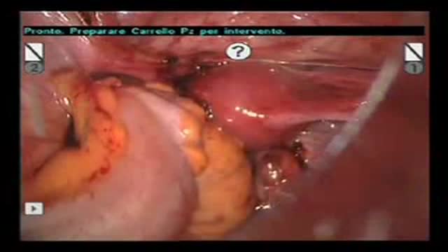

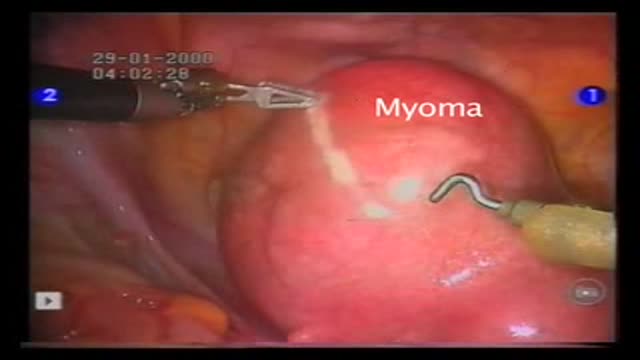
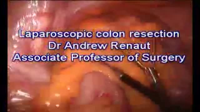
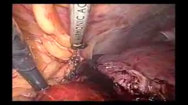




Hand Assisted Laparoscopic Sigmoid Resection and Rectopexy

Educational only: videos on MedicalVideos.com are for general information and are not medical advice. Always consult your own doctor about your health.
Hand assisted laparoscopic sigmoid resection and rectopexy for full thickness rectal prolapse.
This video in 30 seconds
A narrated hand-assisted laparoscopic sigmoid resection and rectopexy for a 49-year-old woman with full-thickness rectal prolapse. The surgeon walks through port placement, mobilization of the sigmoid and rectum, division of the inferior mesenteric artery, the stapled end-to-end anastomosis, and the rectopexy sutures, and closes with a summary of the steps and the three-week cosmetic result.
Key points
- The case is introduced as full-thickness rectal prolapse with constipation, after a negative workup for other causes
- Ports and a hand-assist incision are placed and the patient is positioned in lithotomy
- The sigmoid mesentery and rectum are mobilized, with the left ureter identified during dissection
- The inferior mesenteric artery is isolated and divided with a vascular stapler
- The redundant sigmoid is resected, a stapled end-to-end anastomosis is performed, and rectopexy sutures placed beforehand are tied
Chapters
- 0:05 — Patient case and surgical preparation
- 0:41 — Port and hand-assist placement
- 1:29 — Mobilizing sigmoid mesentery and rectum
- 2:56 — Dividing the inferior mesenteric artery
- 4:53 — Resection and preparing the anastomosis
- 6:10 — Rectopexy stitches placed before anastomosis
- 7:08 — Anastomosis performed and summary of steps
Video Transcript
A narrated hand-assisted laparoscopic sigmoid resection and rectopexy for a 49-year-old woman with full-thickness rectal prolapse. The surgeon walks through port placement, mobilization of the sigmoid and rectum, division of the inferior mesenteric artery, the stapled end-to-end anastomosis, and the rectopexy sutures, and closes with a summary of the steps and the three-week cosmetic result.
Read the full transcript
The patient is a 49-year-old female with full thickness rectal prolapse and constipation. Workup for neoplastic or metabolic ideologies, colonic inertia, and obstructive defecation was negative. Preparation for surgery included IV antibiotics and a mechanical bowel preparation. The patient is positioned in a lithotomy position with both arms tucked and secured to the table with a U-shaped beanbag and heavy tape. A foley catheter is inserted into the bladder, oral gastric tube in the stomach, and venodine boots are placed on the calves. The hip should be as horizontal as possible.
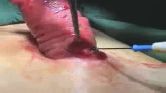
A 12-millimeter Hassan trocar is placed in the infirm bilical position, a 12-millimeter trocar in the right lower quadrant, and a 5-millimeter trocar in the left lower quadrant. An 8-centimeter phanen-stealing scission is placed two finger breaths above the pubic symphysis for the gel port or other hand assist device. The dissection is carried down to the anterior rectus sheath, which is open in a curvilinear fashion. The anterior rectus fascia is mobilized superiorly and inferiorly. The rectus muscles are split in the midline, entering the peritoneum. This picture demonstrates the placement of the ports.
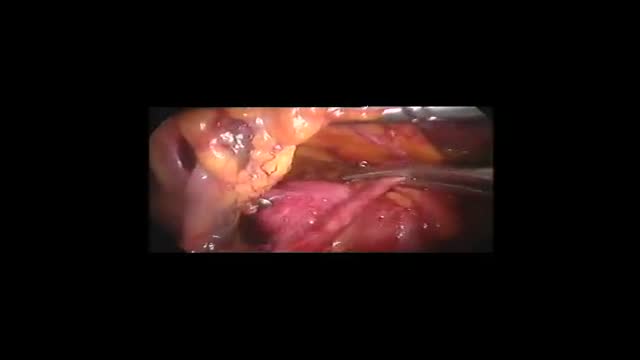
A 30-degree 10-millimeter camera is used through the umbilical port. The mesentery of the sigmoid colon and rectum are mobilized in a medial to lateral fashion. The inferior mesenteric artery is grasped and raised between the first finger and thumb. An incision is made at the base of the mesentery, extending down into the pelvis. The retroperitoneal attachments are gently brushed away. The key is upward traction on the artery. The left ureter is identified before proceeding further with the operation. The incision at the base of the mesenter is extended down to the rectum, exposing the presacral space.
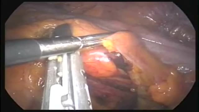
The hand provides upward traction on the sigmoid colon to aid in exposure. Using careful blunt dissection and electric artery, the rectum is mobilized posteriorly down to the tip of the coccyx. Upward traction of the hand posterior to the rectum facilitates the mobilization. The inferior mesenteric artery is isolated and the extra fat cleared using the electrocotery. The vessel is divided using an endo-GIA stapler with a vascular load. An endoloupe should be closed at hand in the case of failure of the staple line. The mesentery of the colon is elevated and the dissection is carried laterally.
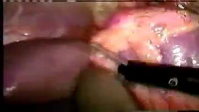
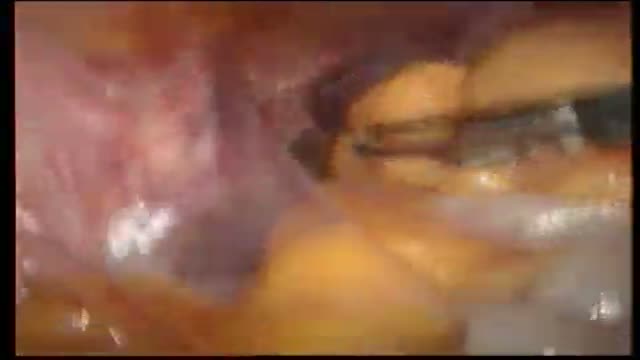
The purple retroperitoneal attachments are swept away. The dissection is carried out to the left sidewall. Next, the lateral attachments are divided. A 5mm left-sided trocar has been placed. Once these attachments are open, the previous area of mobilization is seen. The descending colon does not need to be fully mobilized since the sigmoid colon is redundant. The lateral dissection is carried down toward the rectum. Some of the upper lateral rectal attachments are divided to ensure good mobility of the rectum. The lateral rectal attachments are not fully divided. This view demonstrates the fully mobilized rectum.
The cap of the gel port is removed to perform the resection and rectopaxi. The mesentery of the upper rectum is cleared. Using a right-angle bowel clamp, the rectum is divided sharply. A purse string of 2-0 proline is placed around the rectum. The mesentery of the sigmoid colon is divided. The extent of sigmoid resection depends on the redundancy of the colon. Using a purse string device, the proximal colon is divided. An anvil for a 29mm EEA stapler is secured in the proximal colon. The stapler is passed transannually and the rectal purse string tied around the stem of the stapler.
Before the anastomosis is performed, the rectopaxi stitches are placed. Zero ethobon sutures are placed through the lateral rectal attachments and into the pre-sacral fascia. The stitches are not tied until the anastomosis is completed. A total of four stitches are used, two on either side of the rectum. If there is any bleeding from the pre-sacral area, the sutures are tied immediately. If needed, other maneuvers to control pre-sacral bleeding should be considered at this point in the operation. Next, the EEA anastomosis is performed. This is a single staple, double purse string, end to end colorectal anastomosis.
Finally, the rectopaxi sutures are tied. This shows the excellent cosmetic result afforded by this operation three weeks after the procedure. In summary, this procedure utilizes the following steps. Medial to lateral mobilization of the sigmoid mesentery. Post-year mobilization of the rectum. Bascular division. Lateral mobilization of the sigmoid and upper rectum. Resection of the redundant sigmoid colon and suture rectopaxi.
Transcript auto-generated from the video’s audio for accessibility and study. Educational content only — not medical advice.
Video Overview
From our Surgery Videos collection: Surgery videos: watch real operations step by step — general, laparoscopic and specialist surgery performed in the OR, explained for students and surgeons. Browse all Surgery Videos videos →
SORT BY-
Top Comments
-
Latest comments