
- Procedures & Tests
- Symptoms A-Z
- Symptom Checker
- Kidney Diet
- First Aid
- Anatomy Atlas
- Ask MedicalVideos
- Drug Guide
- Lab Values
- Vital Signs
- Muscles A–Z
- Clinical Signs
- Medical Abbreviations
- Medical Terminology
- Physical Examination
- Surgical Examination
- Ophthalmology
- Clinical Skills
- Orthopedics
- Surgery Videos
- Laparoscopy
- Pediatrics
- Funny Videos
- Cardiothoracic Surgery
- Nursing Videos
- Plastic Surgery
- Otorhinolaryngology
- Histology and Histopathology
- Neurosurgery
- Dermatology
- Pediatric Surgery
- Urology
- Dentistry
- Oncology and Cancers
- Anatomy Videos
- Health and Fitness
- Radiology
- Anaesthesia
- Physical Therapy
- Pharmacology
- Interventional Radiology
- Cardiology
- Endocrinology
- Gynecology
- Emergency Medicine
- Psychiatry and Psychology
- Childbirth Videos
- General Medical Videos
- Nephrology
- Physiology
- Diet and Food Health
- Diabetes Mellitus
- Neurology
- Women Health
- Osteoporosis
- Gastroenterology
- Pulmonology
- Hematology
- Rheumatology
- Toxicology
- Nuclear Medicine
- Infectious Diseases
- Vascular Disease
- Reproductive Health
- Burns and Wound Healing
- Kidney Zone Podcast
- Other
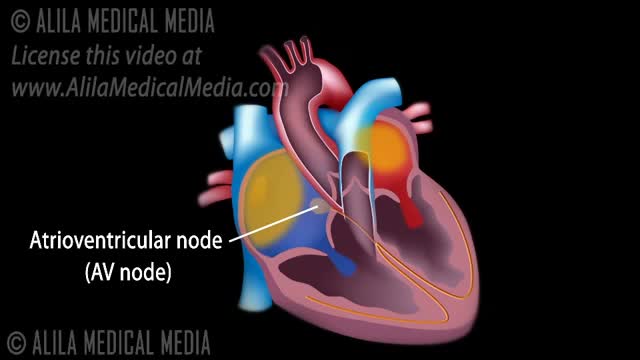
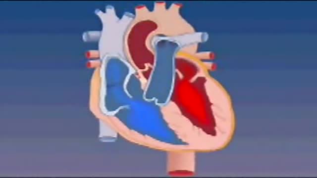

heart dissection

Educational only: videos on MedicalVideos.com are for general information and are not medical advice. Always consult your own doctor about your health.
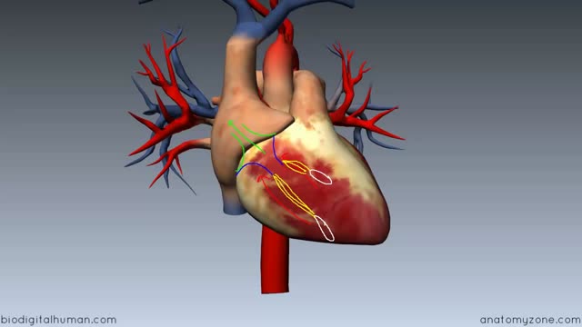
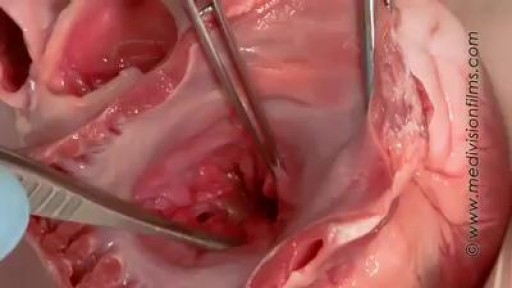
A teaching dissection explores the anatomy of a sheep's heart. The narrator identifies the ventricles and a coronary artery on the surface, then probes the great vessels to show which chambers they connect. The heart used is an animal heart, presented as matching human anatomy, in Anaesthesia teaching.
Video Transcript
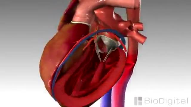
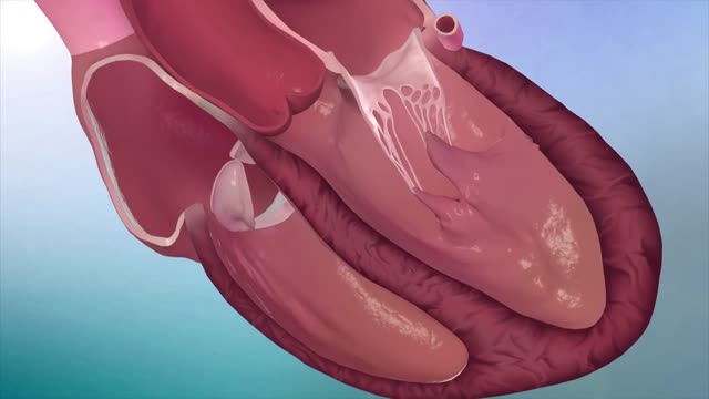
This teaching video presents a dissection of a sheep's heart, explained as closely matching a human heart. The narrator identifies the right and left ventricles and a coronary artery on the surface, then probes the great vessels to show which chambers they connect, noting wall thickness as the dissection proceeds.
Read the full transcript
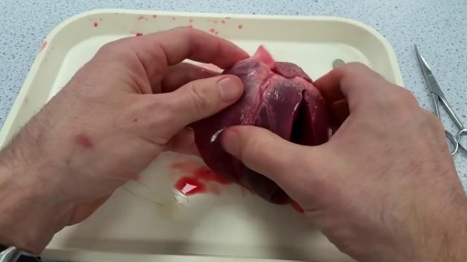
Today we're going to do a heart dissection. This was not a human heart, but a sheep's heart. But it looks and works exactly like a human heart as well. It's just a little smaller. So if you want you can think of this heart like the heart of a child. First of all, let's take a look at the outside. You can see there's some hematoma, some bleeding in here. The heart has obviously received some trauma during or after slaughtering. But that's not going to be an issue for our dissection.
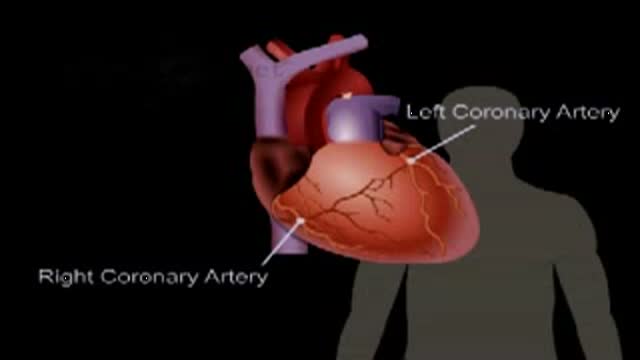
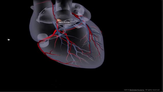
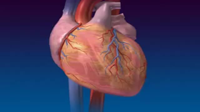
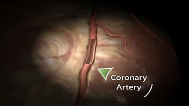
So let's look at the anatomy first. You will find that one bit of the heart is really tender to pressure. Well, the other side is really hard and cannot be pushed in easily. This here is the right ventricle and this is the left ventricle. What you can see here, as lined with fat, is a coronary artery. So that is the blood supply for the heart itself. As you can see, we have a lot of openings within the heart. So let's first of all see what I will need.
Let's start off with this big one here. I will insert a probe in that. I am pushing into the right ventricle. This tells me that I must be in the pulmonary trunk over here. I'm going into the right ventricle. Let's choose another opening like this one over here. You will see that those two are connected. You can see it's connected. All those three parts are connected. So let's see where that one goes. As you can see, you can probably see that the wall is pretty thick. But I am poking into the left ventricle here.
So that is actually the aorta that I am in. And it has different branches. The aortic arch would go down there and supply the body and the organs with blood. The other one would go up to the brain. And the major one on the side would also supply the arms. Let's take a look at what this here is. You can see I am pushing through it. I am obviously inside the atrium here. I am pushing through into also the right ventricle. So this one here must be the vena cava.
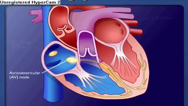
I am going to a heart's atrium and into the ventricle. There must be one on the other side. So let's see. Here I am in the left atrium. I am pushing down into the left ventricle from there. And that's how it would work. The blood would first flow into the atrium with those things here. And then they contract. And then they are pushed into the ventricles. And then the ventricles would contract like that. And push the blood out through the aorta or pulmonary trunk here. That's how it works.
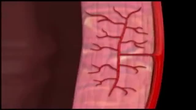
First the aorta would also supply the coronary arteries. If you are over here, see this one very nicely. I will try. There are actually multiple openings as you can see. This is the aorta as I said. The heart supplies itself with blood through the coronary arteries. Because it obviously needs a lot of oxygen to work. It's a very strong muscle. So let's see. We can access that coronary artery. And we are in. Going somewhere. And you can see we are coming out right here in the left ventricle.
We are obviously inside a branch of the coronary artery. That supplies this bit of the left ventricle. Let's see where else we can go. Trying it from this side again. Just a little cup to cheat. Now we opened the coronary artery. I am trying to straighten my catheter. See you in the catheter again. And you can see. I am within the very thin walls of the left coronary artery. You can see it bends down there. A real catheter would of course follow this bend. But I am probably just going to do damage and pinch the walls.
So it can't really go further. But yeah, it gives you an idea. So let's try and open up the right ventricle. The right atrium. By making a decision starting from the vena cava. And there you can see. They opened the atrium over here. And the valve that enters the right ventricle. And they opened the right ventricle over here. That's a little blood clot inside. So let's try and open up the left ventricle in the same way. And as you can see the walls are really thick. Not as easily to be seen on first glimpse.
They are really thick walls on this one. Versus the really thin wall on the right ventricle. That's the easiest way to tell apart the left and right side of the artery. Because you can see the thin wall is really thick wall. You can see this is the valve. This is one part of it. And the other part of it. And you can imagine when it was shut like that. And the blood would flow through, fill the heart. And as the heart beats, contracts, this valve would shut.
And close the ventricle, the left ventricle. Close it off from the left atrium. So the only way for the blood to be ejected is through the aorta. Let's see where that is again. You can see this on the back here. Behind the mitral valve is where the aorta goes off. Let's see if we can open that one up. So the blood goes into the atrium. And there it goes into the left ventricle. The one with the thick walls. And then it goes out that way into the aorta and into the body.
And for the other side of the heart, you'll remember we had an entrance here as well. You can see this over here. And there we're in the pulmonary trunk. And entering the right ventricle. I'll try and cut down on this as well. And there you can even see the valve here. You can see it's a very tiny thin valve that is right on my palm over here. So the blood would on this side go into the right atrium, into the right ventricle. And as the ventricles contract, the valve here closes and shuts off the atrium.
So the only way for the blood to eject to leave the right ventricle is through the pulmonary valve. And pulmonary trunk over here. So again, those is what the colostar would look like sort of.
Transcript auto-generated from the video’s audio for accessibility and study. Educational content only — not medical advice.
Related reading
Medical Videos
Free medical and surgery videos for patients, students and professionals — trusted since 2007.
Founded by Dr. Mohamed Ibrahim, MD, PhD — physician-scientist.
Follow us on Facebook · Instagram
SORT BY-
Top Comments
-
Latest comments