
- Procedures & Tests
- Symptoms A-Z
- Symptom Checker
- Kidney Diet
- First Aid
- Anatomy Atlas
- Ask MedicalVideos
- Drug Guide
- Lab Values
- Vital Signs
- Muscles A–Z
- Clinical Signs
- Medical Abbreviations
- Medical Terminology
- Physical Examination
- Surgical Examination
- Ophthalmology
- Clinical Skills
- Orthopedics
- Surgery Videos
- Laparoscopy
- Pediatrics
- Funny Videos
- Cardiothoracic Surgery
- Nursing Videos
- Plastic Surgery
- Otorhinolaryngology
- Histology and Histopathology
- Neurosurgery
- Dermatology
- Pediatric Surgery
- Urology
- Dentistry
- Oncology and Cancers
- Anatomy Videos
- Health and Fitness
- Radiology
- Anaesthesia
- Physical Therapy
- Pharmacology
- Interventional Radiology
- Cardiology
- Endocrinology
- Gynecology
- Emergency Medicine
- Psychiatry and Psychology
- Childbirth Videos
- General Medical Videos
- Nephrology
- Physiology
- Diet and Food Health
- Diabetes Mellitus
- Neurology
- Women Health
- Osteoporosis
- Gastroenterology
- Pulmonology
- Hematology
- Rheumatology
- Toxicology
- Nuclear Medicine
- Infectious Diseases
- Vascular Disease
- Reproductive Health
- Burns and Wound Healing
- Kidney Zone Podcast
- Other

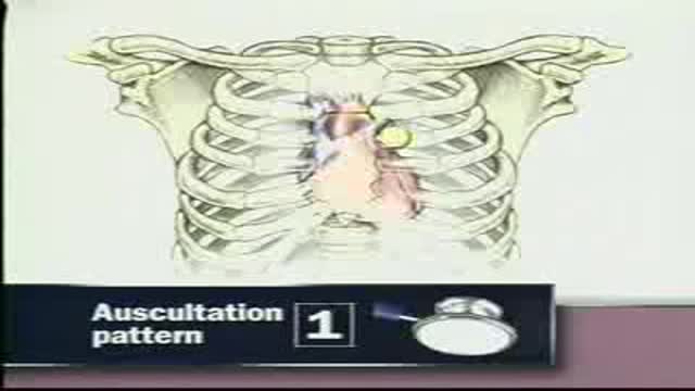
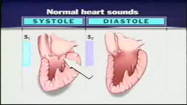
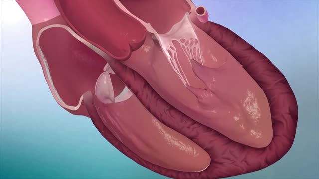

The Best Heart Animation

Educational only: videos on MedicalVideos.com are for general information and are not medical advice. Always consult your own doctor about your health.
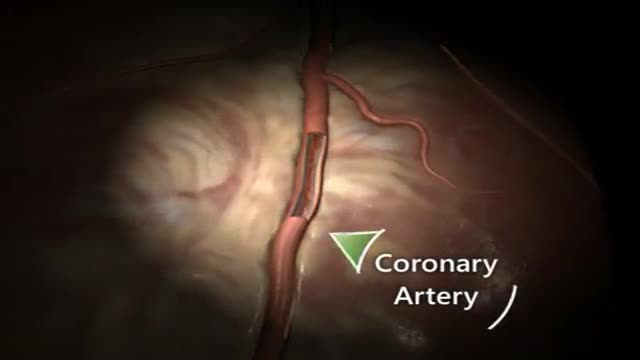
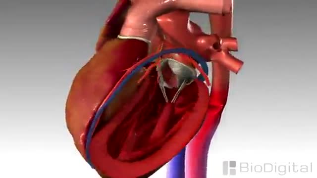
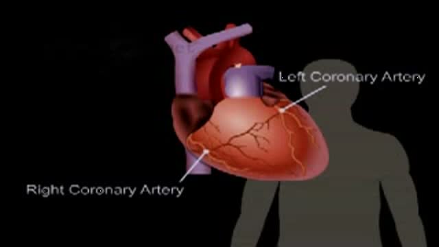
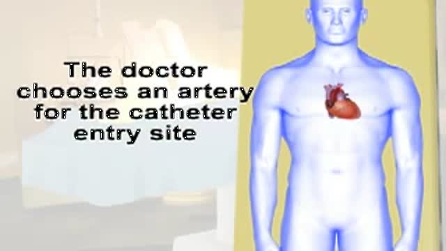
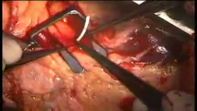
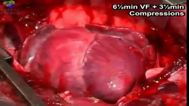
An animation of the heart is presented in this short cardiovascular video. The animated sequence shows the heart's structure and movement as it beats. It is a basic educational visual for students and viewers interested in how the heart works. It is part of the site's library of recorded clinical and educational material.
This video in 30 seconds
A narrated physiology animation of the cardiac cycle. It explains the heart as two pumps divided by a septum, pacemaker activity starting in the SA node, conduction through the AV node, bundle of His and Purkinje fibres, and closes by mapping the P wave, QRS complex and T wave of the ECG onto the electrical and mechanical events they represent.
Key points
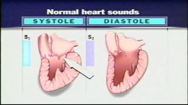
- The heart is described as two pumps whose chambers contract in systole and fill in diastole
- Pacemaker cells in the SA node are shown initiating the impulse
- The AV node delay allows the atria to empty before ventricular contraction
- Impulses spread rapidly through the bundle and Purkinje fibres so ventricles contract as a unit
- The P wave, QRS complex and T wave are matched to depolarisation and repolarisation events
Chapters
- 0:01 — Heart as two coordinated pumps
- 0:57 — SA node pacemaker activity begins
- 1:32 — AV node and ventricular conduction
- 2:25 — ECG waves and cardiac cycle
Video Transcript
A narrated physiology animation of the cardiac cycle. It explains the heart as two pumps divided by a septum, the pacemaker activity of the SA node, conduction through the AV node, the bundle of His and the Purkinje fibers, and how the chambers are excited and contract as units. It closes by mapping the ECG — the P wave, the QRS complex and the T wave — onto the electrical and mechanical events they represent.
Read the full transcript
The heart generates pressure gradients that drive blood flow through the vascular system. Although the heart appears to be a single structure, it is divided by a central septum into two separate pumps, each consisting of two chambers, the atrium and the ventricle, that alternately contract and empty during systole, then relax and fill during diastole. The timing of the electrical and mechanical events in these chambers must be precisely controlled to ensure efficient cardiac function. The timing of atrial and ventricular contractions relies on two primary cell types, cardiac myocytes, which do the mechanical pumping, and auto-rhythmic cells, which initiate and conduct electrical impulses that drive contractile activity.
Auto-rhythmic cells cyclically depolarize on their own, then fire an action potential each time they reach threshold, a process known as pacemaker activity. This activity originates from primary pacemaker cells in the sinoatrial or SA node in the upper right atrial wall. The pacemaker potentials swiftly move through the interatrial pathway to the left atrium and spread throughout the atrial myocardium via gap junctions. As a result, both atria depolarize and contract as a single unit. The impulses generated by the SA node also rapidly spread through the inter-nodal pathway to the atrial ventricular or AV node at the base of the right atrium.
The AV node is the only point of electrical contact between the atria and ventricles. Here, the impulse is briefly slow, allowing the atria to contract and empty their contents into the ventricle before ventricular depolarization and contraction occur. The impulse travels quickly through the bundle of hits within the interventricular septum. The bundle branches into smaller pockinji fibers that radiate throughout the ventricular myocardium. Action potentials travel quickly through the pockinji fibers, then spread to the surrounding ventricular myocytes via gap junctions. Depolarization triggers contraction. Repolarization triggers relaxation. The swift spread of the impulse through the specialized conduction system ensures that the ventricles become excited and contract as a unit.
The spread of cardiac electrical activity may be recorded using electrodes placed on the body's surface. The recording, called the electrocardiogram, or ECG, has several distinct components. The P-Wave corresponds to atrial depolarization. The QRS complex appears with ventricular depolarization. The T-Wave represents ventricular repolarization. The P-Wave initiates atrial contraction, which completes ventricular filling. The QRS complex initiates ventricular contraction, which pumps blood out of the heart. During this time, the atria repolarized and relaxed. The T-Wave initiates ventricular relaxation, during which the ventricles fill once again. One cardiac cycle is complete.
Transcript auto-generated from the video’s audio for accessibility and study. Educational content only — not medical advice.
Medical Videos
Free medical and surgery videos for patients, students and professionals — trusted since 2007.
Founded by Dr. Mohamed Ibrahim, MD, PhD — physician-scientist.
Follow us on Facebook · Instagram
SORT BY-
Top Comments
-
Latest comments