
- Physical Examination
- Surgical Examination
- Ophthalmology
- Clinical Skills
- Orthopedics
- Surgery Videos
- Laparoscopy
- Pediatrics
- Funny Videos
- Cardiothoracic Surgery
- Nursing Videos
- Plastic Surgery
- Otorhinolaryngology
- Histology and Histopathology
- Neurosurgery
- Dermatology
- Pediatric Surgery
- Urology
- Dentistry
- Oncology and Cancers
- Anatomy Videos
- Health and Fitness
- Radiology
- Anaesthesia
- Physical Therapy
- Pharmacology
- Interventional Radiology
- Cardiology
- Endocrinology
- Gynecology
- Emergency Medicine
- Psychiatry and Psychology
- Childbirth Videos
- General Medical Videos
- Nephrology
- Physiology
- Diet and Food Health
- Diabetes Mellitus
- Neurology
- Women Health
- Osteoporosis
- Gastroenterology
- Pulmonology
- Hematology
- Rheumatology
- Toxicology
- Nuclear Medicine
- Infectious Diseases
- Vascular Disease
- Reproductive Health
- Burns and Wound Healing
- Kidney Zone Podcast
- Other
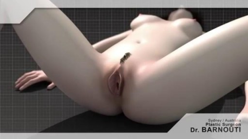









Labiaplasty: Understanding the Anatomy

Educational only: videos on MedicalVideos.com are for general information and are not medical advice. Always consult your own doctor about your health.
Otto Placik MD. a board certified Chicago based plastic surgeon presents Vulvar Vaginal Genital anatomy lesson reviewing the Vulva, Mons Pubis, clitoral hood, prepuce, frenulum, labia minora & majora, vagina, urethra and fourchette with surgical implications and techniques. Photos pictures and video of anatomic models are reviewed in detail on different models. Great for patients thinking about or planning before labiaplasty or vaginal cosmetic surgery
This video in 30 seconds
A plastic surgeon uses anatomical models to explain vulvar anatomy for patients considering labiaplasty. The lesson identifies the mons pubis, clitoral hood, clitoris, frenulum, labia minora and majora, vestibule, urethra, vaginal opening, and associated glands, outlines in general terms how labia and clitoral hood reductions are approached, and shows the same structures in cross-section.
Key points
- External structures are identified on an anatomical model
- The vestibule, urethra, and vaginal opening are located
- Associated glands of the vulvar area are mentioned
- Labia and clitoral hood reduction approaches are outlined generally
- A cross-section model shows the internal arrangement of structures
Chapters
- 0:01 — Purpose of the anatomy lesson
- 0:48 — Mons pubis and clitoral hood
- 1:48 — Vestibule urethra and vaginal opening
- 3:22 — Reduction approaches outlined generally
- 4:34 — Cross-section model of anatomy
Video Transcript
A plastic surgeon uses anatomical models to explain vulvar anatomy for patients considering labiaplasty. The lesson identifies the mons pubis, clitoral hood, clitoris, frenulum, labia minora and majora, vestibule, urethra, vaginal opening and hymen, and mentions associated glands. It then outlines, in general terms, how labia and clitoral hood reductions are approached and shows the same structures in cross-section.
Read the full transcript
When I perform a labiaplasty consultation, one of the most difficult parts of the examination is having the patient relate their desires and goals for the surgery. This is most likely due to the fact that they have a difficult time understanding the anatomy and the last time they learned it was in junior high school. So to that end, I prepared this video so that you can have a better understanding of the relevant female genital anatomy. This is a useful model which was initially designed to help people understand the anatomy
for vulvar piercings, but we'll use it for this anatomical discussion. Right up here would be located the Mons pubis, also known as the Mons venaris or the mound of venus. We as plastic surgeons sometimes perform liposuction in this area if it is too full. This location right here is called the clitoral hood. It has the excessive skin in this location and we can perform a clitoral hood reduction. When it's completely removed it is called an unroofing. Just below this the clitoral hood conceals the clitoris which is marked here with a green
sticker. Beneath the clitoris is the frenulum of the clitoris. Now the frenulum of the clitoris is in direct continuity with the labia minora or the inner lips. The inner lips are here joining together at the bottom at what is called the posterior entroitus, the posterior foreshet or the posterior commissure. The labia minora encircle what is called the vaginal or vulvar vestibule. Within the vestibule is the clitoris and the urethra. The urethra is where urine comes from and it's located here and identified with the yellow sticker. Just beneath this is the vaginal orifice or the opening also called the entroitus.
Just into the entrance to the vagina is where the hymen is located and it's where the vestibule becomes the vagina that we identify the hymen. There are a couple of glands that occasionally can become diseased and one is called the bartholin gland or the vestibular gland and they're located just beneath the vaginal opening and these can occasionally get infected or form cysts. At the level of the urethra or called periurethral glands are also skeins glands. Some people have identified these as being responsible for the G spot.
At this location is where apesiotomies are formed. Sometimes an apesiotomy is performed off the midline on an angle here to avoid damaging the commissure and occasionally it happens spontaneously with birth and it's torn and this is what we call a perinealplasty when that is repaired. Typically a labia minora reduction or a labiaplasty involves reduction of the labia minora in this location. Labia majora which are located here and typically have hair can be either enlarged when they're small utilizing lipo injection or fat injections or can be reduced.
We tend to like to conceal the incisions on the labia majora reduction by concealing them along this location. Labia minora reductions can be done by removing skin along this edge in this direction or in a wedge like section. It's important to have a very good understanding of the labia minora because in essence it sometimes has this tripod configuration with one limb extending down here, one limb extending towards the frenulum of the clitoris and another one contributing to the clitoral hood. So it is not uncommon to perform a labia minora reduction in combination with the
clitoral hood reduction which again is to be distinguished from a clitoral unroofing. This is a three dimensional model of the female anatomy which provides a greater understanding through showing a cross section of the relevant anatomy. This is not exactly identical to the human but provides a good reference source. This is the vulva with the labia majora located right here. This would be the location of the clitoris which is not clearly identified but can be better seen on the cross section. These would be the labia minora, the vaginal vestibule or vulvar vestibule with the vagina
located here and the urethra should be located here. I'm going to separate the two halves of this model to better show you on cross section. In this model the green is again located over the clitoris which is protected here by what would seem to be the clitoral hood and this is the body of the clitoris right here. At this point where the yellow sticker is located is the opening to the urethra which travels back to the bladder in this location. This is the pubic bone located here in this gray color and the bladder is located
just behind that. Just behind the urethra is the vaginal opening and where the vestibule turns into the vaginal canal we identify the hymen and that is where a hymenorophy or a hymenoplasty can be performed. Back here is the rectum. This would be the posterior entroitus or the posterior opening to the vaginal canal which is located here and again is occasionally torn in a pisiotomy. Sometimes in a very severe vaginal trauma during pregnancy the muscles can be torn and these need to be repaired but involve a much more complex procedure.
This may not have answered all your questions but I hope you found it informative. Thank you for tuning in.
Transcript auto-generated from the video’s audio for accessibility and study. Educational content only — not medical advice.
Video Overview
From our Gynecology collection: Pelvic exams, Pap smears, IUD insertion, hysteroscopy and women's health procedures explained step by step. Browse all Gynecology videos →
Medical Videos
Free medical and surgery videos for patients, students and professionals — trusted since 2007.
Founded by Dr. Mohamed Ibrahim, MD, PhD — physician-scientist.
Follow us on Facebook · Instagram
SORT BY-
Top Comments
-
Latest comments