- Physical Examination
- Surgical Examination
- Ophthalmology
- Clinical Skills
- Orthopedics
- Surgery Videos
- Laparoscopy
- Pediatrics
- Funny Videos
- Cardiothoracic Surgery
- Nursing Videos
- Plastic Surgery
- Otorhinolaryngology
- Histology and Histopathology
- Neurosurgery
- Dermatology
- Pediatric Surgery
- Urology
- Dentistry
- Oncology and Cancers
- Anatomy Videos
- Health and Fitness
- Radiology
- Anaesthesia
- Physical Therapy
- Pharmacology
- Interventional Radiology
- Cardiology
- Endocrinology
- Gynecology
- Emergency Medicine
- Psychiatry and Psychology
- Childbirth Videos
- General Medical Videos
- Nephrology
- Physiology
- Diet and Food Health
- Diabetes Mellitus
- Neurology
- Women Health
- Osteoporosis
- Gastroenterology
- Pulmonology
- Hematology
- Rheumatology
- Toxicology
- Nuclear Medicine
- Infectious Diseases
- Vascular Disease
- Reproductive Health
- Burns and Wound Healing
- Other
Latest videos
A video showing how to catheter the male urethra
A video from the New England Journal of Medicine performed by Harvard medical school showing Thoracocentesis
A video from the New England Journal of Medicine performed by Harvard Medical School showing basic lacerations repair

operation on the stomach
a complete discription of the instruments used in laparacopic surgeries and there function
A video from Harvard medical school showing Paracentesis
The video shows how to perform the orotracheal intubation.Performed by harvard medical school
Hip examination by Harvard medical school

How to give a gluteal intra-muscular injection
Positive Pressure Ventilation with a face mask and a bag-valve device
Laparoscopic-assister percutaneous vaginal tape vault suspension, a minimally invasive prolapse repair with post-hysterectomy and uterine-sparing options
This video shows how to insert a chest tube
A video showing abscess incision and drainage
Central Venous Line Placement in the subclavian vein
Examination of the shoulder
Examination of the Shoulder and Elbow
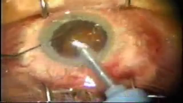
Cataract eye surgery
Examination of different gaits
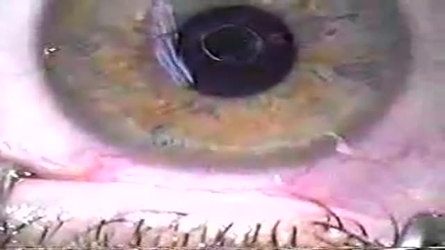
A very bad lasik eye surgery duringwhich the surgeon messed everything
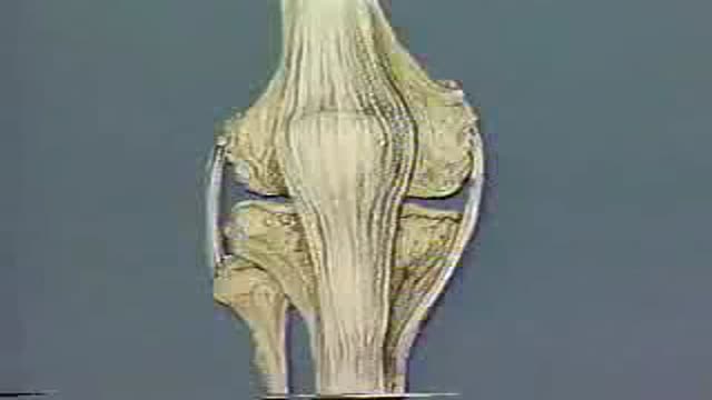
Full clinical and physical assessment of the knee and the knee joint
